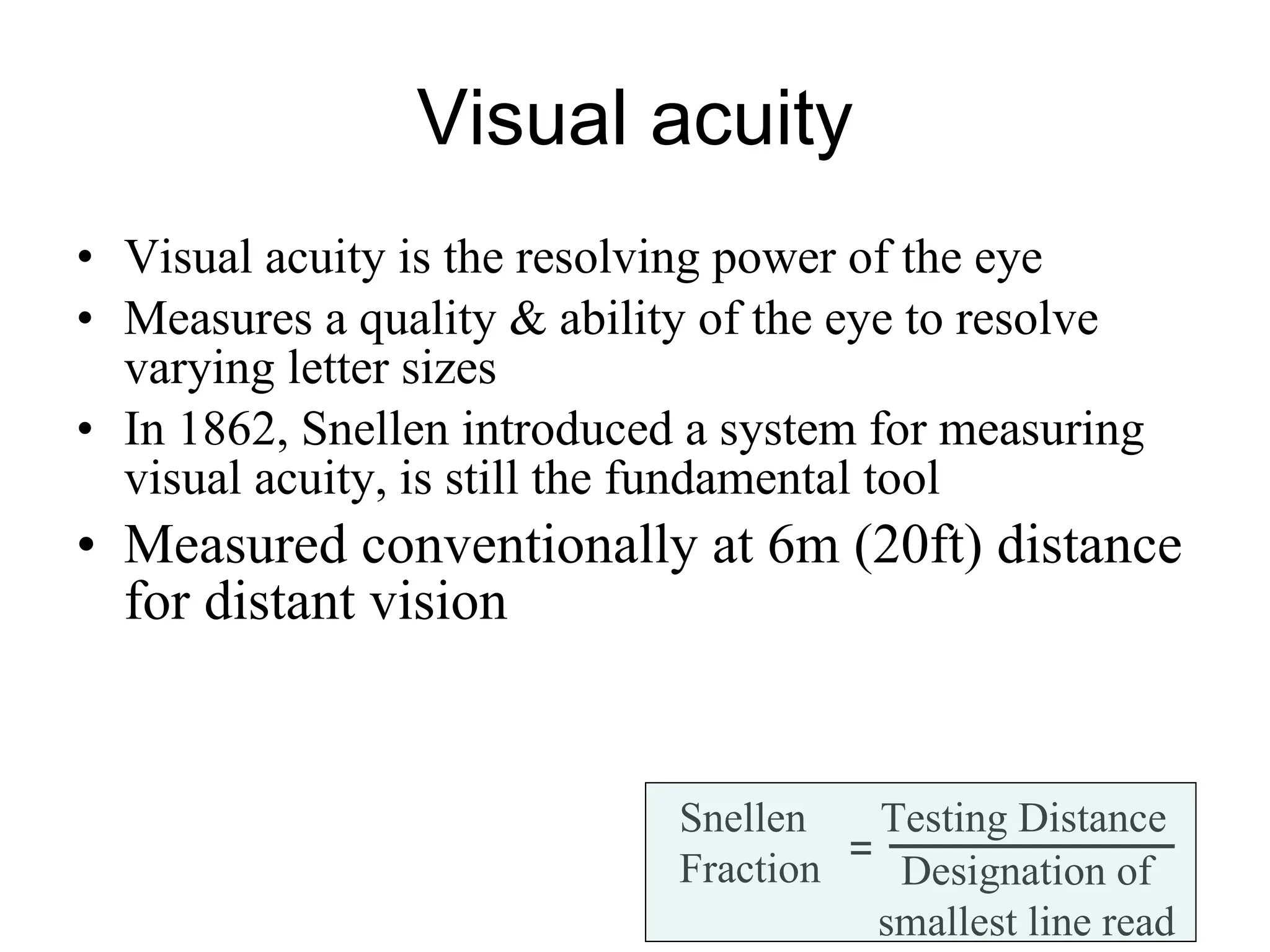
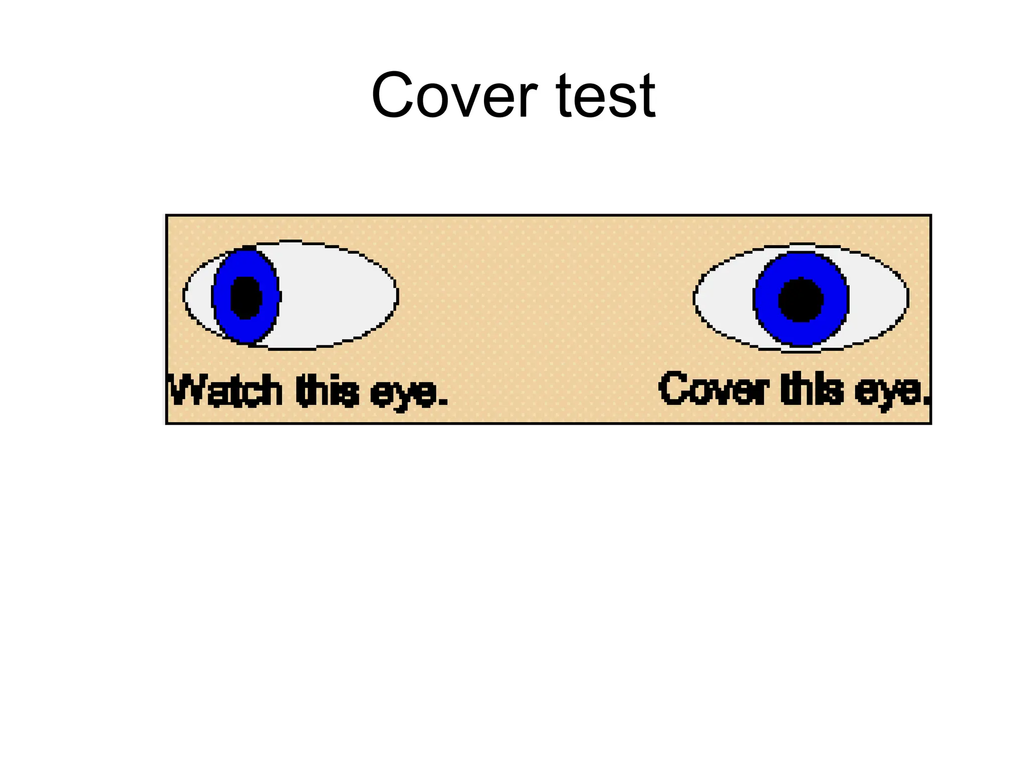
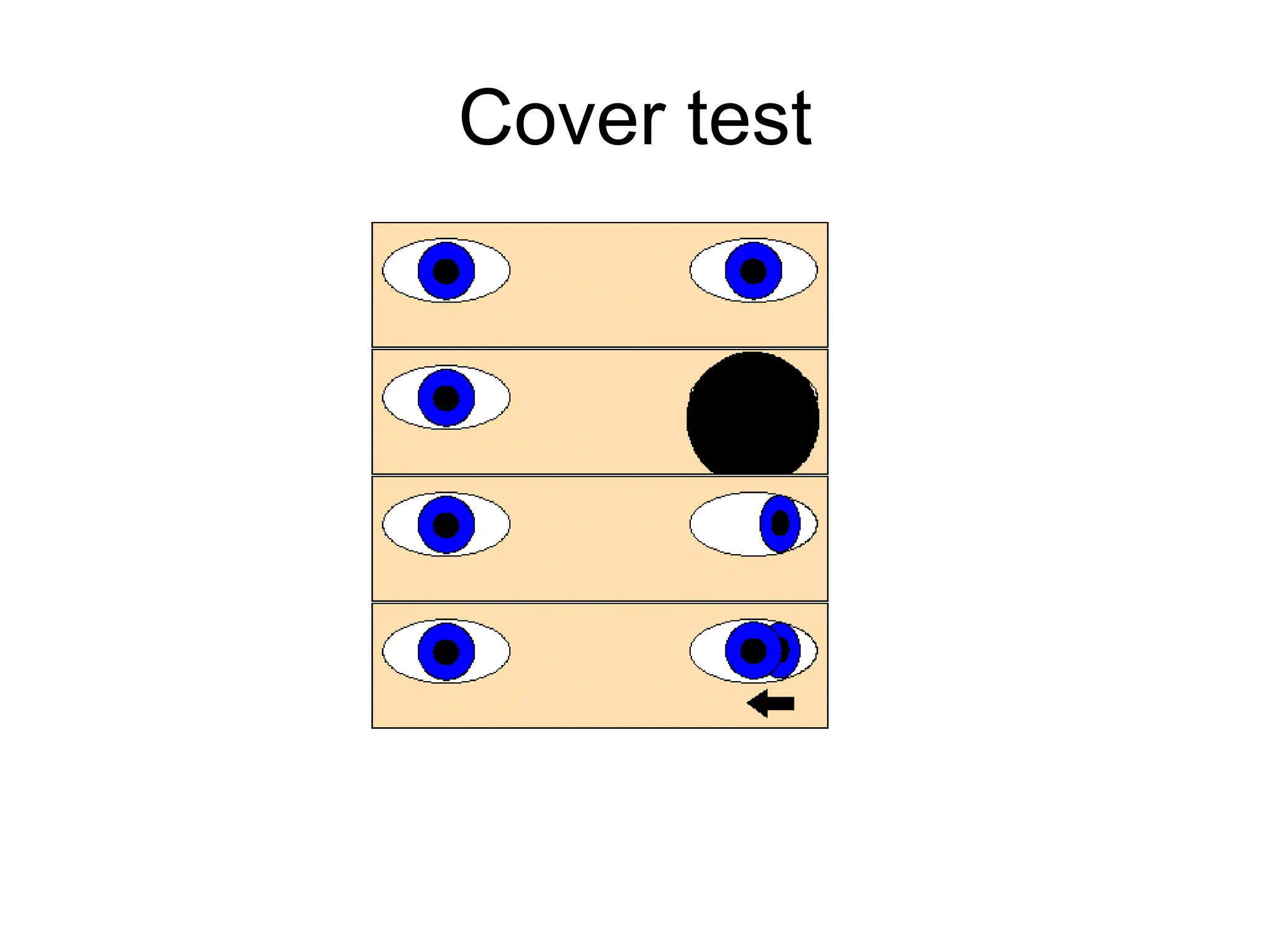
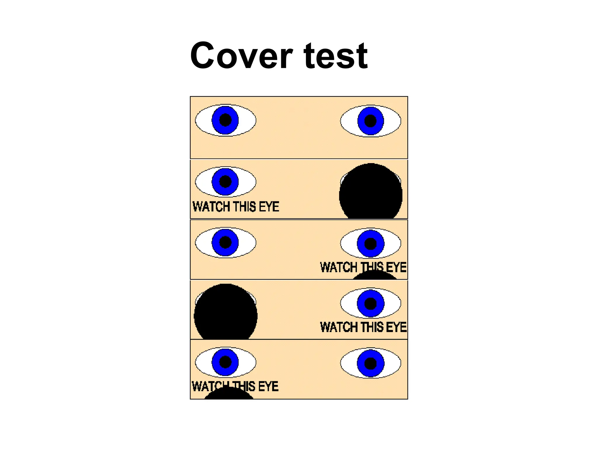
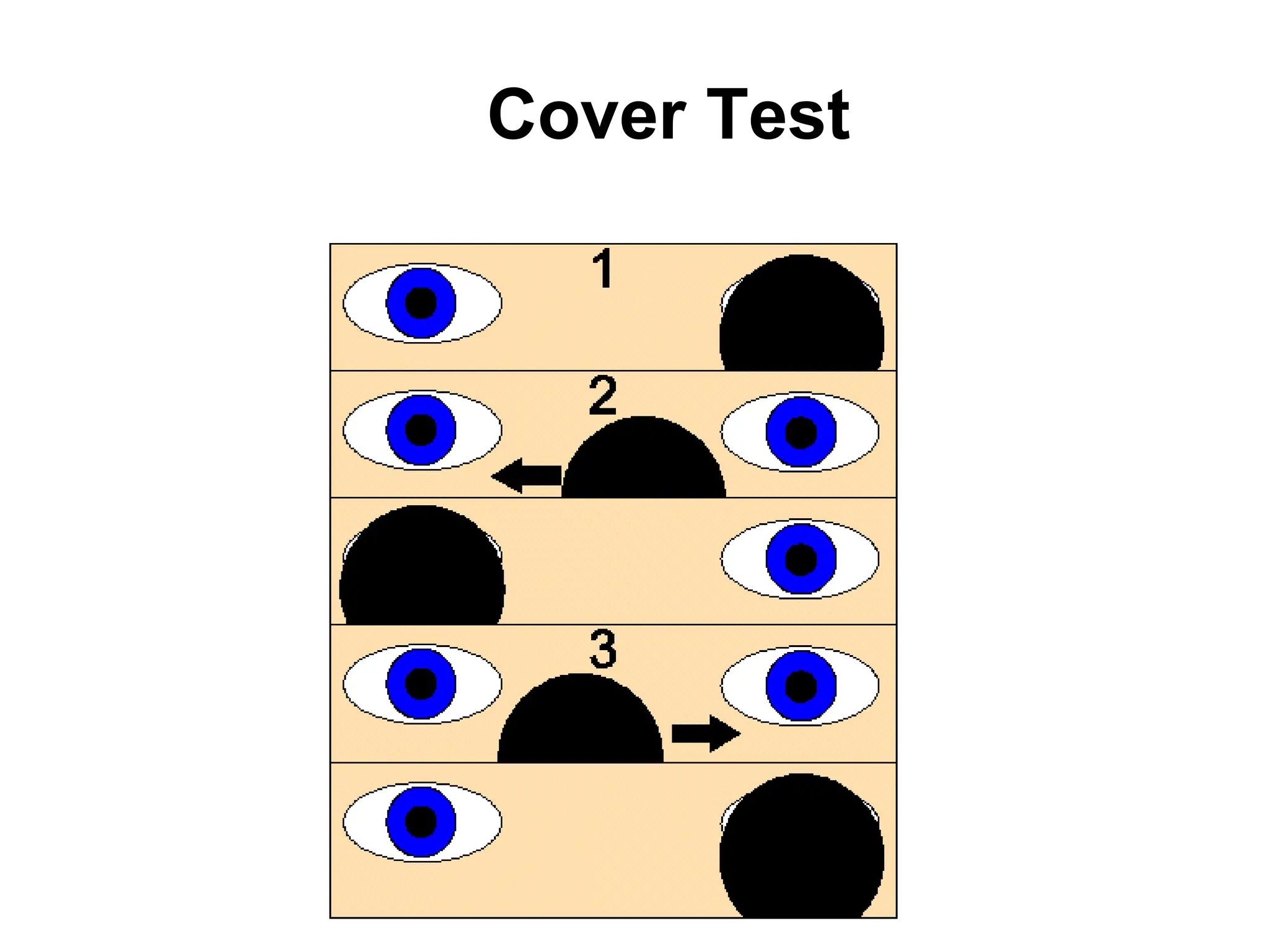
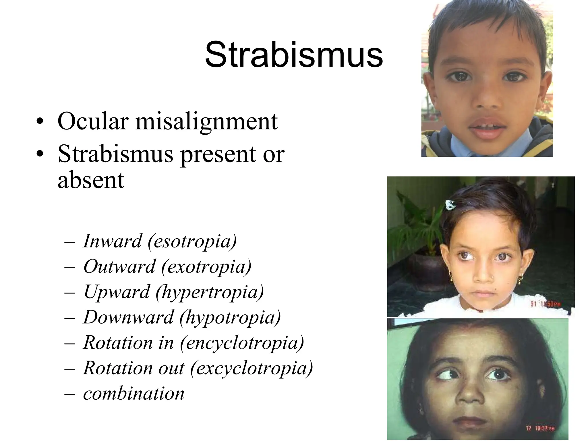
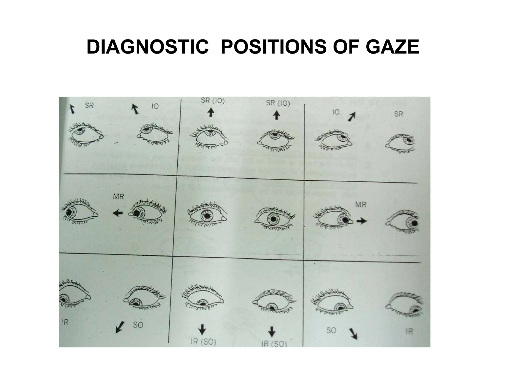
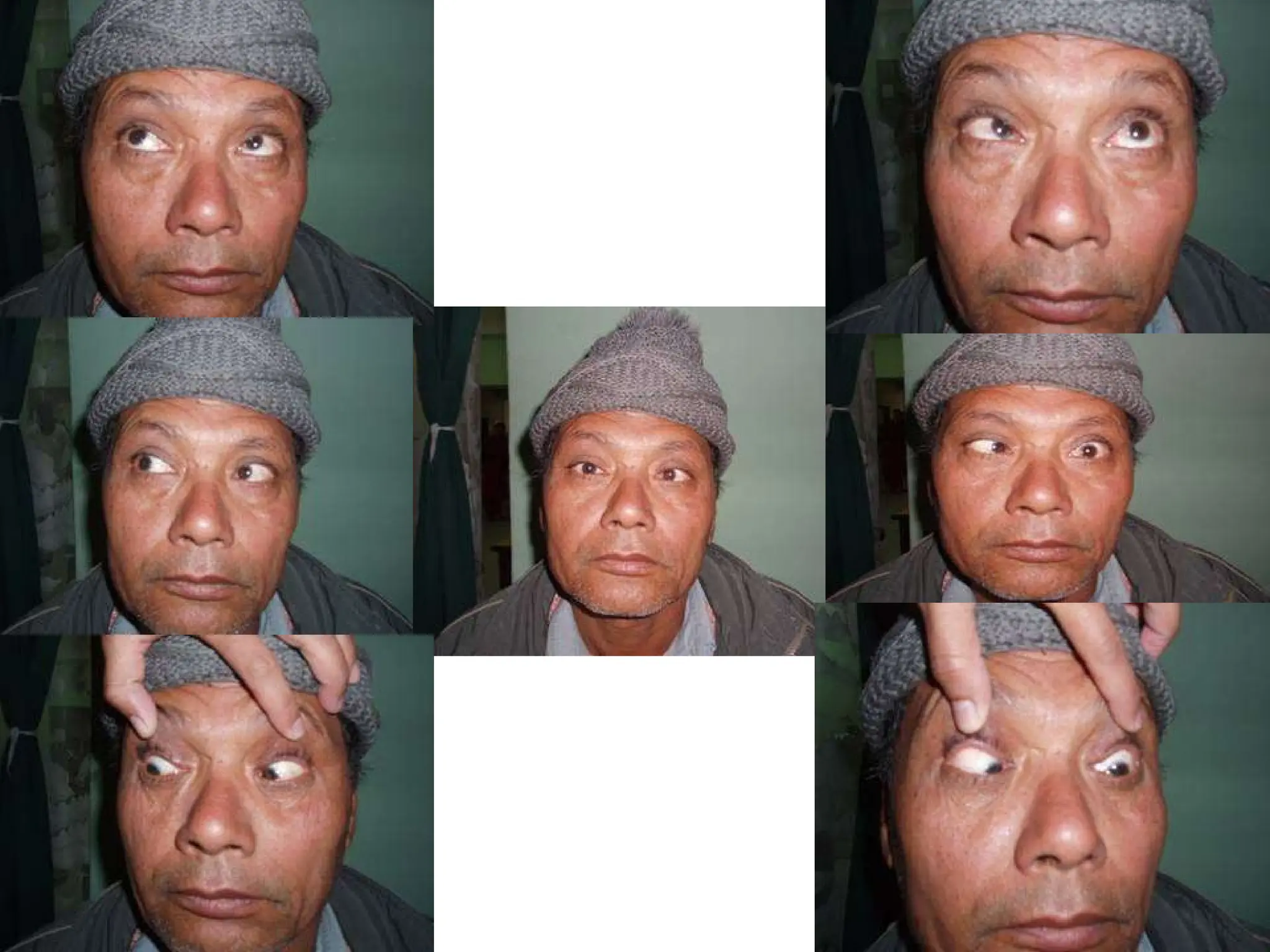
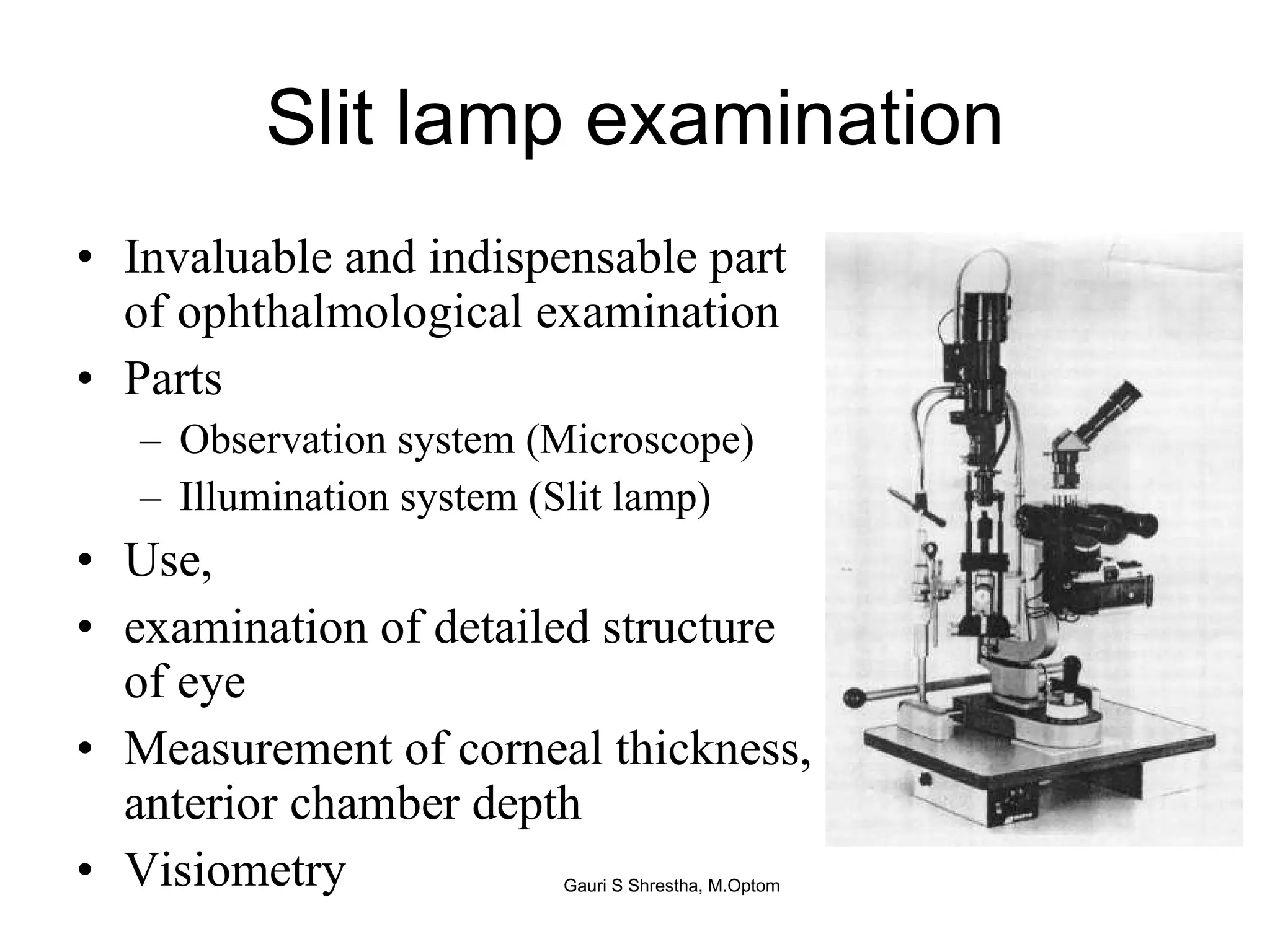
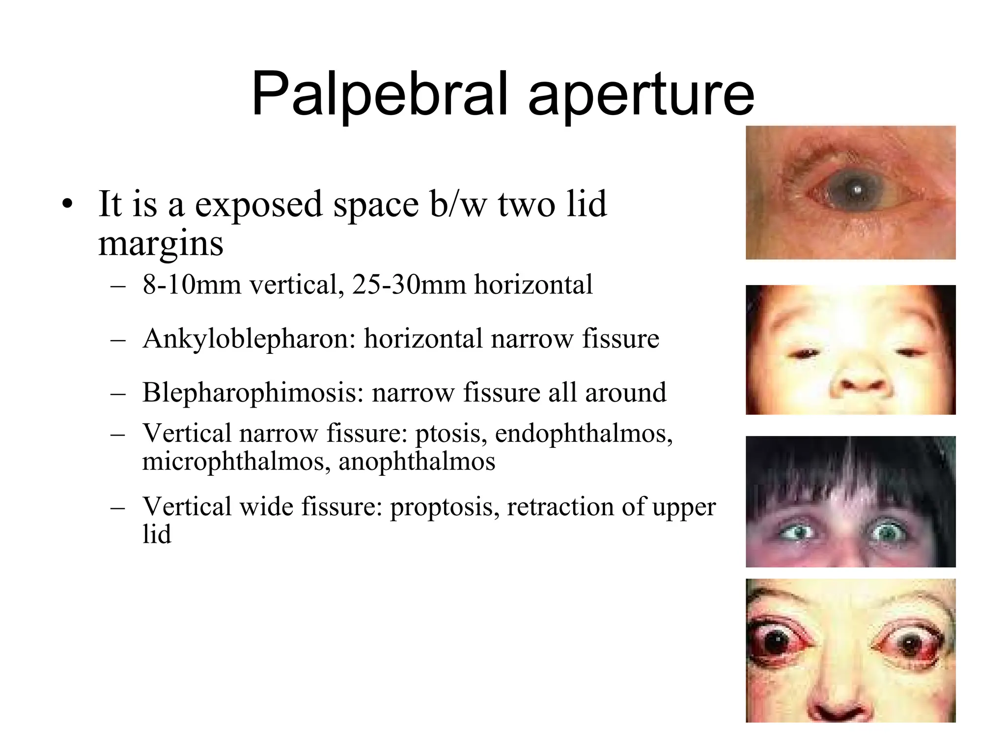
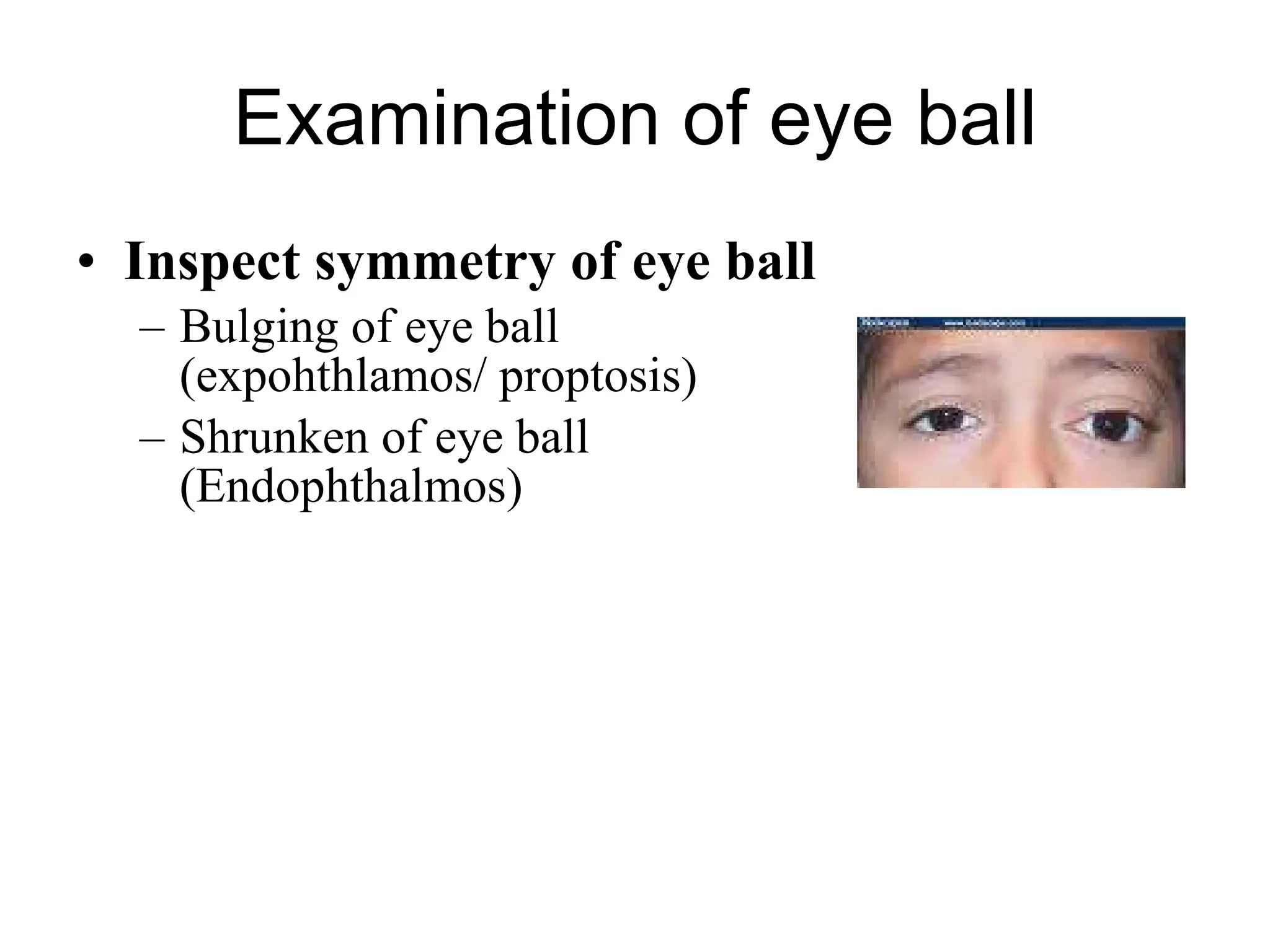
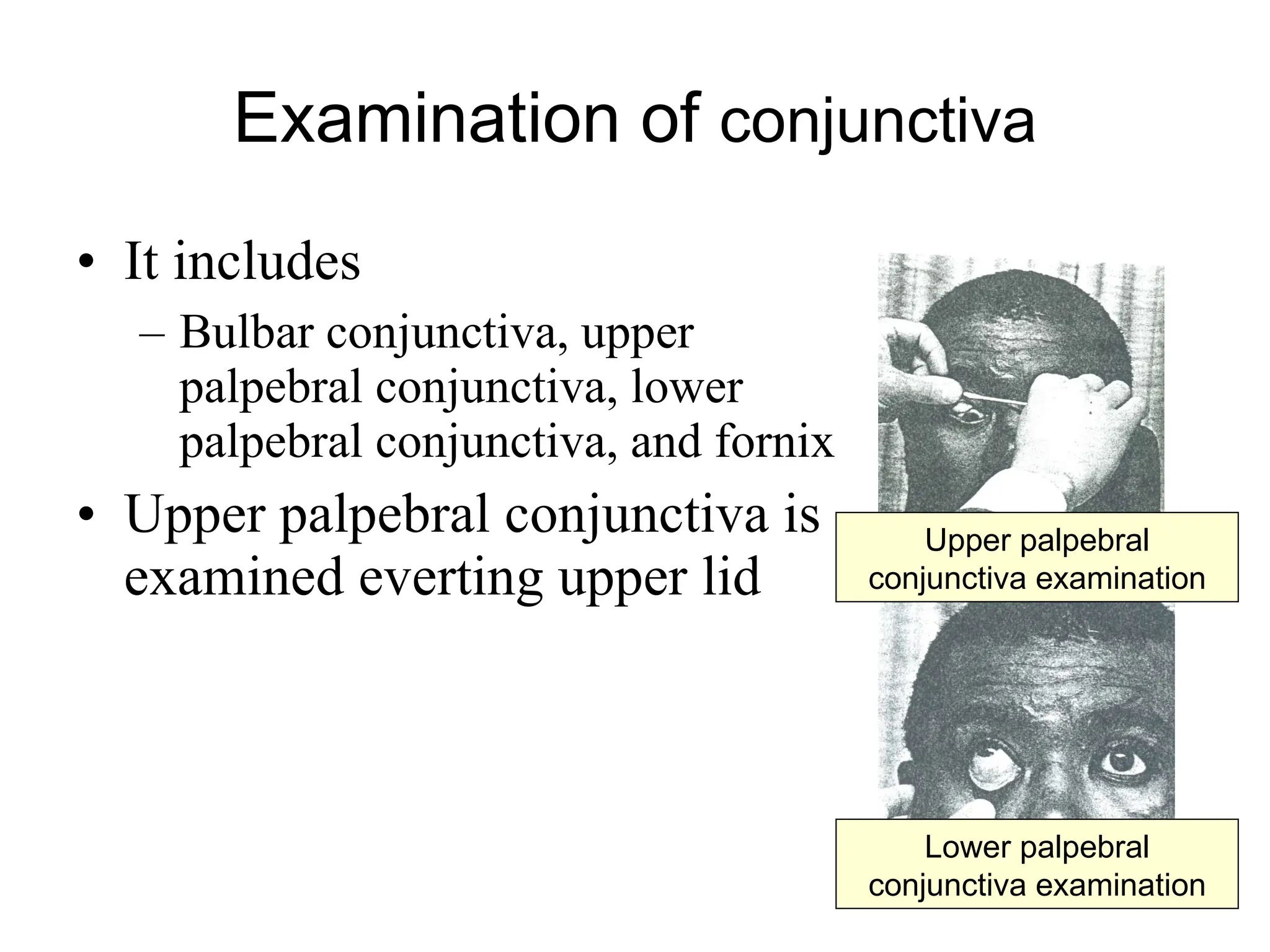
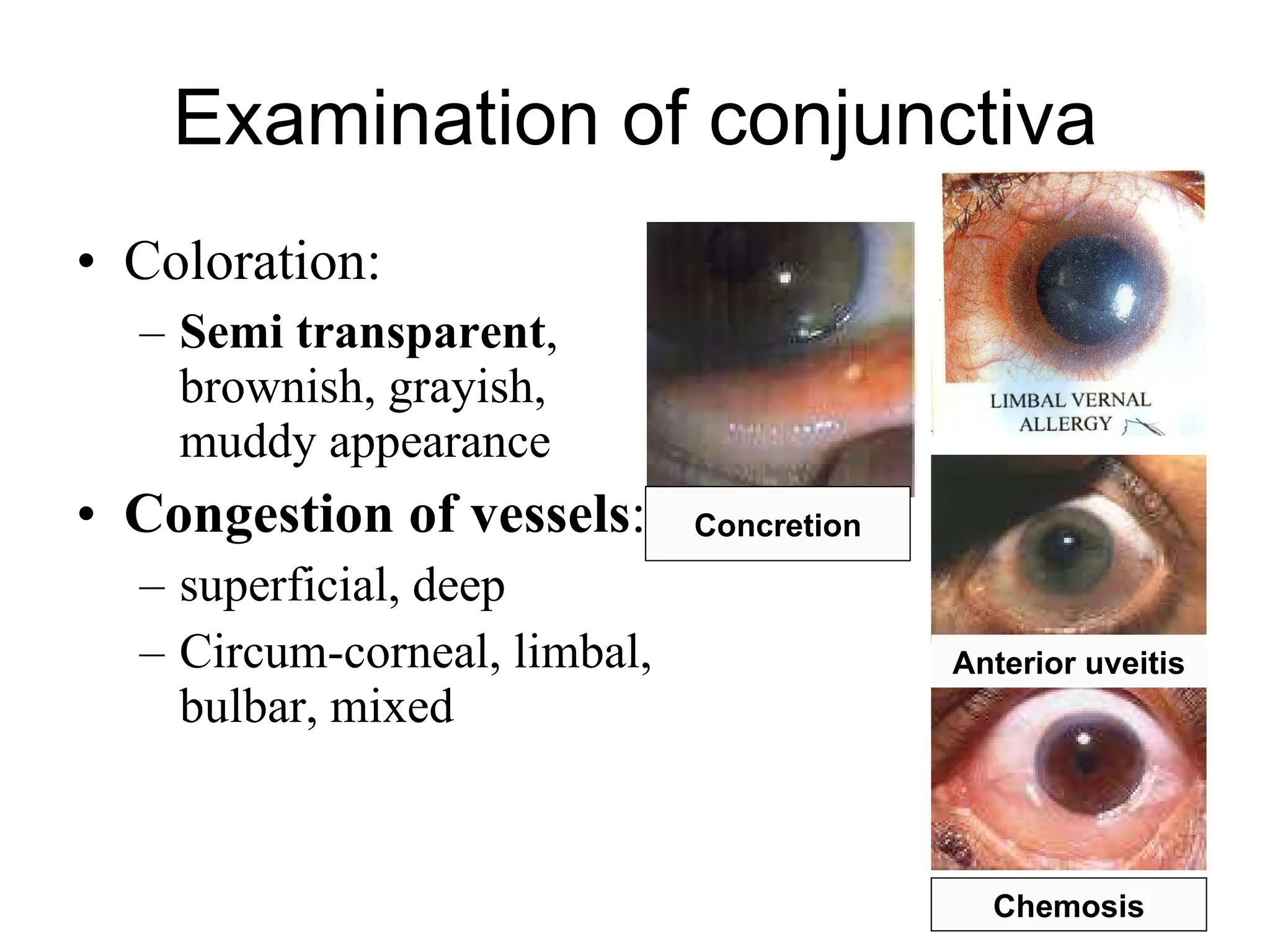
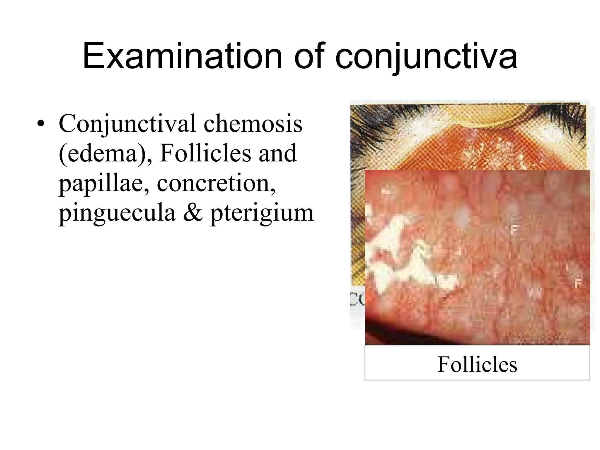
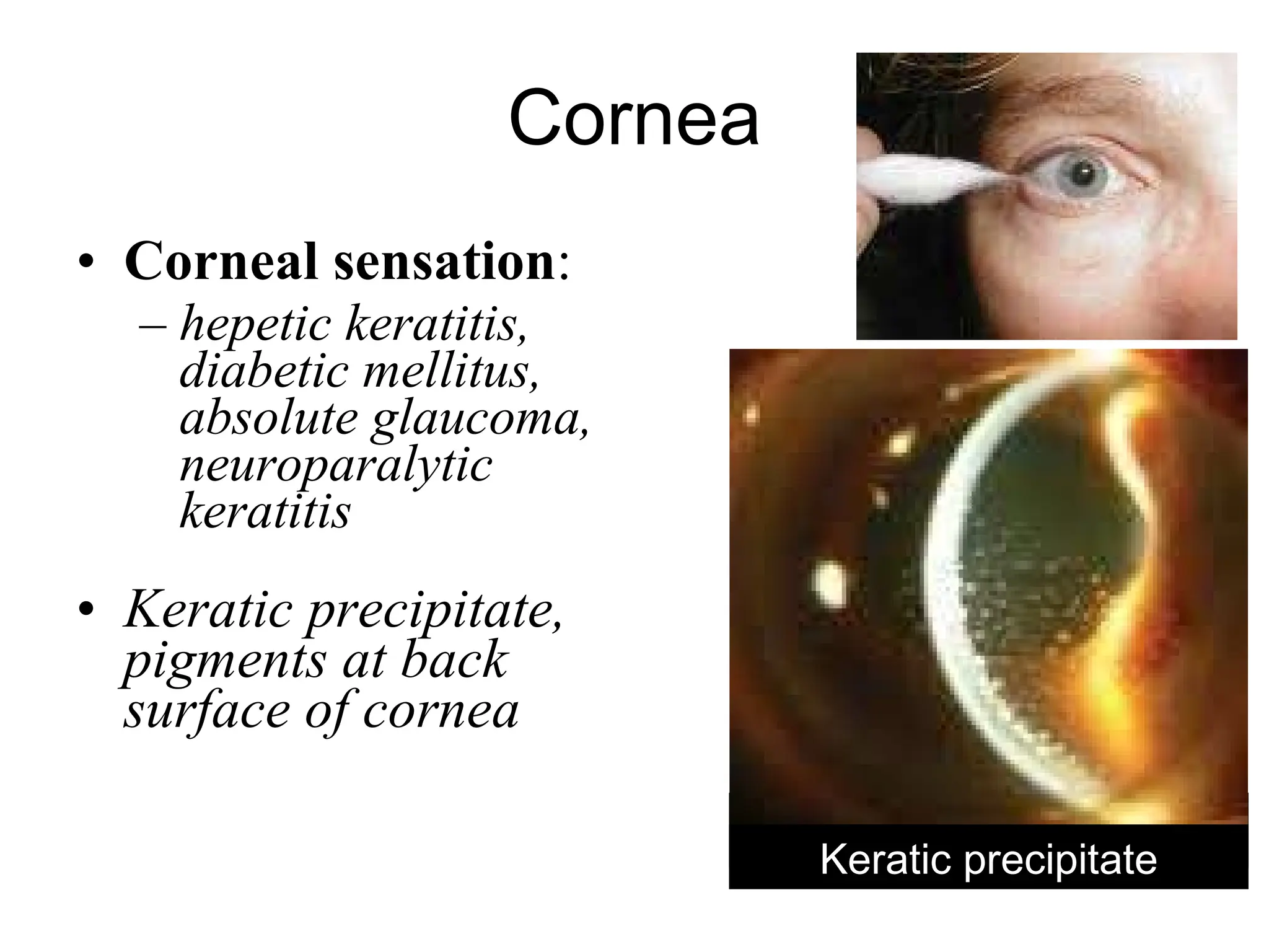
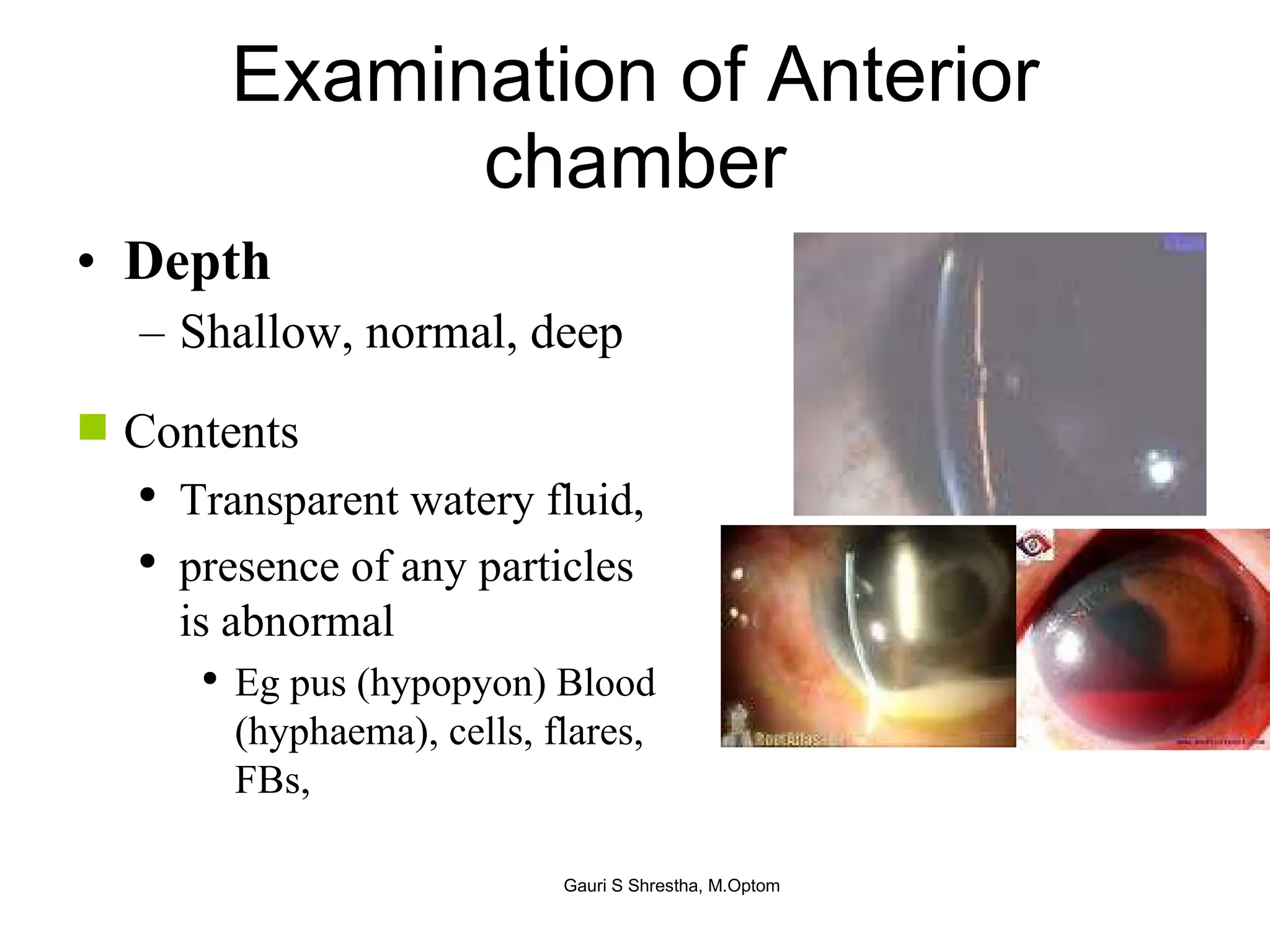
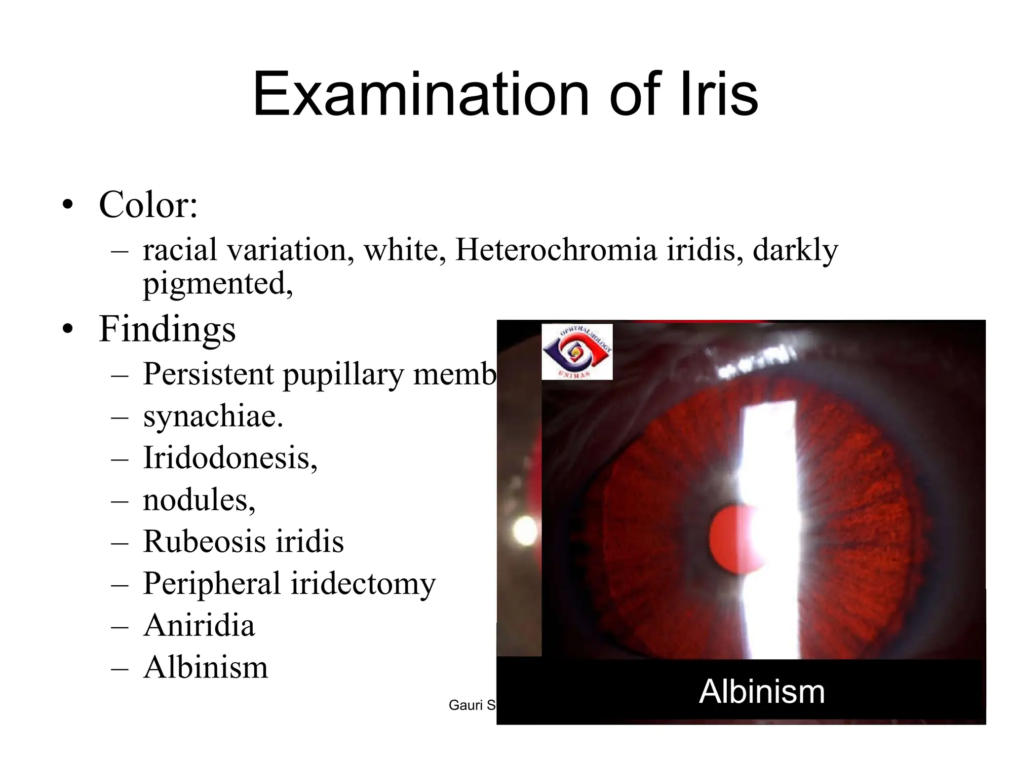
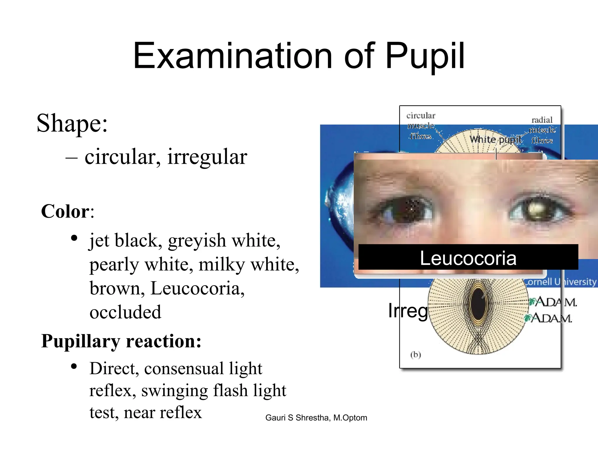
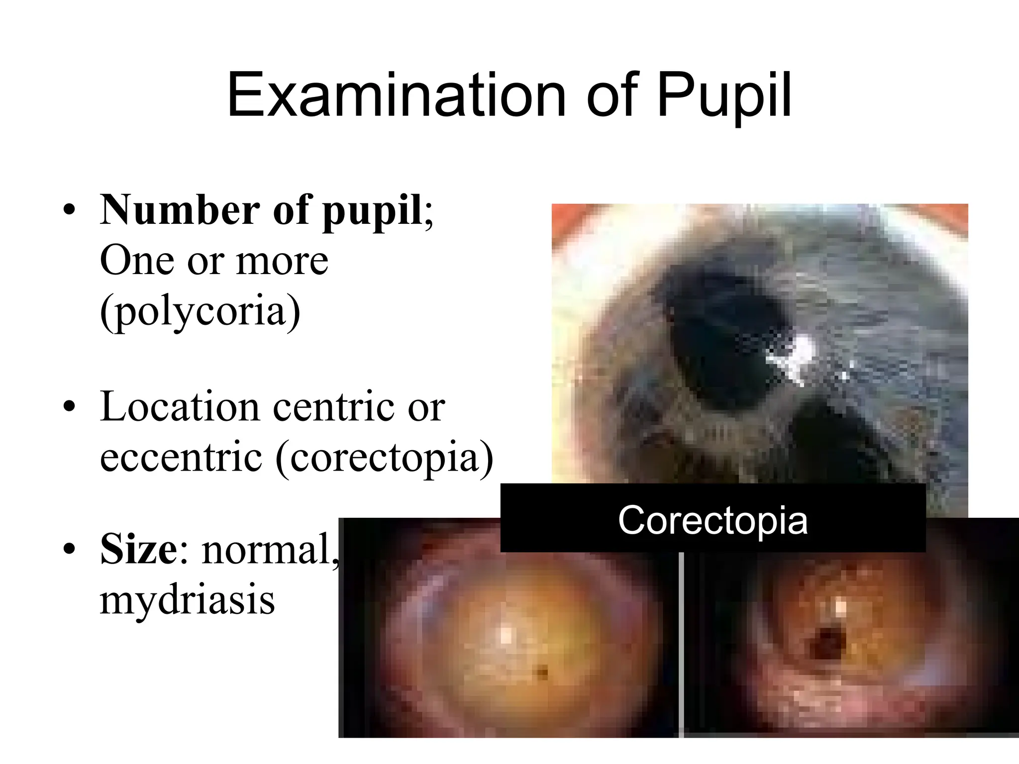
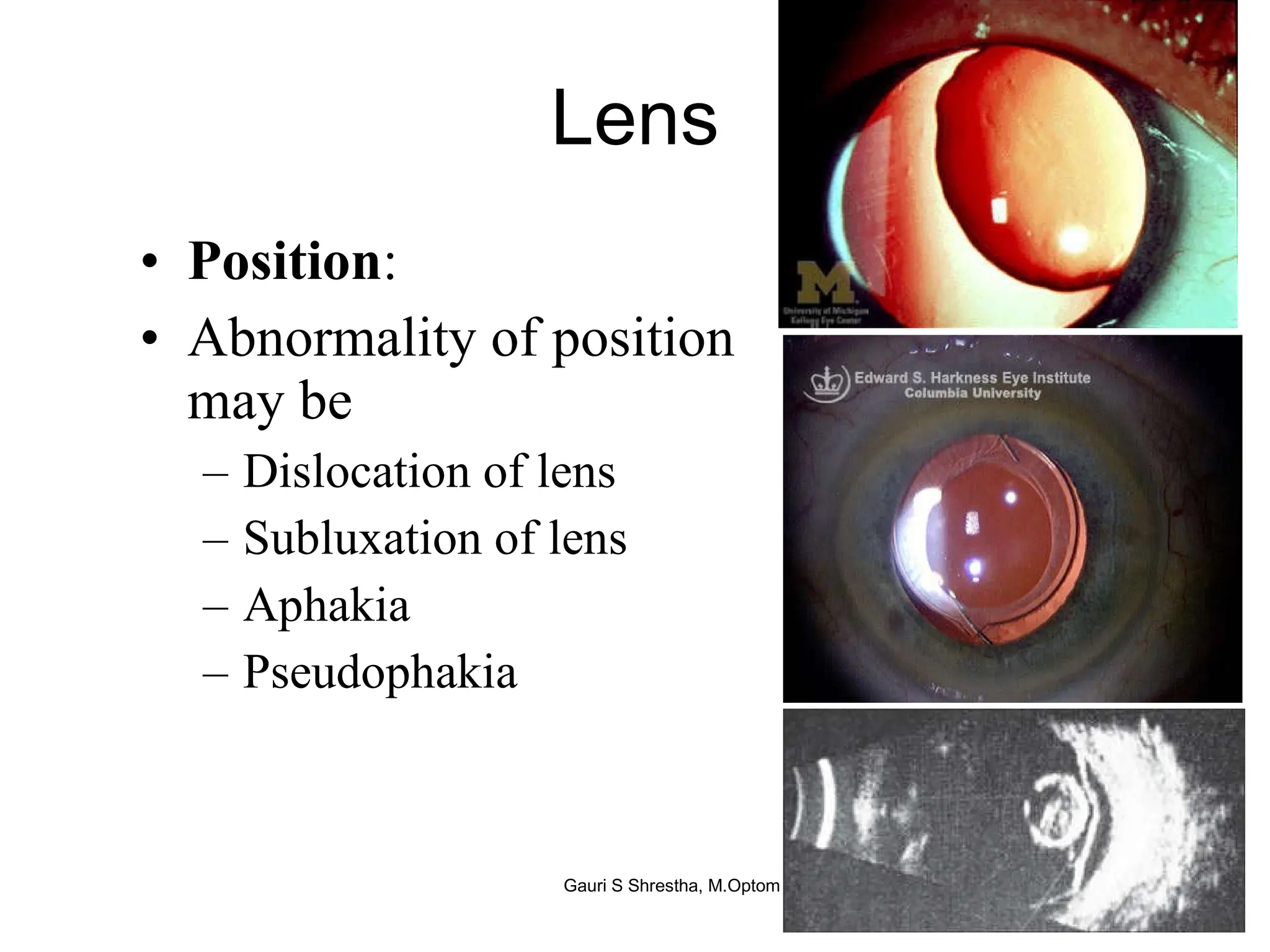
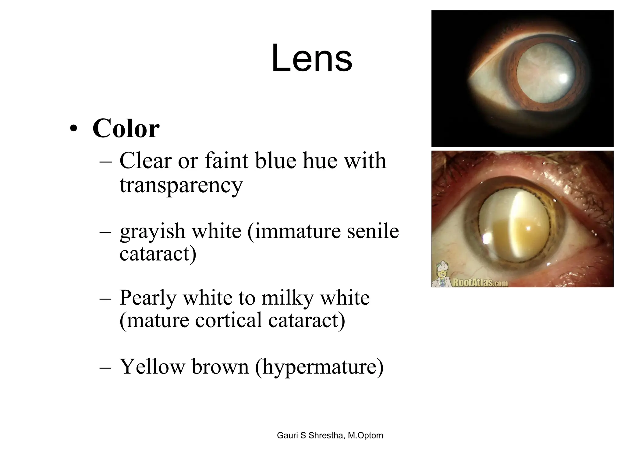
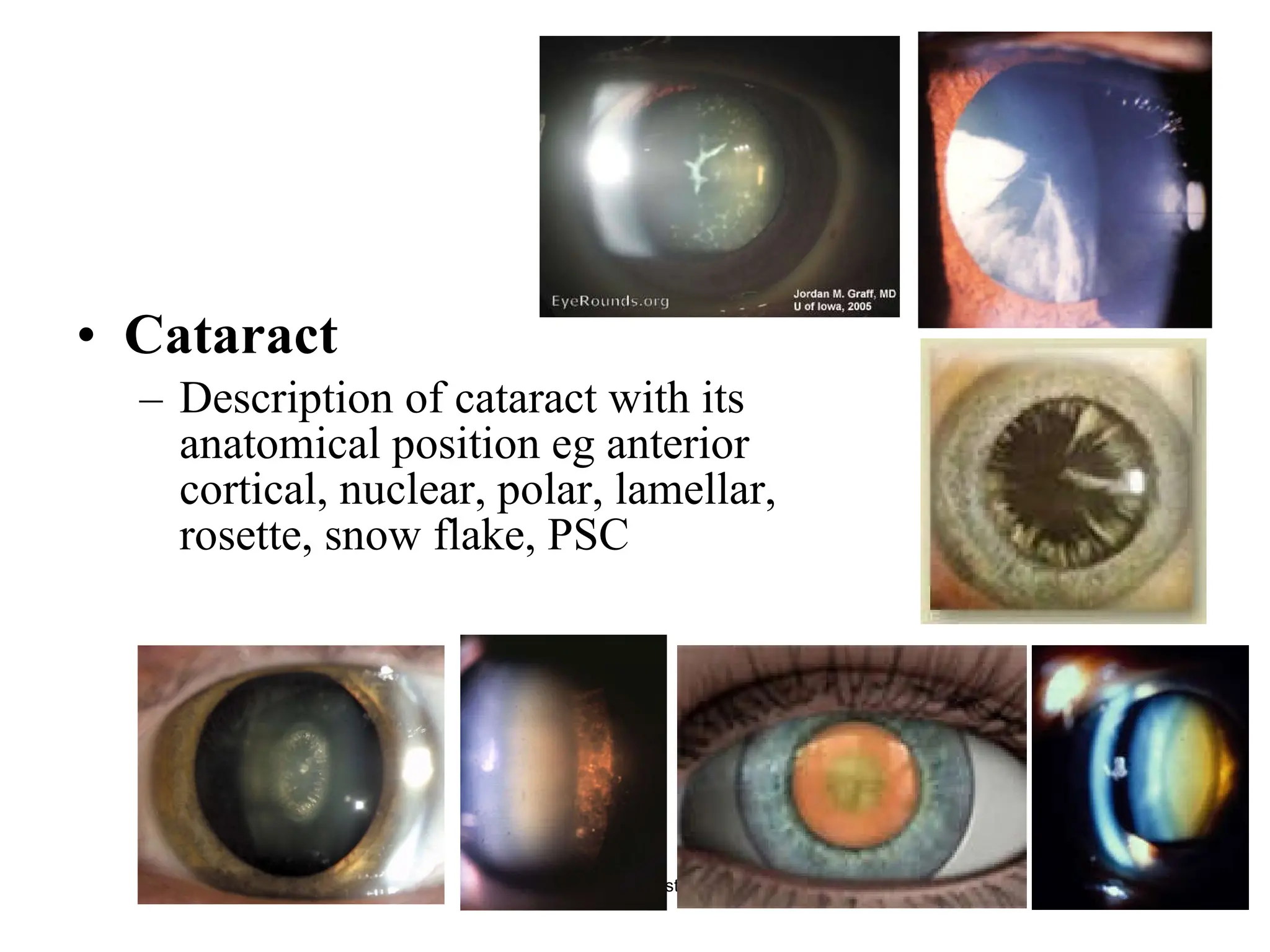
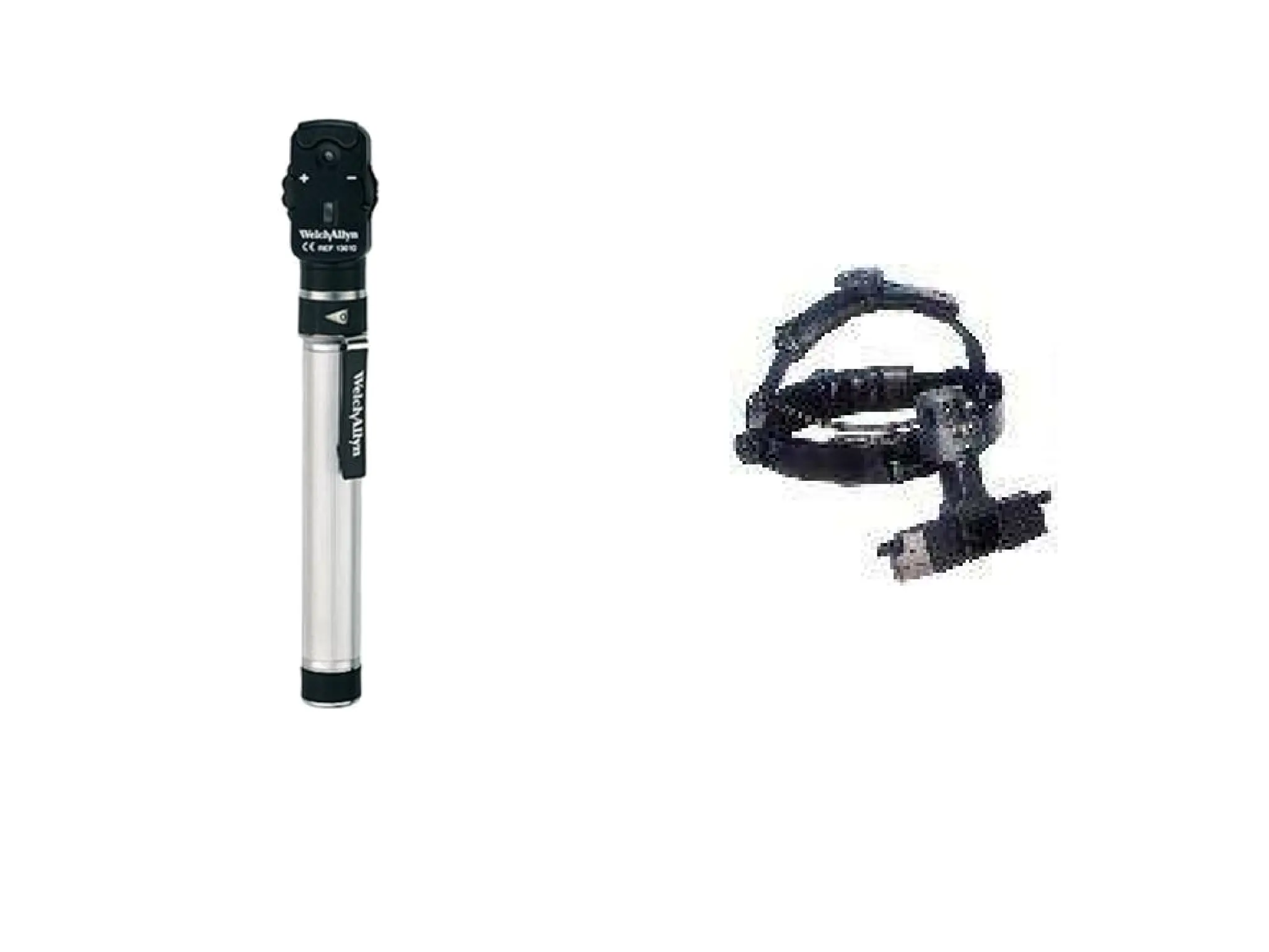
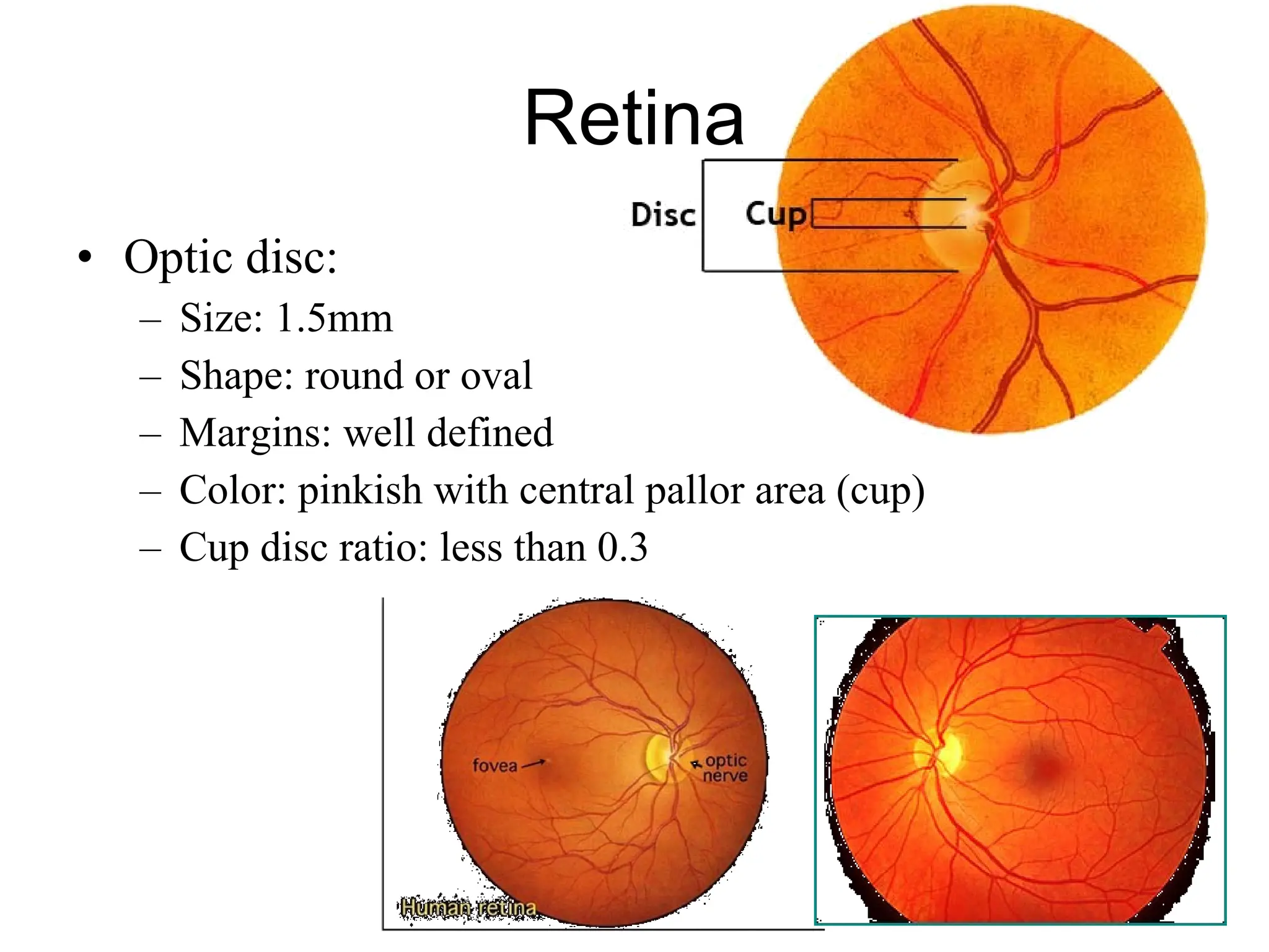
Nursing Assessment of eye Gauri S. Shrestha, M.Optom, FIACLE Part I
Learning Objective Able to describe importance of history taking Able to describe distant visual acuity  Able to describe eye alignment test Able to describe eye movement Able to describe pupillary reaction Able to describe anterior segment examination of eye Able to describe posterior segment of the eye
Patient History Helps acquire a skill necessary to perform an examination The first and the most important aspects that informs us why the patient sought the nursing care
Purpose To acquire a skill necessary to perform an examination. To acquire base line information for the examination. To guide the patients with questions those are most productive and extract relevant information. To determine which specific tests or procedures should be performed during the examination ultimately leading to definitive diagnosis and management plan.
Patient preparation Greet the patient and be polite. Establish an interpersonal relation to build confidence of patient. Make patient comfortable sitting arrangement in the room such as arrange comfortable stool, adjusting room temperature, provide a glass of water etc
Implementation DEMOGRAPHIC DATA In includes patient's full name, birth date, gender, complete address, contact telephone number and occupation.  GENERAL OBSERVATION treat as a person remain aware of all aspects of the patient's personal as well as visual and ocular needs.  Observation Note how patient walks, his stature, gait, head position, facial asymmetry, skin color, speech, and odor.
Implementation  CASE HISTORY  Patient's chief complaint  Assessment Distance or near blur vision, double vision, eye strain, watering, burning, stinging, redness, swelling, floater, ocular injury. Routine examination. The referred cases of headache, diabetes mellitus, hypertension, tuberculosis. Hysterical vision loss.
Implementation VISUAL AND OCULAR HISTORY It tells the importance of eye care.  seeking another opinion.  provides a record of changes that may have occurred over time.  eg. recurrent uveitis, myopia progression.   Assessment Duration of complaint, history of recurrence, associated other ocular symptoms, progression of diseases, duration of treatment received, history of injuries or surgeries, last eye examination.
Implementation  MEDICAL HISTORY rule out ocular manifestation of systemic conditions.  reveals such pertinent systemic condition which patients find not important. Assessment History of hypertension, Diabetes mellitus, Thyroid diseases, or arthritis, multiple sclerosis, arteritis, asthma etc,
Implementation  MEDICATION AND HYPERSENSITIVITY History of medications helps to elicit visual as well as ocular manifestation of medications of systemic medicines .  Assessment History of any systemic medications causing ocular manifestations Antianxiety (diazepam) agents, B-blockers, and antihistamines causes decrease aqueous tear secretion leading to dryness.  Chloroquine - retinal pigment epithelial changes, Visual Field defect, and Color vision defect.  Ethanbutol intake - retrobulbar optic neuritis.  Steroids - glaucoma; delay wound healing, and posterior capsular cataract History of ocular medication which can cause systemic manifestation Cycloplegics can cause hallucination, dry mucosa, and fever.
Implementation  FAMILY OCULAR HISTORY A history of poor vision in the family- definitive link to an accurate diagnosis.  Hereditary- a powerful tool in making the correct diagnosis.  Assessment History of the similar diseases in family. All of corneal, vitreal, retinal, choroidal dystrophies are hereditary.  History of similar type of vision loss or any inflammation particularly in first degree relatives (parents). e.g., glaucoma, strabismus, amblyopia or myopia.  History of the similar episodes in many family members in subsequent generation or in specific gender  Autosomal dominant trait is mostly found in aniridia, congenital cataract, retinoblastoma, cone dystrophy. Autosomal recessive in retinitis pigmentosa. X-linked in ocular albinism, and red-green color deficiency.
Implementation  FAMILY MEDICAL HISTORY Similarly, it is important to know diseases that may exist in other family members.  Assessment History of the similar pattern of diseases in cousins, nephews, uncles and aunt etc., Some diseases such as diabetes may have chances of genetic predisposition where as hypertension and multiple sclerosis may have high frequency in family members yet not be inherited in a known pattern.
Implementation  VOCATIONAL AND RECREATIONAL DEMAND Consideration of the vocational needs of patient  ask about the special visual need of the patient.  Assessment Special demands for visual acuity, potential for eye injury, or use of protective devices.
Summary Upon complete history taking, nurse will be able to develop individualized goals for examination and assessment, identify presence of abnormality, and make patient aware of signs and symptom of eye diseases. History taking helps to gain patient’s confidence, also prepares the patient for further assessment and examination procedure
Visual acuity Visual acuity is the resolving power of the eye  Measures a quality & ability of the eye to resolve varying letter sizes In 1862, Snellen introduced a system for measuring visual acuity, is still the fundamental tool Measured conventionally at 6m (20ft) distance for distant vision Snellen Fraction = Testing Distance Designation of smallest line read
Visual acuity Snellen acuity is based on a minimum angle of resolution of 1 min of arc Testing distance of 6 m θ MAR
Visual acuity Purpose To quantify visual acuity. To determine severity of vision impairing disorders.  To classify visual impairment, low vision and blindness  Assessment of patient Assess vision related history such type of vision loss (gradual, sudden, transient) as time of onset, duration, history of spectacle wear. Assess history of red eye, pain, swelling, diplopia, photophobia, trauma, surgery.  Observe abnormality in posture, capacity to comprehension.
Visual acuity Required Instrument Snellen vision chart Occluder Patient's habitual glasses (if present) Pin hole Patient preparation Position patient at the appropriate distance i.e. 6 meter from the vision chart in adequately illuminated room.  Explain patient about the nature and importance of the test to seek his or her confidence.
 
Distant Visual Acuity Depending upon the distance at which top letter can be read, vision is recorded as 5/60, 3/60, 1/60 Below 1m distance, CF 3ft, CF 2ft, CF 1ft, CFCF HM ± , PR  ±, PL +, NPL Able to read up to 36m line= 6/36 Able to read up to 60m line= 6/60 Able to read up to 6m line= 6/6
Evaluation Record the score of Snellen test as visual acuity unaided, with correction, and pin hole. Visual acuity of 6/6 is considered normal and visual acuity of less than 6/6 is clinically considered abnormal. Functionally, visual acuity less than 6/12 is considered abnormal.
EYE ALIGNMENT TEST   Introduction Proper eye alignment is necessary for clear, comfortable, binocular vision. Poor eye alignment can lead to eyestrain, blurred vision, double vision, strabismus, and amblyopia.  Purpose To determine the presence or the absence of deviation in eye. To detect latent nystagmus.  To detect fixation preference in infants and toddlers.
Assessment of patient Assess history of eyestrain, headache, double vision Assess family history of squint and amblyopia. Assess history of eye and head injury.  Notice abnormal head adaptations such as head tilt and face turn. Required Instrument Occluder Pen torch light Fixation target Patient preparation Explain patient about the nature of the test to seek his or her confidence and co-operation. Seat patient comfortably in dimly illuminated room.
Procedure Comfortable sitting arrangement  Dim room light. Flash a penlight onto the bridge of the patient's nose from 40-50cm away. Look for the spot on both corneas where the light shines. Light shines symmetrically at equal distance at the pupillary center
Cover test
Cover test
Cover test
Cover test   
Cover Test  
Left eye exotropia
Exophoria
 
Evaluation Symmetry in corneal light reflex in two eyes generally indicates that there is no deviation. Asymmetry in corneal light reflex suggests that there is a squint.
Strabismus Ocular misalignment Strabismus present or absent Inward (esotropia) Outward (exotropia) Upward (hypertropia) Downward (hypotropia) Rotation in (encyclotropia) Rotation out (excyclotropia) combination
EXTRAOCULAR MUSCLE MOVEMENT   Introduction Extraocular muscle movement is tested in different gazes to investigate their integrity and integrity of their associated nerves.  Defect in ocular motor systems may result from muscular as well as neurological causes.  Purpose To investigate integrity of extraocular muscles. To investigate integrity of associated nerves of extraocular muscles. To measure extent of the movement of extraocular muscles.
Assessment of patient Assess history of trauma to eye, diplopia, headache associated with nausea and vomiting, scotoma at the fixation, and scintillation. Notice any abnormal head posture such head tilt right or left, chin up or down, face turn to left or right, and dropping of eye lids.  Notice patient feeling pain on movement of eyeball.  Assess best corrected or pin hole visual acuity to notice decrease in visual acuity. Required Instruments A pen light or transilluminator Fixation target
Patient preparation If the patient wears spectacle or contact lenses, make sure their removal. Explain patient about the nature of the test to seek his or her confidence and co-operation. Seat patient comfortably in adequately illuminated room.
DIAGNOSTIC  POSITIONS OF GAZE
Ocular motility test RSR RLR RIR RSO MR RIO LIO LSR LLR LIR LSO
 
Evaluation The key observations to note are overaction or restriction in specific gaze.  Recorded on basically six gazes: Right, right up, right down, left, left up, and left down.
TESTS OF PUPILLARY FUNCTION   Introduction Testing a patient's pupillary function involves  measurement of pupil size and measurement of light reflexes.  Purpose To measure and compare size of the pupil for detecting anisocoria (unequal pupil size). To measure and compare shape of the pupil related to trauma, surgery, and/ or inflammation of the eye. To assess pupil's reaction to light for detecting abnormality in function of iris, optic nerve, and visual pathway.
Assessment of patient Same as extraocular movement assessment. Required Instruments A pen light or transilluminator Millimeter ruler A fixation target Patient preparation If the patient wears spectacle or contact lenses, make sure their removal. Explain patient about the nature of the test to seek his or her confidence and co-operation. Seat patient comfortably in dimly illuminated room.
PUPIL SIZE MEASUREMENT Have a patient fixate a distance target in a room illuminated enough to see the pupil.  Place the ruler by nurse's non-dominant hand on measuring eye on the supraorbital ridge and position it so that half of the pupil is covered. Measure and record the size of the pupil of each eye with the help of millimeter ruler. Record the
Pupillary reaction Direct and consensual Whenever pupil is shine with light should constrict. Absence of constriction indicates the presence of abnormality. When direct and indirect pupillary response is absent, indicate that direct pupillary light response is negative.
Pupillary reaction Swinging flash light
Pupillary reaction Swinging flash light If the rate of constriction of pupil is the same in each eye when the light is shine in the eye, the result should be recorded as negative. This is the normal response. If the rate of constriction of the two pupils is different, or the pupil of the eye receiving the light dilates instead of constriction, the results are recorded as a positive response. This is abnormal finding. It is recorded as RAPD (relative afferent pupillary defect) positive or negative.
ASSESS EXTERNAL (ANTERIOR) EYE STRUCTURES   Introduction The external examination includes a gross inspection of the external structures of the eye by means of magnifying loupe and slit lamp biomicroscopy.  Purpose To examine anterior eye structure in routine. To detect any abnormality such as infection, inflammation, swelling, opacity in anterior eye structures.  To detect diseases of lids, conjunctiva, sclera, episclera, cornea, and lens. To assess anterior chamber angle depth.
ASSESS EXTERNAL (ANTERIOR) EYE STRUCTURES   Assessment of patient Assessment includes brief history related to eye and systemic health. Problem identified during history and the preliminary eye examination should be further evaluated. Assess history of trauma or swelling of eye and adnexa. Preliminary eye examination should include, ectropion, intropion, pupillary reaction, redness, swelling of anterior segment of eye, watering, color of conjunctiva, color of cornea (transparent). Assess visual acuity unaided, with glasses and pin hole.
ASSESS EXTERNAL (ANTERIOR) EYE STRUCTURES   Instrumentation Magnifying loupe. Slit lamp biomicroscope Patient preparation If the patient wears spectacle or contact lenses, make sure their removal. Explain patient about the nature of the test to seek his or her confidence and co-operation. Seat patient comfortably in dimly illuminated room in front of slit lamp.  Set the goal of anterior eye examination.
Slit lamp examination Invaluable and indispensable part of ophthalmological examination Parts Observation system (Microscope) Illumination system (Slit lamp) Use,  examination of detailed structure of eye Measurement of corneal thickness, anterior chamber depth Visiometry Gauri S Shrestha, M.Optom
Examination of eye lids and eye brows Inspect the level of two eye brows, symmetry in lid crease eg ptosis Inspect eye lashes:  absent  (madarosis)  whitening  (poliosis)  irregular red lid margins  (pediculosis) Position of eye lids Upper eye lid covers 1/6 th  of cornea Lower eye lids just touches limbus Movement:  follow the movement of eye ball in down ward gaze  (lags behind in graves diseases)
Examination of eye lids Blinking: Inspect whether normal blinking present Excessive blinking : FB in eye, lid irritation, dry eye Blinking reduced or absent :  trigeminal anesthesia, 7 th  nerve praresis Lagophthalmos  (incomplete closure of eye lids)  eg proptosis, congenital ptosis, facial nerve palsy Lid margin mark any of the following Entropion, ectropion, Trichiasis, districhiasis, madarosis, poliosis, scales LAGOPHTHALMOS
Palpebral aperture It is a exposed space b/w two lid margins 8-10mm vertical, 25-30mm horizontal Ankyloblepharon: horizontal narrow fissure Blepharophimosis: narrow fissure all around Vertical narrow fissure: ptosis, endophthalmos, microphthalmos, anophthalmos Vertical wide fissure: proptosis, retraction of upper lid
Examinations of lacrimal apparatus Examination is important in cases with epiphora in corneal ulcers and before surgery Inspect lacrimal puncta  for eversion, stenosis, absent, or discharge Lacrimal sac area inspection :  redness,  swelling, fistula Regurgitation test   press over the lacrimal sac at medial canthus to inspect regurgitation of discharge from the puncta
Examination of eye ball Inspect symmetry of eye ball Bulging of eye ball (expohthlamos/ proptosis) Shrunken of eye ball (Endophthalmos)
Examination of  conjunctiva It includes Bulbar conjunctiva, upper palpebral conjunctiva, lower palpebral conjunctiva, and fornix Upper palpebral conjunctiva is examined everting upper lid Upper palpebral conjunctiva examination Lower palpebral conjunctiva examination
Examination of conjunctiva Coloration:  Semi transparent , brownish, grayish, muddy appearance Congestion of vessels :  superficial, deep Circum-corneal, limbal, bulbar, mixed Anterior uveitis Chemosis Concretion
Examination of conjunctiva Conjunctival chemosis (edema), Follicles and papillae, concretion, pinguecula & pterigium Follicles
Examination of sclera Color White, yellow, bluish, pigments Inflammation, Staphyloma, foreign body entraped (transparent), perforation Yellow sclera Blue sclera Pigments  Staphyloma
Cornea Diameter Shape :  corneal curvature Surface :  ulceration, abrasion, vascularization, distortion,  Transparency:  bright shining, cornea edema, opacity Corneal Ulcer Corneal abrasion Corneal vascularization Corneal distortion Corneal opacity Corneal edema
Cornea Corneal sensation :  hepetic keratitis, diabetic mellitus, absolute glaucoma, neuroparalytic keratitis Keratic precipitate, pigments at back surface of cornea Herpetic keratitis Keratic precipitate
Examination of Anterior chamber Depth   Shallow, normal, deep Gauri S Shrestha, M.Optom Contents Transparent watery fluid,  presence of any particles is abnormal Eg pus (hypopyon) Blood (hyphaema), cells, flares, FBs,
Examination of Iris Color:  racial variation, white, Heterochromia iridis, darkly pigmented,  Findings Persistent pupillary membrane,  synachiae.  Iridodonesis,  nodules,  Rubeosis iridis Peripheral iridectomy Aniridia Albinism Gauri S Shrestha, M.Optom Heterochromia iridis Persistent pupillary membrane synachiae Nodules Rubeosis Iridis Peripheral iridectomy Aniridia Albinism
Examination of Pupil Shape:  circular, irregular  Gauri S Shrestha, M.Optom Irregular pupil Color :  jet black, greyish white, pearly white, milky white, brown, Leucocoria, occluded Pupillary reaction: Direct, consensual light reflex, swinging flash light test, near reflex Leucocoria
Examination of Pupil Number of pupil ; One or more (polycoria) Location centric or eccentric (corectopia) Size : normal, miosis, mydriasis Gauri S Shrestha, M.Optom Polycoria Corectopia
Lens Position :  Abnormality of position may be Dislocation of lens Subluxation of lens Aphakia Pseudophakia Gauri S Shrestha, M.Optom
Lens Shape biconvex structure Abnormal lens shape may be Spherophakia Lenticonus Coloboma of lens Gauri S Shrestha, M.Optom
Lens Color Clear or faint blue hue with transparency grayish white (immature senile cataract) Pearly white to milky white (mature cortical cataract) Yellow brown (hypermature) Gauri S Shrestha, M.Optom
Lens Cataract Description of cataract with its anatomical position eg anterior cortical, nuclear, polar, lamellar, rosette, snow flake, PSC Gauri S Shrestha, M.Optom
Summary With a practice and experience, slit lamp examination requires only a few minutes of examination time. It will yield invaluable information that will be used to document the health and integrity of the anterior ocular structures.
ASSESSMENT OF INTERNAL EYE STRUCTURE   Assessment of internal eye structure includes  assessment of middle vitreous,  posterior vitreous,  fundus (retina) and optic disc.  Purpose To examine internal eye structure in routine. To detect any abnormality such as opacity in vitreous. To detect any abnormality in retina such as cupping of disc, hemorrhage, exudates, scars, and tear.
Assessment of patient Assess history of decrease in vision, sudden loss of vision, diabetes mellitus, hypertension, high myopia, glaucoma, and other systemic conditions. Evaluate presence of any media opacities or cataract, squint, ocular movement, and diseases of anterior segments.  Assess best corrected visual acuity. Assess history of trauma or swelling of eye and adnexa. Required Instrument Monocular direct ophthalmoscope Patient preparation Explain patient about the nature of the test to seek his or her confidence and co-operation. Seat patient comfortably in dimly illuminated room in front nurse.  Check the purpose of the test. Make sure removal of patient's spectacle.
 
Vitreous examination A transparent and inert cavity Look for Any cells, opacities, hemorrhage, fibrosis Gauri S Shrestha, M.Optom
Retina Optic disc:  Size: 1.5mm Shape: round or oval Margins: well defined Color: pinkish with central pallor area (cup) Cup disc ratio: less than 0.3
Retina Abnormal:  blurred disc margin,  large cup disc ratio,  haemorrhage,  neovascularization,  myopic crescent Gauri S Shrestha, M.Optom
Retina Macula Situated at the posterior pole 2 disc diameter lateral to disc margin It is the darkly pigmented area in retina Fovea is the vessels free zone visualized as bright reflex Gauri S Shrestha, M.Optom
Retina Examine Macular hole, cherry red spot, macular edema, exudates, drusens, scarring  Gauri S Shrestha, M.Optom
Retina Retinal blood vessels Arterioles are smaller, brighter than vein running parallel A:V :: 2:3 Examine for Narrowing of arterioles, tortuosity of vessels, sheathing, pulsation Gauri S Shrestha, M.Optom
Retina General background Pinkish red in color (Physiologic variation possible) Gauri S Shrestha, M.Optom Tessellated  and tigroid fundus:  visibility of choroidal pigments Retinal hemorrhage Exudates Retinitis pigmentosa microaneurisms
Summary List in sequential order of components of history taking. Lists the things we observe during history taking and examination. How is the visual acuity denoted? List the anterior ocular structures under examination List the posterior ocular structures under examination. Gauri S Shrestha, M.Optom
Thank you

Nursing assessment of eye part1

  • 1.
    Nursing Assessment ofeye Gauri S. Shrestha, M.Optom, FIACLE Part I
  • 2.
    Learning Objective Ableto describe importance of history taking Able to describe distant visual acuity Able to describe eye alignment test Able to describe eye movement Able to describe pupillary reaction Able to describe anterior segment examination of eye Able to describe posterior segment of the eye
  • 3.
    Patient History Helpsacquire a skill necessary to perform an examination The first and the most important aspects that informs us why the patient sought the nursing care
  • 4.
    Purpose To acquirea skill necessary to perform an examination. To acquire base line information for the examination. To guide the patients with questions those are most productive and extract relevant information. To determine which specific tests or procedures should be performed during the examination ultimately leading to definitive diagnosis and management plan.
  • 5.
    Patient preparation Greetthe patient and be polite. Establish an interpersonal relation to build confidence of patient. Make patient comfortable sitting arrangement in the room such as arrange comfortable stool, adjusting room temperature, provide a glass of water etc
  • 6.
    Implementation DEMOGRAPHIC DATAIn includes patient's full name, birth date, gender, complete address, contact telephone number and occupation. GENERAL OBSERVATION treat as a person remain aware of all aspects of the patient's personal as well as visual and ocular needs. Observation Note how patient walks, his stature, gait, head position, facial asymmetry, skin color, speech, and odor.
  • 7.
    Implementation CASEHISTORY Patient's chief complaint Assessment Distance or near blur vision, double vision, eye strain, watering, burning, stinging, redness, swelling, floater, ocular injury. Routine examination. The referred cases of headache, diabetes mellitus, hypertension, tuberculosis. Hysterical vision loss.
  • 8.
    Implementation VISUAL ANDOCULAR HISTORY It tells the importance of eye care. seeking another opinion. provides a record of changes that may have occurred over time. eg. recurrent uveitis, myopia progression. Assessment Duration of complaint, history of recurrence, associated other ocular symptoms, progression of diseases, duration of treatment received, history of injuries or surgeries, last eye examination.
  • 9.
    Implementation MEDICALHISTORY rule out ocular manifestation of systemic conditions. reveals such pertinent systemic condition which patients find not important. Assessment History of hypertension, Diabetes mellitus, Thyroid diseases, or arthritis, multiple sclerosis, arteritis, asthma etc,
  • 10.
    Implementation MEDICATIONAND HYPERSENSITIVITY History of medications helps to elicit visual as well as ocular manifestation of medications of systemic medicines . Assessment History of any systemic medications causing ocular manifestations Antianxiety (diazepam) agents, B-blockers, and antihistamines causes decrease aqueous tear secretion leading to dryness. Chloroquine - retinal pigment epithelial changes, Visual Field defect, and Color vision defect. Ethanbutol intake - retrobulbar optic neuritis. Steroids - glaucoma; delay wound healing, and posterior capsular cataract History of ocular medication which can cause systemic manifestation Cycloplegics can cause hallucination, dry mucosa, and fever.
  • 11.
    Implementation FAMILYOCULAR HISTORY A history of poor vision in the family- definitive link to an accurate diagnosis. Hereditary- a powerful tool in making the correct diagnosis. Assessment History of the similar diseases in family. All of corneal, vitreal, retinal, choroidal dystrophies are hereditary. History of similar type of vision loss or any inflammation particularly in first degree relatives (parents). e.g., glaucoma, strabismus, amblyopia or myopia. History of the similar episodes in many family members in subsequent generation or in specific gender Autosomal dominant trait is mostly found in aniridia, congenital cataract, retinoblastoma, cone dystrophy. Autosomal recessive in retinitis pigmentosa. X-linked in ocular albinism, and red-green color deficiency.
  • 12.
    Implementation FAMILYMEDICAL HISTORY Similarly, it is important to know diseases that may exist in other family members. Assessment History of the similar pattern of diseases in cousins, nephews, uncles and aunt etc., Some diseases such as diabetes may have chances of genetic predisposition where as hypertension and multiple sclerosis may have high frequency in family members yet not be inherited in a known pattern.
  • 13.
    Implementation VOCATIONALAND RECREATIONAL DEMAND Consideration of the vocational needs of patient ask about the special visual need of the patient. Assessment Special demands for visual acuity, potential for eye injury, or use of protective devices.
  • 14.
    Summary Upon completehistory taking, nurse will be able to develop individualized goals for examination and assessment, identify presence of abnormality, and make patient aware of signs and symptom of eye diseases. History taking helps to gain patient’s confidence, also prepares the patient for further assessment and examination procedure
  • 15.
    Visual acuity Visualacuity is the resolving power of the eye Measures a quality & ability of the eye to resolve varying letter sizes In 1862, Snellen introduced a system for measuring visual acuity, is still the fundamental tool Measured conventionally at 6m (20ft) distance for distant vision Snellen Fraction = Testing Distance Designation of smallest line read
  • 16.
    Visual acuity Snellenacuity is based on a minimum angle of resolution of 1 min of arc Testing distance of 6 m θ MAR
  • 17.
    Visual acuity PurposeTo quantify visual acuity. To determine severity of vision impairing disorders. To classify visual impairment, low vision and blindness Assessment of patient Assess vision related history such type of vision loss (gradual, sudden, transient) as time of onset, duration, history of spectacle wear. Assess history of red eye, pain, swelling, diplopia, photophobia, trauma, surgery. Observe abnormality in posture, capacity to comprehension.
  • 18.
    Visual acuity RequiredInstrument Snellen vision chart Occluder Patient's habitual glasses (if present) Pin hole Patient preparation Position patient at the appropriate distance i.e. 6 meter from the vision chart in adequately illuminated room. Explain patient about the nature and importance of the test to seek his or her confidence.
  • 19.
  • 20.
    Distant Visual AcuityDepending upon the distance at which top letter can be read, vision is recorded as 5/60, 3/60, 1/60 Below 1m distance, CF 3ft, CF 2ft, CF 1ft, CFCF HM ± , PR ±, PL +, NPL Able to read up to 36m line= 6/36 Able to read up to 60m line= 6/60 Able to read up to 6m line= 6/6
  • 21.
    Evaluation Record thescore of Snellen test as visual acuity unaided, with correction, and pin hole. Visual acuity of 6/6 is considered normal and visual acuity of less than 6/6 is clinically considered abnormal. Functionally, visual acuity less than 6/12 is considered abnormal.
  • 22.
    EYE ALIGNMENT TEST Introduction Proper eye alignment is necessary for clear, comfortable, binocular vision. Poor eye alignment can lead to eyestrain, blurred vision, double vision, strabismus, and amblyopia. Purpose To determine the presence or the absence of deviation in eye. To detect latent nystagmus. To detect fixation preference in infants and toddlers.
  • 23.
    Assessment of patientAssess history of eyestrain, headache, double vision Assess family history of squint and amblyopia. Assess history of eye and head injury. Notice abnormal head adaptations such as head tilt and face turn. Required Instrument Occluder Pen torch light Fixation target Patient preparation Explain patient about the nature of the test to seek his or her confidence and co-operation. Seat patient comfortably in dimly illuminated room.
  • 24.
    Procedure Comfortable sittingarrangement Dim room light. Flash a penlight onto the bridge of the patient's nose from 40-50cm away. Look for the spot on both corneas where the light shines. Light shines symmetrically at equal distance at the pupillary center
  • 25.
  • 26.
  • 27.
  • 28.
  • 29.
  • 30.
  • 31.
  • 32.
  • 33.
    Evaluation Symmetry incorneal light reflex in two eyes generally indicates that there is no deviation. Asymmetry in corneal light reflex suggests that there is a squint.
  • 34.
    Strabismus Ocular misalignmentStrabismus present or absent Inward (esotropia) Outward (exotropia) Upward (hypertropia) Downward (hypotropia) Rotation in (encyclotropia) Rotation out (excyclotropia) combination
  • 35.
    EXTRAOCULAR MUSCLE MOVEMENT Introduction Extraocular muscle movement is tested in different gazes to investigate their integrity and integrity of their associated nerves. Defect in ocular motor systems may result from muscular as well as neurological causes. Purpose To investigate integrity of extraocular muscles. To investigate integrity of associated nerves of extraocular muscles. To measure extent of the movement of extraocular muscles.
  • 36.
    Assessment of patientAssess history of trauma to eye, diplopia, headache associated with nausea and vomiting, scotoma at the fixation, and scintillation. Notice any abnormal head posture such head tilt right or left, chin up or down, face turn to left or right, and dropping of eye lids. Notice patient feeling pain on movement of eyeball. Assess best corrected or pin hole visual acuity to notice decrease in visual acuity. Required Instruments A pen light or transilluminator Fixation target
  • 37.
    Patient preparation Ifthe patient wears spectacle or contact lenses, make sure their removal. Explain patient about the nature of the test to seek his or her confidence and co-operation. Seat patient comfortably in adequately illuminated room.
  • 38.
  • 39.
    Ocular motility testRSR RLR RIR RSO MR RIO LIO LSR LLR LIR LSO
  • 40.
  • 41.
    Evaluation The keyobservations to note are overaction or restriction in specific gaze. Recorded on basically six gazes: Right, right up, right down, left, left up, and left down.
  • 42.
    TESTS OF PUPILLARYFUNCTION Introduction Testing a patient's pupillary function involves measurement of pupil size and measurement of light reflexes. Purpose To measure and compare size of the pupil for detecting anisocoria (unequal pupil size). To measure and compare shape of the pupil related to trauma, surgery, and/ or inflammation of the eye. To assess pupil's reaction to light for detecting abnormality in function of iris, optic nerve, and visual pathway.
  • 43.
    Assessment of patientSame as extraocular movement assessment. Required Instruments A pen light or transilluminator Millimeter ruler A fixation target Patient preparation If the patient wears spectacle or contact lenses, make sure their removal. Explain patient about the nature of the test to seek his or her confidence and co-operation. Seat patient comfortably in dimly illuminated room.
  • 44.
    PUPIL SIZE MEASUREMENTHave a patient fixate a distance target in a room illuminated enough to see the pupil. Place the ruler by nurse's non-dominant hand on measuring eye on the supraorbital ridge and position it so that half of the pupil is covered. Measure and record the size of the pupil of each eye with the help of millimeter ruler. Record the
  • 45.
    Pupillary reaction Directand consensual Whenever pupil is shine with light should constrict. Absence of constriction indicates the presence of abnormality. When direct and indirect pupillary response is absent, indicate that direct pupillary light response is negative.
  • 46.
  • 47.
    Pupillary reaction Swingingflash light If the rate of constriction of pupil is the same in each eye when the light is shine in the eye, the result should be recorded as negative. This is the normal response. If the rate of constriction of the two pupils is different, or the pupil of the eye receiving the light dilates instead of constriction, the results are recorded as a positive response. This is abnormal finding. It is recorded as RAPD (relative afferent pupillary defect) positive or negative.
  • 48.
    ASSESS EXTERNAL (ANTERIOR)EYE STRUCTURES Introduction The external examination includes a gross inspection of the external structures of the eye by means of magnifying loupe and slit lamp biomicroscopy. Purpose To examine anterior eye structure in routine. To detect any abnormality such as infection, inflammation, swelling, opacity in anterior eye structures. To detect diseases of lids, conjunctiva, sclera, episclera, cornea, and lens. To assess anterior chamber angle depth.
  • 49.
    ASSESS EXTERNAL (ANTERIOR)EYE STRUCTURES Assessment of patient Assessment includes brief history related to eye and systemic health. Problem identified during history and the preliminary eye examination should be further evaluated. Assess history of trauma or swelling of eye and adnexa. Preliminary eye examination should include, ectropion, intropion, pupillary reaction, redness, swelling of anterior segment of eye, watering, color of conjunctiva, color of cornea (transparent). Assess visual acuity unaided, with glasses and pin hole.
  • 50.
    ASSESS EXTERNAL (ANTERIOR)EYE STRUCTURES Instrumentation Magnifying loupe. Slit lamp biomicroscope Patient preparation If the patient wears spectacle or contact lenses, make sure their removal. Explain patient about the nature of the test to seek his or her confidence and co-operation. Seat patient comfortably in dimly illuminated room in front of slit lamp. Set the goal of anterior eye examination.
  • 51.
    Slit lamp examinationInvaluable and indispensable part of ophthalmological examination Parts Observation system (Microscope) Illumination system (Slit lamp) Use, examination of detailed structure of eye Measurement of corneal thickness, anterior chamber depth Visiometry Gauri S Shrestha, M.Optom
  • 52.
    Examination of eyelids and eye brows Inspect the level of two eye brows, symmetry in lid crease eg ptosis Inspect eye lashes: absent (madarosis) whitening (poliosis) irregular red lid margins (pediculosis) Position of eye lids Upper eye lid covers 1/6 th of cornea Lower eye lids just touches limbus Movement: follow the movement of eye ball in down ward gaze (lags behind in graves diseases)
  • 53.
    Examination of eyelids Blinking: Inspect whether normal blinking present Excessive blinking : FB in eye, lid irritation, dry eye Blinking reduced or absent : trigeminal anesthesia, 7 th nerve praresis Lagophthalmos (incomplete closure of eye lids) eg proptosis, congenital ptosis, facial nerve palsy Lid margin mark any of the following Entropion, ectropion, Trichiasis, districhiasis, madarosis, poliosis, scales LAGOPHTHALMOS
  • 54.
    Palpebral aperture Itis a exposed space b/w two lid margins 8-10mm vertical, 25-30mm horizontal Ankyloblepharon: horizontal narrow fissure Blepharophimosis: narrow fissure all around Vertical narrow fissure: ptosis, endophthalmos, microphthalmos, anophthalmos Vertical wide fissure: proptosis, retraction of upper lid
  • 55.
    Examinations of lacrimalapparatus Examination is important in cases with epiphora in corneal ulcers and before surgery Inspect lacrimal puncta for eversion, stenosis, absent, or discharge Lacrimal sac area inspection : redness, swelling, fistula Regurgitation test press over the lacrimal sac at medial canthus to inspect regurgitation of discharge from the puncta
  • 56.
    Examination of eyeball Inspect symmetry of eye ball Bulging of eye ball (expohthlamos/ proptosis) Shrunken of eye ball (Endophthalmos)
  • 57.
    Examination of conjunctiva It includes Bulbar conjunctiva, upper palpebral conjunctiva, lower palpebral conjunctiva, and fornix Upper palpebral conjunctiva is examined everting upper lid Upper palpebral conjunctiva examination Lower palpebral conjunctiva examination
  • 58.
    Examination of conjunctivaColoration: Semi transparent , brownish, grayish, muddy appearance Congestion of vessels : superficial, deep Circum-corneal, limbal, bulbar, mixed Anterior uveitis Chemosis Concretion
  • 59.
    Examination of conjunctivaConjunctival chemosis (edema), Follicles and papillae, concretion, pinguecula & pterigium Follicles
  • 60.
    Examination of scleraColor White, yellow, bluish, pigments Inflammation, Staphyloma, foreign body entraped (transparent), perforation Yellow sclera Blue sclera Pigments Staphyloma
  • 61.
    Cornea Diameter Shape: corneal curvature Surface : ulceration, abrasion, vascularization, distortion, Transparency: bright shining, cornea edema, opacity Corneal Ulcer Corneal abrasion Corneal vascularization Corneal distortion Corneal opacity Corneal edema
  • 62.
    Cornea Corneal sensation: hepetic keratitis, diabetic mellitus, absolute glaucoma, neuroparalytic keratitis Keratic precipitate, pigments at back surface of cornea Herpetic keratitis Keratic precipitate
  • 63.
    Examination of Anteriorchamber Depth Shallow, normal, deep Gauri S Shrestha, M.Optom Contents Transparent watery fluid, presence of any particles is abnormal Eg pus (hypopyon) Blood (hyphaema), cells, flares, FBs,
  • 64.
    Examination of IrisColor: racial variation, white, Heterochromia iridis, darkly pigmented, Findings Persistent pupillary membrane, synachiae. Iridodonesis, nodules, Rubeosis iridis Peripheral iridectomy Aniridia Albinism Gauri S Shrestha, M.Optom Heterochromia iridis Persistent pupillary membrane synachiae Nodules Rubeosis Iridis Peripheral iridectomy Aniridia Albinism
  • 65.
    Examination of PupilShape: circular, irregular Gauri S Shrestha, M.Optom Irregular pupil Color : jet black, greyish white, pearly white, milky white, brown, Leucocoria, occluded Pupillary reaction: Direct, consensual light reflex, swinging flash light test, near reflex Leucocoria
  • 66.
    Examination of PupilNumber of pupil ; One or more (polycoria) Location centric or eccentric (corectopia) Size : normal, miosis, mydriasis Gauri S Shrestha, M.Optom Polycoria Corectopia
  • 67.
    Lens Position : Abnormality of position may be Dislocation of lens Subluxation of lens Aphakia Pseudophakia Gauri S Shrestha, M.Optom
  • 68.
    Lens Shape biconvexstructure Abnormal lens shape may be Spherophakia Lenticonus Coloboma of lens Gauri S Shrestha, M.Optom
  • 69.
    Lens Color Clearor faint blue hue with transparency grayish white (immature senile cataract) Pearly white to milky white (mature cortical cataract) Yellow brown (hypermature) Gauri S Shrestha, M.Optom
  • 70.
    Lens Cataract Descriptionof cataract with its anatomical position eg anterior cortical, nuclear, polar, lamellar, rosette, snow flake, PSC Gauri S Shrestha, M.Optom
  • 71.
    Summary With apractice and experience, slit lamp examination requires only a few minutes of examination time. It will yield invaluable information that will be used to document the health and integrity of the anterior ocular structures.
  • 72.
    ASSESSMENT OF INTERNALEYE STRUCTURE Assessment of internal eye structure includes assessment of middle vitreous, posterior vitreous, fundus (retina) and optic disc. Purpose To examine internal eye structure in routine. To detect any abnormality such as opacity in vitreous. To detect any abnormality in retina such as cupping of disc, hemorrhage, exudates, scars, and tear.
  • 73.
    Assessment of patientAssess history of decrease in vision, sudden loss of vision, diabetes mellitus, hypertension, high myopia, glaucoma, and other systemic conditions. Evaluate presence of any media opacities or cataract, squint, ocular movement, and diseases of anterior segments. Assess best corrected visual acuity. Assess history of trauma or swelling of eye and adnexa. Required Instrument Monocular direct ophthalmoscope Patient preparation Explain patient about the nature of the test to seek his or her confidence and co-operation. Seat patient comfortably in dimly illuminated room in front nurse. Check the purpose of the test. Make sure removal of patient's spectacle.
  • 74.
  • 75.
    Vitreous examination Atransparent and inert cavity Look for Any cells, opacities, hemorrhage, fibrosis Gauri S Shrestha, M.Optom
  • 76.
    Retina Optic disc: Size: 1.5mm Shape: round or oval Margins: well defined Color: pinkish with central pallor area (cup) Cup disc ratio: less than 0.3
  • 77.
    Retina Abnormal: blurred disc margin, large cup disc ratio, haemorrhage, neovascularization, myopic crescent Gauri S Shrestha, M.Optom
  • 78.
    Retina Macula Situatedat the posterior pole 2 disc diameter lateral to disc margin It is the darkly pigmented area in retina Fovea is the vessels free zone visualized as bright reflex Gauri S Shrestha, M.Optom
  • 79.
    Retina Examine Macularhole, cherry red spot, macular edema, exudates, drusens, scarring Gauri S Shrestha, M.Optom
  • 80.
    Retina Retinal bloodvessels Arterioles are smaller, brighter than vein running parallel A:V :: 2:3 Examine for Narrowing of arterioles, tortuosity of vessels, sheathing, pulsation Gauri S Shrestha, M.Optom
  • 81.
    Retina General backgroundPinkish red in color (Physiologic variation possible) Gauri S Shrestha, M.Optom Tessellated and tigroid fundus: visibility of choroidal pigments Retinal hemorrhage Exudates Retinitis pigmentosa microaneurisms
  • 82.
    Summary List insequential order of components of history taking. Lists the things we observe during history taking and examination. How is the visual acuity denoted? List the anterior ocular structures under examination List the posterior ocular structures under examination. Gauri S Shrestha, M.Optom
  • 83.