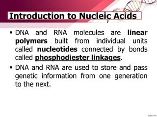
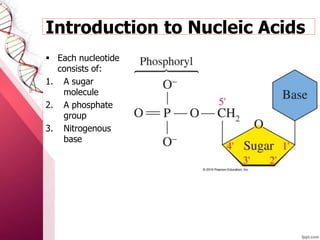
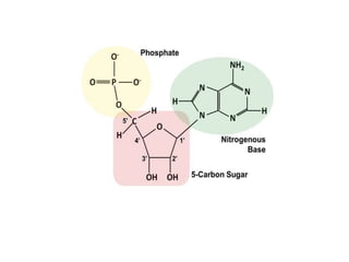
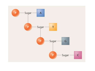
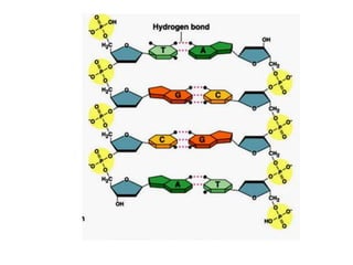
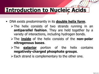
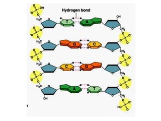
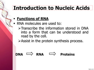
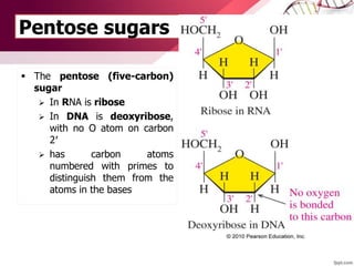
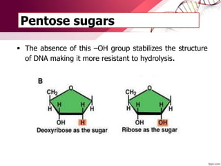
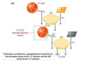
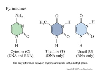
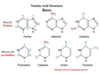
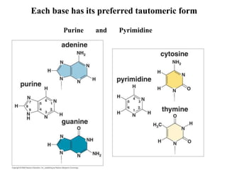
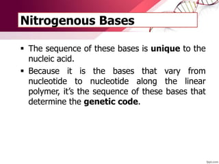
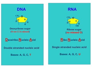
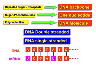
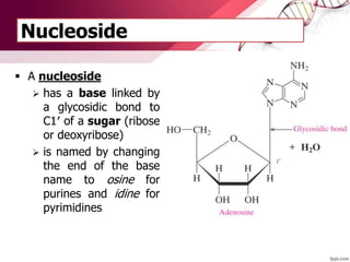
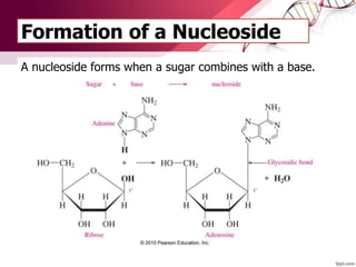
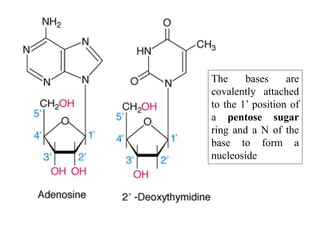
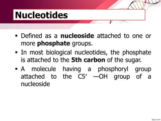
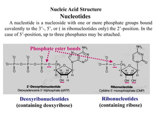
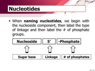
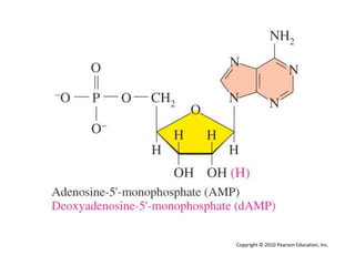
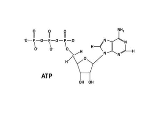
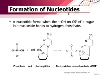
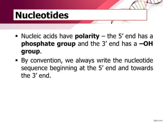
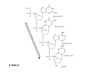
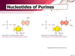
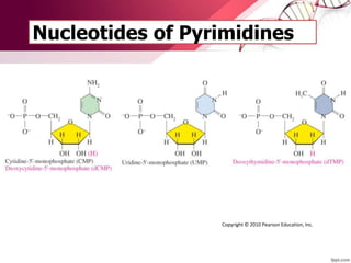
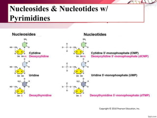
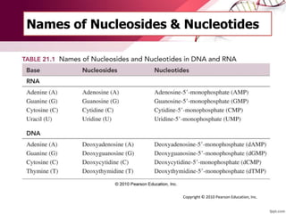
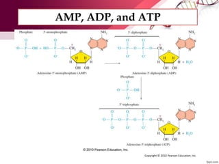
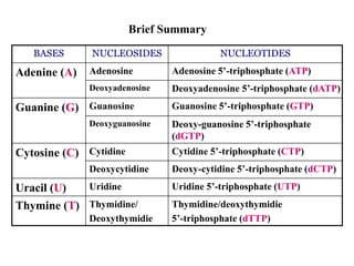
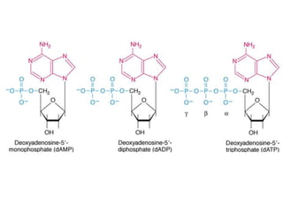
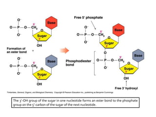
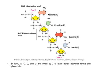
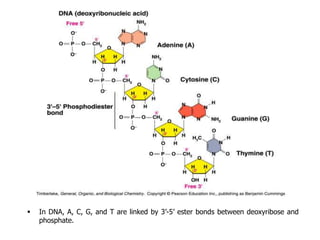
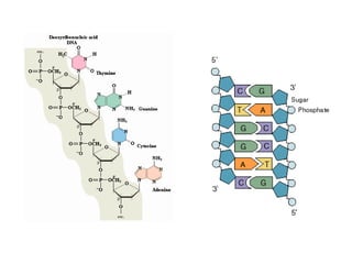
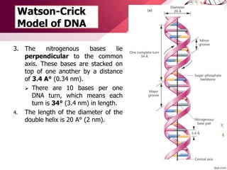
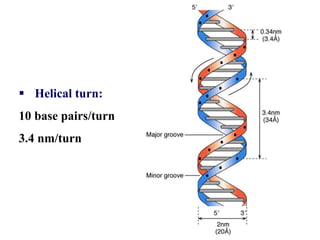
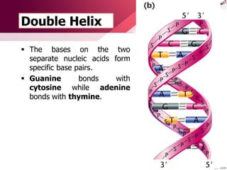
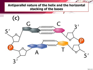
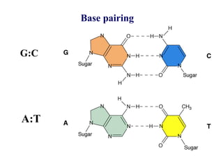
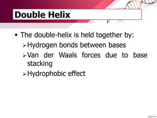
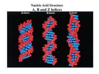
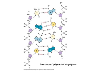
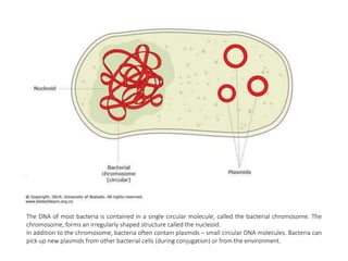
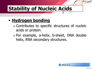
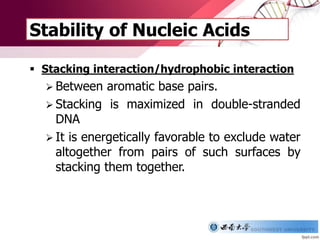
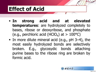
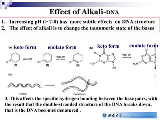
NUCLEIC ACIDS
Garry D. Lasaga
VPHY 241
Introduction to Nucleic Acids
 DNA and RNA molecules are linear
polymers built from individual units
called nucleotides connected by bonds
called phosphodiester linkages.
 DNA and RNA are used to store and pass
genetic information from one generation
to the next.
 Each nucleotide
consists of:
1. A sugar
molecule
2. A phosphate
group
3. Nitrogenous
base
Introduction to Nucleic Acids
 In both DNA and RNA, each nucleotide consists
of a linear backbone that consists of repeating
sugar-phosphate units.
 However, it’s the nitrogenous bases that are
responsible for storing the genetic information.
Introduction to Nucleic Acids
 DNA exists predominantly in its double helix form.
The helix consists of two strands running in an
antiparallel fashion. They are held together by a
variety of interactions, including hydrogen bonds.
The inside of the helix consists of the non-polar
nitrogenous bases.
The exterior portion of the helix contains
negatively-charged phosphate groups.
Each strand is complementary to the other one.
 Functions of DNA
 DNA molecules are used to:
 Store genetic information that can be
accessed by the organism and used to
build proteins.
 Pass down genetic information to
offspring.
Introduction to Nucleic Acids
 Functions of RNA
 RNA molecules are used to:
Transcribe the information stored in DNA
into a form that can be understood and
read by the cell.
Assist in the protein synthesis process.
DNA RNA Proteins
Introduction to Nucleic Acids
Composition of Nucleic
Acids
Pentose sugars
 The pentose (five-carbon)
sugar
 In RNA is ribose
 In DNA is deoxyribose,
with no O atom on carbon
2′
 has carbon atoms
numbered with primes to
distinguish them from the
atoms in the bases
Pentose sugars
 The absence of this –OH group stabilizes the structure
of DNA making it more resistant to hydrolysis.
 The sugars of nucleic acids are connected
to one another by 3’- to -5’
phosphodiester linkages.
 The –OH group on the 3rd carbon of one
sugar is connected to the –OH group on the
5th carbon of an adjacent sugar via a
phosphate group.
Backbone and Phosphodiester Bond
Nucleotides are linked by a phosphodiester bond between
the phosphate group at the C-5' position and the OH
group on the C-3' position
 This chain of repeating sugar-phosphate
units makes the backbone of the nucleic
acids.
 This backbone remains constant and
unchanged.
Backbone and Phosphodiester Bond
 The phosphate groups contain a negative
charge. This means that:
In an aqueous environment, these hydrophilic
regions interact with the polar H2O molecules
to stabilize the structure of DNA.
The phosphodiester linkages are much less
likely to be attacked by nucleophilic agents,
which means that they are less susceptible to
hydrolysis.
Backbone and Phosphodiester Bond
Nitrogenous Bases
 The bases vary from one nucleotide to the
next.
 There are two categories of bases – purines
and pyrimidines.
Nitrogenous Bases
 PURINES
 Consist of two fused ring structures.
 Both DNA and RNA have two types of
purines – adenine and guanine.
Nitrogenous Bases
 PYRIMIDINES
 Pyrimidines contain a single ring.
 DNA contain two pyrimidines – thymine
and cytosine.
 In RNA, the thymine is replaced by uracil.
The only difference between thymine and uracil is the methyl group.
Copyright © 2010 Pearson Education, Inc.
Bicyclic
Purines:
Thymine (T) is a 5-methyluracil (U)
Nucleic Acid Structure
Bases
Monocyclic
pyrimidine:
Each base has its preferred tautomeric form
Purine and Pyrimidine
Nitrogenous Bases
 The sequence of these bases is unique to the
nucleic acid.
 Because it is the bases that vary from
nucleotide to nucleotide along the linear
polymer, it’s the sequence of these bases that
determine the genetic code.
DNA RNA
o
H
H
H
H
H
CH2
Deoxyribose sugar
(O on C2 is missed)
o
OH
H
H
H
H
CH2
Ribose sugar
(no missed O)
Ribo-Nucleic-AcidDeoxiribo-Nucleic-Acid
Single stranded nucleic acidDouble stranded nucleic acid
Bases: A, G, C, T Bases: A, G, C, U
Repeated Sugar - Phosphate
Sugar–Phosphate-Base
Polynucleotide
DNA backbone
One nucleotide
DNA Molecule
DNA Double stranded
RNA single stranded
T C G A T A G
A G TC T A C
UUmRNA
DNA
Nucleosides and
Nucleotides
Nucleoside
 A nucleoside
 has a base linked by
a glycosidic bond to
C1′ of a sugar (ribose
or deoxyribose)
 is named by changing
the end of the base
name to osine for
purines and idine for
pyrimidines
Formation of a Nucleoside
A nucleoside forms when a sugar combines with a base.
The bases are
covalently attached
to the 1’ position of
a pentose sugar
ring and a N of the
base to form a
nucleoside
Nucleotides
 Defined as a nucleoside attached to one or
more phosphate groups.
 In most biological nucleotides, the phosphate
is attached to the 5th carbon of the sugar.
 A molecule having a phosphoryl group
attached to the C5′ —OH group of a
nucleoside
A nucleotide is a nucleoside with one or more phosphate groups bound
covalently to the 3’-, 5’, or ( in ribonucleotides only) the 2’-position. In the
case of 5’-position, up to three phosphates may be attached.
Deoxyribonucleotides
(containing deoxyribose)
Ribonucleotides
(containing ribose)
Phosphate ester bonds
Nucleic Acid Structure
Nucleotides
Nucleotides
 When naming nucleotides, we begin with
the nucleoside component, then label the type
of linkage and then label the # of phosphate
groups.
Nucleoside 5’ -Phosphate
Sugar base Linkage # of phosphates
Copyright © 2010 Pearson Education, Inc.
Formation of Nucleotides
 A nucleotide forms when the —OH on C5′ of a sugar
in a nucleoside bonds to hydrogen phosphate.
O
N
N
NH2
O
CH2HO
O
O OHP
O-
- +
O
N
N
NH2
O
CH2O
O
O-
P-
O
Phosphate and deoxycytidine Deoxycytidine monophosphate (dCMP)
Copyright © 2010 Pearson Education, Inc.
Formation of Nucleotides
Nucleotides
 Nucleic acids have polarity – the 5’ end has a
phosphate group and the 3’ end has a –OH
group.
 By convention, we always write the nucleotide
sequence beginning at the 5’ end and towards
the 3’ end.
5’-GTAC-3’
Nucleotides of Purines
Copyright © 2010 Pearson Education, Inc.
Nucleotides of Pyrimidines
Copyright © 2010 Pearson Education, Inc.
Nucleosides & Nucleotides w/
Pyrimidines
Copyright © 2010 Pearson Education, Inc.
Names of Nucleosides & Nucleotides
Copyright © 2010 Pearson Education, Inc.
AMP, ADP, and ATP
Copyright © 2010 Pearson Education, Inc.
BASES NUCLEOSIDES NUCLEOTIDES
Adenine (A) Adenosine Adenosine 5’-triphosphate (ATP)
Deoxyadenosine Deoxyadenosine 5’-triphosphate (dATP)
Guanine (G) Guanosine Guanosine 5’-triphosphate (GTP)
Deoxyguanosine Deoxy-guanosine 5’-triphosphate
(dGTP)
Cytosine (C) Cytidine Cytidine 5’-triphosphate (CTP)
Deoxycytidine Deoxy-cytidine 5’-triphosphate (dCTP)
Uracil (U) Uridine Uridine 5’-triphosphate (UTP)
Thymine (T) Thymidine/
Deoxythymidie
Thymidine/deoxythymidie
5’-triphosphate (dTTP)
Brief Summary
PRIMARY STRUCTURE OF
NUCLEIC ACIDS
Primary Structure of Nucleic Acids
In the primary structure of nucleic acids:
 Nucleotides are joined by phosphodiester
bonds.
The 3’-OH group of the sugar in one nucleotide forms an ester bond to the phosphate
group on the 5’-carbon of the sugar of the next nucleotide.
Structure of Nucleic Acids
A nucleic acid polymer:
 Has a free 5’-phosphate
group at one end and a free
3’-OH group at the other
end.
 Is read from the free 5’-end
using the letters of the
bases.
 This example reads
5’—A—C—G—T—3’.
 In RNA, A, C, G, and U are linked by 3’-5’ ester bonds between ribose and
phosphate.
 In DNA, A, C, G, and T are linked by 3’-5’ ester bonds between deoxyribose and
phosphate.
WATSON-CRICK MODEL OF DNA
Watson-Crick Model of DNA
 Maurice Wilkins and
Rosalind Franklin were
able to obtain x-ray
diffraction
photographs of a DNA
fiber.
 These diffraction patterns
indicated that DNA had a
helical structure.
Watson-Crick Model of DNA
 Deduced by James
Watson and Francis Crick
in 1953.
 DNA is the genetic
material of all organisms
except for some viruses.
 Essential for replicating
DNA and transcribing RNA
 The foundation of the
molecular biology
Watson-Crick
Model of DNA
 James Watson and
Francis Crick deduced
the features of the
structure of DNA.
1. A single DNA molecule
consists of two
individual nucleic acid
chains that wind
about a common
axis. These two
polynucleotide chains
run in opposite
directions.
The two strands of the double helix are held together by base
pairing in an anti-parallel orientation
A:T & G:C base
pairs
Watson-Crick
Model of DNA
2. The backbone of the
nucleic acids is found
on the exterior of the
DNA while the
nitrogenous bases
are found on the
inside.
Watson-Crick
Model of DNA
3. The nitrogenous bases lie
perpendicular to the common
axis. These bases are stacked on
top of one another by a distance
of 3.4 A° (0.34 nm).
 There are 10 bases per one
DNA turn, which means each
turn is 34° (3.4 nm) in length.
4. The length of the diameter of the
double helix is 20 A° (2 nm).
 Helical turn:
10 base pairs/turn
3.4 nm/turn
Double Helix
 The bases on the two
separate nucleic acids form
specific base pairs.
 Guanine bonds with
cytosine while adenine
bonds with thymine.
Antiparallel nature of the helix and the horizontal
stacking of the bases
Base pairing
A:T
G:C
Double Helix
 The double-helix is held together by:
Hydrogen bonds between bases
Van der Waals forces due to base
stacking
Hydrophobic effect
A-form B-form Z-form
Nucleic Acid Structure
A, B and Z helices
Structure of polynucleotide polymer
The DNA of most bacteria is contained in a single circular molecule, called the bacterial chromosome. The
chromosome, forms an irregularly shaped structure called the nucleoid.
In addition to the chromosome, bacteria often contain plasmids – small circular DNA molecules. Bacteria can
pick up new plasmids from other bacterial cells (during conjugation) or from the environment.
CHEMICAL & PHYSICAL PROPERTIES
OF NUCLEIC ACIDS
Stability of Nucleic Acids
 Hydrogen bonding
 Contributes to specific structures of nucleic
acids or protein.
 For example, a-helix, b-sheet, DNA double
helix, RNA secondary structures.
Stability of Nucleic Acids
 Stacking interaction/hydrophobic interaction
 Between aromatic base pairs.
 Stacking is maximized in double-stranded
DNA
 It is energetically favorable to exclude water
altogether from pairs of such surfaces by
stacking them together.
Effect of Acid
 In strong acid and at elevated
temperatures: are hydrolyzed completely to
bases, ribose or deoxyribose, and phosphate
(e.g., perchloric acid (HClO4) at > 100°C)
 In more dilute mineral acid (e.g., pH 3–4), the
most easily hydrolyzed bonds are selectively
broken. E.g., glycosydic bonds attaching
purine bases to the ribose ring are broken by
formic acid.
Effect of Alkali-DNA
keto form enolate form keto form enolate form
3. This affects the specific hydrogen bonding between the base pairs, with
the result that the double-stranded structure of the DNA breaks down;
that is the DNA becomes denatured .
1. Increasing pH (> 7-8) has more subtle effects on DNA structure
2. The effect of alkali is to change the tautomeric state of the bases
Chemical Denaturation
A number of chemical agents can cause the denaturation of
DNA or RNA at neutral pH, e.g. Urea (H2NCONH2) is used in
denaturing PAGE; Formamide (HCONH2) is used Southern
and Northern blotting.
Disrupting the hydrogen bonding of the bulk water
solution
Hydrophobic effect (stacking interaction) is reduced
Denaturation of the strands
Mechanism
Spectroscopic and Thermal
Properties of Nucleic Acids
 Nucleic acids absorb UV light due to the conjugated
aromatic nature of the bases
 The wavelength of maximum absorption of light by
both DNA and RNA is 260 nm (lmax = 260 nm)
 Applications: can be used for detection, quantitation
and assessment of purity (A260/A280)
UV Absorption
Thermal denaturation/melting
Heating also leads to the destruction of double-stranded
hydrogen-bonded regions of DNA and RNA.
RNA: the absorbance increases gradually and irregularly
DNA: the absorbance increases cooperatively.
melting temperature (Tm): the temperature at the mid-point of the
smooth transition, which has a 20% increase in absorbance. 80-100 °C for
long DNA molecules

Nucleic Acids and DNA

  • 1.
    NUCLEIC ACIDS Garry D.Lasaga VPHY 241
  • 2.
    Introduction to NucleicAcids  DNA and RNA molecules are linear polymers built from individual units called nucleotides connected by bonds called phosphodiester linkages.  DNA and RNA are used to store and pass genetic information from one generation to the next.
  • 3.
     Each nucleotide consistsof: 1. A sugar molecule 2. A phosphate group 3. Nitrogenous base Introduction to Nucleic Acids
  • 6.
    Introduction to NucleicAcids  In both DNA and RNA, each nucleotide consists of a linear backbone that consists of repeating sugar-phosphate units.  However, it’s the nitrogenous bases that are responsible for storing the genetic information.
  • 8.
    Introduction to NucleicAcids  DNA exists predominantly in its double helix form. The helix consists of two strands running in an antiparallel fashion. They are held together by a variety of interactions, including hydrogen bonds. The inside of the helix consists of the non-polar nitrogenous bases. The exterior portion of the helix contains negatively-charged phosphate groups. Each strand is complementary to the other one.
  • 10.
     Functions ofDNA  DNA molecules are used to:  Store genetic information that can be accessed by the organism and used to build proteins.  Pass down genetic information to offspring. Introduction to Nucleic Acids
  • 11.
     Functions ofRNA  RNA molecules are used to: Transcribe the information stored in DNA into a form that can be understood and read by the cell. Assist in the protein synthesis process. DNA RNA Proteins Introduction to Nucleic Acids
  • 12.
  • 13.
    Pentose sugars  Thepentose (five-carbon) sugar  In RNA is ribose  In DNA is deoxyribose, with no O atom on carbon 2′  has carbon atoms numbered with primes to distinguish them from the atoms in the bases
  • 14.
    Pentose sugars  Theabsence of this –OH group stabilizes the structure of DNA making it more resistant to hydrolysis.
  • 15.
     The sugarsof nucleic acids are connected to one another by 3’- to -5’ phosphodiester linkages.  The –OH group on the 3rd carbon of one sugar is connected to the –OH group on the 5th carbon of an adjacent sugar via a phosphate group. Backbone and Phosphodiester Bond
  • 16.
    Nucleotides are linkedby a phosphodiester bond between the phosphate group at the C-5' position and the OH group on the C-3' position
  • 17.
     This chainof repeating sugar-phosphate units makes the backbone of the nucleic acids.  This backbone remains constant and unchanged. Backbone and Phosphodiester Bond
  • 18.
     The phosphategroups contain a negative charge. This means that: In an aqueous environment, these hydrophilic regions interact with the polar H2O molecules to stabilize the structure of DNA. The phosphodiester linkages are much less likely to be attacked by nucleophilic agents, which means that they are less susceptible to hydrolysis. Backbone and Phosphodiester Bond
  • 19.
    Nitrogenous Bases  Thebases vary from one nucleotide to the next.  There are two categories of bases – purines and pyrimidines.
  • 20.
    Nitrogenous Bases  PURINES Consist of two fused ring structures.  Both DNA and RNA have two types of purines – adenine and guanine.
  • 22.
    Nitrogenous Bases  PYRIMIDINES Pyrimidines contain a single ring.  DNA contain two pyrimidines – thymine and cytosine.  In RNA, the thymine is replaced by uracil.
  • 23.
    The only differencebetween thymine and uracil is the methyl group. Copyright © 2010 Pearson Education, Inc.
  • 24.
    Bicyclic Purines: Thymine (T) isa 5-methyluracil (U) Nucleic Acid Structure Bases Monocyclic pyrimidine:
  • 25.
    Each base hasits preferred tautomeric form Purine and Pyrimidine
  • 26.
    Nitrogenous Bases  Thesequence of these bases is unique to the nucleic acid.  Because it is the bases that vary from nucleotide to nucleotide along the linear polymer, it’s the sequence of these bases that determine the genetic code.
  • 27.
    DNA RNA o H H H H H CH2 Deoxyribose sugar (Oon C2 is missed) o OH H H H H CH2 Ribose sugar (no missed O) Ribo-Nucleic-AcidDeoxiribo-Nucleic-Acid Single stranded nucleic acidDouble stranded nucleic acid Bases: A, G, C, T Bases: A, G, C, U
  • 28.
    Repeated Sugar -Phosphate Sugar–Phosphate-Base Polynucleotide DNA backbone One nucleotide DNA Molecule DNA Double stranded RNA single stranded T C G A T A G A G TC T A C UUmRNA DNA
  • 29.
  • 30.
    Nucleoside  A nucleoside has a base linked by a glycosidic bond to C1′ of a sugar (ribose or deoxyribose)  is named by changing the end of the base name to osine for purines and idine for pyrimidines
  • 31.
    Formation of aNucleoside A nucleoside forms when a sugar combines with a base.
  • 32.
    The bases are covalentlyattached to the 1’ position of a pentose sugar ring and a N of the base to form a nucleoside
  • 33.
    Nucleotides  Defined asa nucleoside attached to one or more phosphate groups.  In most biological nucleotides, the phosphate is attached to the 5th carbon of the sugar.  A molecule having a phosphoryl group attached to the C5′ —OH group of a nucleoside
  • 34.
    A nucleotide isa nucleoside with one or more phosphate groups bound covalently to the 3’-, 5’, or ( in ribonucleotides only) the 2’-position. In the case of 5’-position, up to three phosphates may be attached. Deoxyribonucleotides (containing deoxyribose) Ribonucleotides (containing ribose) Phosphate ester bonds Nucleic Acid Structure Nucleotides
  • 35.
    Nucleotides  When namingnucleotides, we begin with the nucleoside component, then label the type of linkage and then label the # of phosphate groups. Nucleoside 5’ -Phosphate Sugar base Linkage # of phosphates
  • 36.
    Copyright © 2010Pearson Education, Inc.
  • 38.
    Formation of Nucleotides A nucleotide forms when the —OH on C5′ of a sugar in a nucleoside bonds to hydrogen phosphate. O N N NH2 O CH2HO O O OHP O- - + O N N NH2 O CH2O O O- P- O Phosphate and deoxycytidine Deoxycytidine monophosphate (dCMP) Copyright © 2010 Pearson Education, Inc.
  • 39.
  • 40.
    Nucleotides  Nucleic acidshave polarity – the 5’ end has a phosphate group and the 3’ end has a –OH group.  By convention, we always write the nucleotide sequence beginning at the 5’ end and towards the 3’ end.
  • 41.
  • 42.
    Nucleotides of Purines Copyright© 2010 Pearson Education, Inc.
  • 43.
    Nucleotides of Pyrimidines Copyright© 2010 Pearson Education, Inc.
  • 44.
    Nucleosides & Nucleotidesw/ Pyrimidines Copyright © 2010 Pearson Education, Inc.
  • 45.
    Names of Nucleosides& Nucleotides Copyright © 2010 Pearson Education, Inc.
  • 46.
    AMP, ADP, andATP Copyright © 2010 Pearson Education, Inc.
  • 47.
    BASES NUCLEOSIDES NUCLEOTIDES Adenine(A) Adenosine Adenosine 5’-triphosphate (ATP) Deoxyadenosine Deoxyadenosine 5’-triphosphate (dATP) Guanine (G) Guanosine Guanosine 5’-triphosphate (GTP) Deoxyguanosine Deoxy-guanosine 5’-triphosphate (dGTP) Cytosine (C) Cytidine Cytidine 5’-triphosphate (CTP) Deoxycytidine Deoxy-cytidine 5’-triphosphate (dCTP) Uracil (U) Uridine Uridine 5’-triphosphate (UTP) Thymine (T) Thymidine/ Deoxythymidie Thymidine/deoxythymidie 5’-triphosphate (dTTP) Brief Summary
  • 49.
  • 50.
    Primary Structure ofNucleic Acids In the primary structure of nucleic acids:  Nucleotides are joined by phosphodiester bonds.
  • 51.
    The 3’-OH groupof the sugar in one nucleotide forms an ester bond to the phosphate group on the 5’-carbon of the sugar of the next nucleotide.
  • 52.
    Structure of NucleicAcids A nucleic acid polymer:  Has a free 5’-phosphate group at one end and a free 3’-OH group at the other end.  Is read from the free 5’-end using the letters of the bases.  This example reads 5’—A—C—G—T—3’.
  • 53.
     In RNA,A, C, G, and U are linked by 3’-5’ ester bonds between ribose and phosphate.
  • 54.
     In DNA,A, C, G, and T are linked by 3’-5’ ester bonds between deoxyribose and phosphate.
  • 55.
  • 56.
    Watson-Crick Model ofDNA  Maurice Wilkins and Rosalind Franklin were able to obtain x-ray diffraction photographs of a DNA fiber.  These diffraction patterns indicated that DNA had a helical structure.
  • 57.
    Watson-Crick Model ofDNA  Deduced by James Watson and Francis Crick in 1953.  DNA is the genetic material of all organisms except for some viruses.  Essential for replicating DNA and transcribing RNA  The foundation of the molecular biology
  • 58.
    Watson-Crick Model of DNA James Watson and Francis Crick deduced the features of the structure of DNA. 1. A single DNA molecule consists of two individual nucleic acid chains that wind about a common axis. These two polynucleotide chains run in opposite directions.
  • 59.
    The two strandsof the double helix are held together by base pairing in an anti-parallel orientation A:T & G:C base pairs
  • 60.
    Watson-Crick Model of DNA 2.The backbone of the nucleic acids is found on the exterior of the DNA while the nitrogenous bases are found on the inside.
  • 62.
    Watson-Crick Model of DNA 3.The nitrogenous bases lie perpendicular to the common axis. These bases are stacked on top of one another by a distance of 3.4 A° (0.34 nm).  There are 10 bases per one DNA turn, which means each turn is 34° (3.4 nm) in length. 4. The length of the diameter of the double helix is 20 A° (2 nm).
  • 63.
     Helical turn: 10base pairs/turn 3.4 nm/turn
  • 64.
    Double Helix  Thebases on the two separate nucleic acids form specific base pairs.  Guanine bonds with cytosine while adenine bonds with thymine.
  • 65.
    Antiparallel nature ofthe helix and the horizontal stacking of the bases
  • 66.
  • 67.
    Double Helix  Thedouble-helix is held together by: Hydrogen bonds between bases Van der Waals forces due to base stacking Hydrophobic effect
  • 68.
    A-form B-form Z-form NucleicAcid Structure A, B and Z helices
  • 69.
  • 70.
    The DNA ofmost bacteria is contained in a single circular molecule, called the bacterial chromosome. The chromosome, forms an irregularly shaped structure called the nucleoid. In addition to the chromosome, bacteria often contain plasmids – small circular DNA molecules. Bacteria can pick up new plasmids from other bacterial cells (during conjugation) or from the environment.
  • 72.
    CHEMICAL & PHYSICALPROPERTIES OF NUCLEIC ACIDS
  • 73.
    Stability of NucleicAcids  Hydrogen bonding  Contributes to specific structures of nucleic acids or protein.  For example, a-helix, b-sheet, DNA double helix, RNA secondary structures.
  • 74.
    Stability of NucleicAcids  Stacking interaction/hydrophobic interaction  Between aromatic base pairs.  Stacking is maximized in double-stranded DNA  It is energetically favorable to exclude water altogether from pairs of such surfaces by stacking them together.
  • 75.
    Effect of Acid In strong acid and at elevated temperatures: are hydrolyzed completely to bases, ribose or deoxyribose, and phosphate (e.g., perchloric acid (HClO4) at > 100°C)  In more dilute mineral acid (e.g., pH 3–4), the most easily hydrolyzed bonds are selectively broken. E.g., glycosydic bonds attaching purine bases to the ribose ring are broken by formic acid.
  • 76.
    Effect of Alkali-DNA ketoform enolate form keto form enolate form 3. This affects the specific hydrogen bonding between the base pairs, with the result that the double-stranded structure of the DNA breaks down; that is the DNA becomes denatured . 1. Increasing pH (> 7-8) has more subtle effects on DNA structure 2. The effect of alkali is to change the tautomeric state of the bases
  • 77.
    Chemical Denaturation A numberof chemical agents can cause the denaturation of DNA or RNA at neutral pH, e.g. Urea (H2NCONH2) is used in denaturing PAGE; Formamide (HCONH2) is used Southern and Northern blotting. Disrupting the hydrogen bonding of the bulk water solution Hydrophobic effect (stacking interaction) is reduced Denaturation of the strands Mechanism
  • 78.
  • 79.
     Nucleic acidsabsorb UV light due to the conjugated aromatic nature of the bases  The wavelength of maximum absorption of light by both DNA and RNA is 260 nm (lmax = 260 nm)  Applications: can be used for detection, quantitation and assessment of purity (A260/A280) UV Absorption
  • 80.
    Thermal denaturation/melting Heating alsoleads to the destruction of double-stranded hydrogen-bonded regions of DNA and RNA. RNA: the absorbance increases gradually and irregularly DNA: the absorbance increases cooperatively. melting temperature (Tm): the temperature at the mid-point of the smooth transition, which has a 20% increase in absorbance. 80-100 °C for long DNA molecules

Editor's Notes

  • #3 Molecules that store information for cellular growth and reproduction Large molecules consisting of long chains of monomers called nucleotides
  • #17 Phosphodiester means we have the phosphate group in between these 2 ester bonds. The bases are attached to the carbon 1 our the sugar molecule.
  • #20 Changes in bases allow the DNA to store genetic information.
  • #22 NH2 (Adenine) – C=O (Guanine); NH2 (Guanine) *Play an important role in determining the types and number of interactions that are formed between the different bases in the DNA molecule.
  • #31 • The bond holding the sugar to the base is a β-glycosidic bond. Notice that the base group lies above the plane of the sugar molecule.
  • #47 Adding phosphate groups to AMP forms the diphosphate ADP and the triphosphate ATP.
  • #52 The 3’-OH group of the sugar in one nucleotide forms an ester bond to the phosphate group on the 5’-carbon of the sugar of the next nucleotide.
  • #54 Example of RNA
  • #55 Example of DNA
  • #59 The DNA double helix as proposed by Watson and Crick
  • #75 Even in single-stranded DNA, the bases have a tendency to stack on top of each other.