NUCLEIC ACIDS
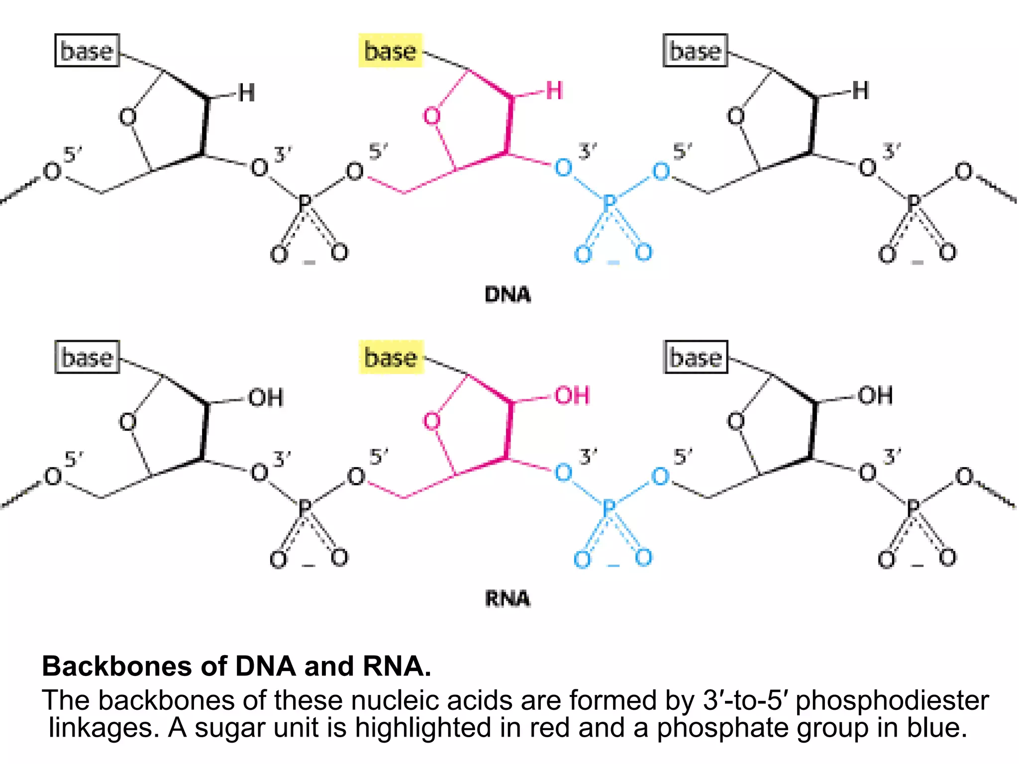
 Nucleic acids are made up of
nucleotides.
 Nucleic acids are RNA and DNA
–Ribonucleic Acid (RNA) and
–Deoxyribonucleic Acid (DNA)
 Nucleotides are essential for all cells since
they make DNA and RNA
 DNA and RNA are the genetic material for the
cells
 Without DNA and RNA
 Protein can not be synthesized
 Cells can not proliferate
 Nucleotides serve as carriers of activated
intermediates in the synthesis of some:
–Carbohydrate
–Lipids and
–Proteins
 Nucleotides are structural components of
several essential co-enzymes:
–Coenzyme A
–NAD+
–NADP+
 Serve as the second messengers in signal
transduction pathways:
 cyclic adenosine monophosphate (cAMP)
 cyclic guanosine monophosphate (cGMP).
 Nucleotide plays an important role as Energy
currency in the cell:
–ATP
–GTP
 Nucleotides are important regulatory
compounds for many of the pathways of
intermediary metabolism, inhibiting or
activating key enzymes
Nitrogen Bases
 There are two kinds of nitrogen-containing
bases Purines and Pyrimidines
 Purines -2 rings (Six-membered and a Five-
membered nitrogen- containing rings fused
together.)
 Pyridmidines –One ring (Six-membered
nitrogen-containing ring.)
 There are 4 Purines and 4 Pyrimidines that
are important in Biochemistry
Purines
 Adenine = 6-amino purine
 Guanine = 2-amino-6-oxy purine
 Hypoxanthine = 6-oxy purine
 Xanthine = 2,6-dioxy purine
Adenine and Guanine are found in
both DNA and RNA
Hypoxanthine and Xanthine are not
found in nucleic acids, but are
important intermediates in the
synthesis and degradation of purine
nucleotides
Pyrimidines
 Uracil = 2,4-dioxy pyrimidine
 Thymine = 2,4-dioxy-5-methyl pyrimidine
 Cytosine = 2-oxy-4-amino pyrimidine
 Orotic acid = 2,4-dioxy-6-carboxy pyrimidine
 Cytosine is found in both DNA and RNA
 Uracil is found only in RNA
 Thymine is normally found in DNA and not in
RNA
 Sometimes tRNA may contain some Thymine
as well as Uracil
Nucleosides
 Results from addition of a sugar to the nitrogen
base, the sugar may be either ribose or 2-
deoxyribose
 Carbon 1 of the sugar is attached to nitrogen 9
of a purine base, or to nitrogen 1 of a
pyrimidine base
• Names of purine nucleosides end in osine
and examples are: Adenosine, Guanosine
and Inosine (from Hypoxanthine)
• The names of pyrimidine nucleosides end in
–idine eg: Uridine, Thymidine, Cytidine
 The numbering of the ring atoms of the base is
written normally, while we use a number and a
prime sign eg. 1', 2', to distinguish the ring
atoms of the sugar.
Nucleotides
 Results from addition of 1 or more phosphates
to the sugar portion of a nucleoside
 Generally, the phosphate is in ester linkage to
carbon 5' of the sugar
 E.g, 3'-5' cAMP indicates that a phosphate is in
ester linkage to both the 3' and 5' hydroxyl
groups of an adenosine molecule and forms a
cyclic structure.
 Some representative names are:
- AMP = Adenosine Monophosphate
- CDP = Cytidine Diphosphate
- dGTP = deoxy Guanosine Triphosphate
- dTTP = deoxy Thymidine Triphosphate (TTP)
- cAMP = 3'-5' cyclic Adenosine Monophosphate
Polynucleotides
 Nucleotides are joined together by 3'-5'
phosphodiester bonds to form
polynucleotides.
 Polymerization of ribonucleotides will
produce an RNA while polymerization of
deoxyribonucleotides leads to DNA.
DNA, RNA, and the Flow of Genetic
Information
 DNA and RNA are long linear polymers that
carry information in a form that can be passed
from one generation to the next
 These macromolecules consist of a large
number of linked nucleotides
 Sugars linked by phosphates form a common
backbone whereas the bases vary among four
kinds
 Genetic information is stored in the sequence
of bases along a nucleic acid chain
 The bases have an additional special property
of forming specific pairs with one another that
are stabilized by hydrogen bonds
 The base pairing results in the formation of a
double helix structure consisting of 2 strands
 These base pairs provide a mechanism for
copying the genetic information in an existing
nucleic acid chain to form a new chain
 DNA is replicated by the action of DNA
polymerase enzymes.
 DNA also is the template for synthesis of
RNAs
 RNA, especially messenger RNA (mRNA)
forms the template for protein synthesis
 Other RNA molecules, such as transfer RNA
(tRNA) and ribosomal RNA (rRNA), are part of
the protein-synthesizing machinery
Polymeric Structure of Nucleic Acids
 A monomeric unit is a nucleotide consisting 3
components: a sugar, a phosphate, and a base
 The phosphate and a sugar does not change from
one nucleotide to the other but the sequence of bases
changes.
 The change in base sequences represents a form of
linear information.
RNA and DNA Differ in the Sugar Component
and One of the Bases
 The sugar in deoxyribonucleic acid (DNA) is
deoxyribose while the sugar in RNA is ribose
Ribose and
Deoxyribose
Atoms in the sugar
are numbered with
primes to distinguish
them from atoms in
bases
 The sugars in nucleic acids are linked to one
another by phosphodiester bridges
 Specifically, the 3′-hydroxyl (3′-OH) group of
the sugar moiety of one nucleotide is esterified
to a phosphate group,
 which is joined to the 5′-hydroxyl group of the
adjacent sugar.
 In addition to the standard 3′→5′ linkage, a
2′→5′ linkage is possible for RNA, which is
important for the formation of mature RNA
Backbones of DNA and RNA.
The backbones of these nucleic acids are formed by 3′-to-5′ phosphodiester
linkages. A sugar unit is highlighted in red and a phosphate group in blue.
Backbones of DNA
and RNA.
The backbones of
these nucleic acids
are formed by 3′-to-5′
phosphodiester
linkages. A sugar
unit is highlighted in
red and a phosphate
group in blue.
 Whereas the backbone is constant in DNA and
RNA, the bases vary from one monomer to the
next
 Two of the bases are derivatives of purine
— Adenine (A) and Guanine (G)
 and two of pyrimidine
— Cytosine (C) and Thymine (T, DNA only) or
Uracil (U, RNA only),
 Note that each phosphodiester bridge has a
negative charge.
 This negative charge repels nucleophilic
species such as hydroxide ion;
 As a result, phosphodiester linkages are much
less susceptible to hydrolytic attack than are
other esters such as carboxylic acid esters.
 This resistance is crucial for maintaining the
integrity of information stored in nucleic acids.
 The absence of the 2′-hydroxyl group in DNA
further increases its resistance to hydrolysis
 The greater stability of DNA accounts for its
use rather than RNA as the hereditary material
in all modern cells and in many viruses
 DIRECTIONALITY OF NUCLEOTIDE CHAINS
 Note that, a DNA chain has polarity
 One end of the chain has a free 5′-OH group
(or a 5′-OH group attached to a phosphate),
whereas the other end has a 3′-OH group,
neither of which is linked to another nucleotide
 By convention, the base sequence is written in
the 5′-to-3′ direction.
 Thus, the symbol ACG indicates that the
unlinked 5′-OH group is on deoxyadenylate,
whereas the unlinked 3′-OH group is on
deoxyguanylate.
 Because of this polarity, ACG and GCA
correspond to different compounds.
 A striking characteristic of naturally occurring
DNA molecules is their length.
 A DNA molecule must comprise many
nucleotides to carry the genetic information
necessary for even the simplest organisms
 E.g, the RNA of a virus such as HIV, which
causes AIDS, is 9719 nucleotides in length
 The E. coli genome is a single DNA molecule
consisting of 4.6 million nucleotides( 4.6 x 10⁶
base pairs =4.6 x 10³ kilo base pairs) kbp
DNA molecules from higher organisms can be
much larger
The human genome comprises of 3 billion
nucleotides [3 x 10⁹], divided among 23
distinct DNA molecules (chromosomes) of
different sizes.
One of the largest known DNA molecules is
found in the Indian muntjak, an Asiatic deer; its
genome is nearly as large as the human
genome but is distributed on only 3
chromosomes
The Indian Muntjak
and Its
Chromosomes.
Cells from a female
Indian muntjak
contain three pairs
of very large
chromosomes
(stained orange).
The cell shown is a
hybrid containing a
pair of human
chromosomes
(stained green) for
comparison.
 The largest of these chromosomes has chains
of more than 1 billion nucleotides
 If such a DNA molecule could be fully
extended, it would stretch more than 1 foot in
length
The Double Helix is Stabilized by Hydrogen
Bonds and Hydrophobic Interactions
 In Nucleic acids, there is specific base-pairing
interactions
 This was discovered in the studies directed at
determining the three-dimensional structure of
DNA
 It was shown that DNA was formed of two
chains that wound in a regular helical structure
Watson-Crick Model of
Double-Helical DNA.
One polynucleotide chain
is shown in blue and the
other in red.
The features of the Watson-Crick model of DNA
deduced from the diffraction patterns are:
1. Two helical polynucleotide chains are coiled
around a common axis. The chains run in
opposite directions.
2. The sugar-phosphate backbones are on the
outside and, therefore, the purine and
pyrimidine bases lie on the inside of the helix.
3. The bases are nearly perpendicular to the helix
axis, and adjacent bases are separated by
3.4 Å.
– The helical structure repeats every 34 Å, so there
are 10 bases (= 34 Å per repeat or 3.4 Å per base)
per turn of helix.
– There is a rotation of 36 degrees per base (360
degrees per full turn or 10 bases per turn).
4. The diameter of the helix is 20 Å.
 How is such a regular structure able to
accommodate the sequence of bases, given
the different sizes and shapes of the purines
and pyrimidines?
 Watson and Crick discovered that:
–Guanine can be paired with Cytosine and
–Adenine with Thymine to form base pairs that
have essentially the same shape
 A=T and G=C
Structures of
the Base Pairs
Proposed by
Watson and
Crick
 The bases are held together by hydrogen
bonds.
 Erwin Chargaff reported, The ratios of Adenine
to Thymine and of Guanine to Cytosine were
nearly the same in all species studied.
 All the Adenine:Thymine and
Guanine:Cytosine ratios are close to 1,
 whereas the adenine-to-guanine ratio varies.
Base compositions experimentally determined
for a variety of organisms
Species A:T G:C A:G
Human being 1.00 1.00 1.56
Salmon 1.02 1.02 1.43
Wheat 1.00 0.97 1.22
Yeast 1.03 1.02 1.67
Escherichia coli 1.09 0.99 1.05
Serratia
marcescens
0.95 0.86 0.70
Axial View of
DNA.
Base pairs
are stacked
nearly one on
top of
another in
the double
helix.

Nucleic acids 2016 Dodoma Nursing-1.ppt

  • 1.
  • 2.
     Nucleic acidsare made up of nucleotides.  Nucleic acids are RNA and DNA –Ribonucleic Acid (RNA) and –Deoxyribonucleic Acid (DNA)
  • 3.
     Nucleotides areessential for all cells since they make DNA and RNA  DNA and RNA are the genetic material for the cells  Without DNA and RNA  Protein can not be synthesized  Cells can not proliferate
  • 4.
     Nucleotides serveas carriers of activated intermediates in the synthesis of some: –Carbohydrate –Lipids and –Proteins  Nucleotides are structural components of several essential co-enzymes: –Coenzyme A –NAD+ –NADP+
  • 6.
     Serve asthe second messengers in signal transduction pathways:  cyclic adenosine monophosphate (cAMP)  cyclic guanosine monophosphate (cGMP).
  • 7.
     Nucleotide playsan important role as Energy currency in the cell: –ATP –GTP  Nucleotides are important regulatory compounds for many of the pathways of intermediary metabolism, inhibiting or activating key enzymes
  • 8.
    Nitrogen Bases  Thereare two kinds of nitrogen-containing bases Purines and Pyrimidines  Purines -2 rings (Six-membered and a Five- membered nitrogen- containing rings fused together.)  Pyridmidines –One ring (Six-membered nitrogen-containing ring.)
  • 9.
     There are4 Purines and 4 Pyrimidines that are important in Biochemistry Purines  Adenine = 6-amino purine  Guanine = 2-amino-6-oxy purine  Hypoxanthine = 6-oxy purine  Xanthine = 2,6-dioxy purine
  • 11.
    Adenine and Guanineare found in both DNA and RNA Hypoxanthine and Xanthine are not found in nucleic acids, but are important intermediates in the synthesis and degradation of purine nucleotides
  • 12.
    Pyrimidines  Uracil =2,4-dioxy pyrimidine  Thymine = 2,4-dioxy-5-methyl pyrimidine  Cytosine = 2-oxy-4-amino pyrimidine  Orotic acid = 2,4-dioxy-6-carboxy pyrimidine
  • 14.
     Cytosine isfound in both DNA and RNA  Uracil is found only in RNA  Thymine is normally found in DNA and not in RNA  Sometimes tRNA may contain some Thymine as well as Uracil
  • 15.
    Nucleosides  Results fromaddition of a sugar to the nitrogen base, the sugar may be either ribose or 2- deoxyribose  Carbon 1 of the sugar is attached to nitrogen 9 of a purine base, or to nitrogen 1 of a pyrimidine base
  • 17.
    • Names ofpurine nucleosides end in osine and examples are: Adenosine, Guanosine and Inosine (from Hypoxanthine) • The names of pyrimidine nucleosides end in –idine eg: Uridine, Thymidine, Cytidine  The numbering of the ring atoms of the base is written normally, while we use a number and a prime sign eg. 1', 2', to distinguish the ring atoms of the sugar.
  • 18.
    Nucleotides  Results fromaddition of 1 or more phosphates to the sugar portion of a nucleoside  Generally, the phosphate is in ester linkage to carbon 5' of the sugar
  • 19.
     E.g, 3'-5'cAMP indicates that a phosphate is in ester linkage to both the 3' and 5' hydroxyl groups of an adenosine molecule and forms a cyclic structure.  Some representative names are: - AMP = Adenosine Monophosphate - CDP = Cytidine Diphosphate - dGTP = deoxy Guanosine Triphosphate - dTTP = deoxy Thymidine Triphosphate (TTP) - cAMP = 3'-5' cyclic Adenosine Monophosphate
  • 21.
    Polynucleotides  Nucleotides arejoined together by 3'-5' phosphodiester bonds to form polynucleotides.  Polymerization of ribonucleotides will produce an RNA while polymerization of deoxyribonucleotides leads to DNA.
  • 23.
    DNA, RNA, andthe Flow of Genetic Information  DNA and RNA are long linear polymers that carry information in a form that can be passed from one generation to the next  These macromolecules consist of a large number of linked nucleotides
  • 24.
     Sugars linkedby phosphates form a common backbone whereas the bases vary among four kinds  Genetic information is stored in the sequence of bases along a nucleic acid chain
  • 25.
     The baseshave an additional special property of forming specific pairs with one another that are stabilized by hydrogen bonds  The base pairing results in the formation of a double helix structure consisting of 2 strands  These base pairs provide a mechanism for copying the genetic information in an existing nucleic acid chain to form a new chain
  • 26.
     DNA isreplicated by the action of DNA polymerase enzymes.  DNA also is the template for synthesis of RNAs  RNA, especially messenger RNA (mRNA) forms the template for protein synthesis  Other RNA molecules, such as transfer RNA (tRNA) and ribosomal RNA (rRNA), are part of the protein-synthesizing machinery
  • 27.
    Polymeric Structure ofNucleic Acids  A monomeric unit is a nucleotide consisting 3 components: a sugar, a phosphate, and a base  The phosphate and a sugar does not change from one nucleotide to the other but the sequence of bases changes.  The change in base sequences represents a form of linear information.
  • 28.
    RNA and DNADiffer in the Sugar Component and One of the Bases  The sugar in deoxyribonucleic acid (DNA) is deoxyribose while the sugar in RNA is ribose
  • 29.
    Ribose and Deoxyribose Atoms inthe sugar are numbered with primes to distinguish them from atoms in bases
  • 30.
     The sugarsin nucleic acids are linked to one another by phosphodiester bridges  Specifically, the 3′-hydroxyl (3′-OH) group of the sugar moiety of one nucleotide is esterified to a phosphate group,  which is joined to the 5′-hydroxyl group of the adjacent sugar.  In addition to the standard 3′→5′ linkage, a 2′→5′ linkage is possible for RNA, which is important for the formation of mature RNA
  • 31.
    Backbones of DNAand RNA. The backbones of these nucleic acids are formed by 3′-to-5′ phosphodiester linkages. A sugar unit is highlighted in red and a phosphate group in blue.
  • 32.
    Backbones of DNA andRNA. The backbones of these nucleic acids are formed by 3′-to-5′ phosphodiester linkages. A sugar unit is highlighted in red and a phosphate group in blue.
  • 33.
     Whereas thebackbone is constant in DNA and RNA, the bases vary from one monomer to the next  Two of the bases are derivatives of purine — Adenine (A) and Guanine (G)  and two of pyrimidine — Cytosine (C) and Thymine (T, DNA only) or Uracil (U, RNA only),
  • 34.
     Note thateach phosphodiester bridge has a negative charge.  This negative charge repels nucleophilic species such as hydroxide ion;  As a result, phosphodiester linkages are much less susceptible to hydrolytic attack than are other esters such as carboxylic acid esters.
  • 35.
     This resistanceis crucial for maintaining the integrity of information stored in nucleic acids.  The absence of the 2′-hydroxyl group in DNA further increases its resistance to hydrolysis  The greater stability of DNA accounts for its use rather than RNA as the hereditary material in all modern cells and in many viruses
  • 36.
     DIRECTIONALITY OFNUCLEOTIDE CHAINS  Note that, a DNA chain has polarity  One end of the chain has a free 5′-OH group (or a 5′-OH group attached to a phosphate), whereas the other end has a 3′-OH group, neither of which is linked to another nucleotide
  • 38.
     By convention,the base sequence is written in the 5′-to-3′ direction.  Thus, the symbol ACG indicates that the unlinked 5′-OH group is on deoxyadenylate, whereas the unlinked 3′-OH group is on deoxyguanylate.  Because of this polarity, ACG and GCA correspond to different compounds.  A striking characteristic of naturally occurring DNA molecules is their length.
  • 39.
     A DNAmolecule must comprise many nucleotides to carry the genetic information necessary for even the simplest organisms  E.g, the RNA of a virus such as HIV, which causes AIDS, is 9719 nucleotides in length  The E. coli genome is a single DNA molecule consisting of 4.6 million nucleotides( 4.6 x 10⁶ base pairs =4.6 x 10³ kilo base pairs) kbp
  • 40.
    DNA molecules fromhigher organisms can be much larger The human genome comprises of 3 billion nucleotides [3 x 10⁹], divided among 23 distinct DNA molecules (chromosomes) of different sizes. One of the largest known DNA molecules is found in the Indian muntjak, an Asiatic deer; its genome is nearly as large as the human genome but is distributed on only 3 chromosomes
  • 41.
    The Indian Muntjak andIts Chromosomes. Cells from a female Indian muntjak contain three pairs of very large chromosomes (stained orange). The cell shown is a hybrid containing a pair of human chromosomes (stained green) for comparison.
  • 42.
     The largestof these chromosomes has chains of more than 1 billion nucleotides  If such a DNA molecule could be fully extended, it would stretch more than 1 foot in length
  • 43.
    The Double Helixis Stabilized by Hydrogen Bonds and Hydrophobic Interactions  In Nucleic acids, there is specific base-pairing interactions  This was discovered in the studies directed at determining the three-dimensional structure of DNA  It was shown that DNA was formed of two chains that wound in a regular helical structure
  • 44.
    Watson-Crick Model of Double-HelicalDNA. One polynucleotide chain is shown in blue and the other in red.
  • 45.
    The features ofthe Watson-Crick model of DNA deduced from the diffraction patterns are: 1. Two helical polynucleotide chains are coiled around a common axis. The chains run in opposite directions. 2. The sugar-phosphate backbones are on the outside and, therefore, the purine and pyrimidine bases lie on the inside of the helix.
  • 46.
    3. The basesare nearly perpendicular to the helix axis, and adjacent bases are separated by 3.4 Å. – The helical structure repeats every 34 Å, so there are 10 bases (= 34 Å per repeat or 3.4 Å per base) per turn of helix. – There is a rotation of 36 degrees per base (360 degrees per full turn or 10 bases per turn). 4. The diameter of the helix is 20 Å.
  • 47.
     How issuch a regular structure able to accommodate the sequence of bases, given the different sizes and shapes of the purines and pyrimidines?  Watson and Crick discovered that: –Guanine can be paired with Cytosine and –Adenine with Thymine to form base pairs that have essentially the same shape  A=T and G=C
  • 48.
    Structures of the BasePairs Proposed by Watson and Crick
  • 49.
     The basesare held together by hydrogen bonds.  Erwin Chargaff reported, The ratios of Adenine to Thymine and of Guanine to Cytosine were nearly the same in all species studied.  All the Adenine:Thymine and Guanine:Cytosine ratios are close to 1,  whereas the adenine-to-guanine ratio varies.
  • 50.
    Base compositions experimentallydetermined for a variety of organisms Species A:T G:C A:G Human being 1.00 1.00 1.56 Salmon 1.02 1.02 1.43 Wheat 1.00 0.97 1.22 Yeast 1.03 1.02 1.67 Escherichia coli 1.09 0.99 1.05 Serratia marcescens 0.95 0.86 0.70
  • 51.
    Axial View of DNA. Basepairs are stacked nearly one on top of another in the double helix.