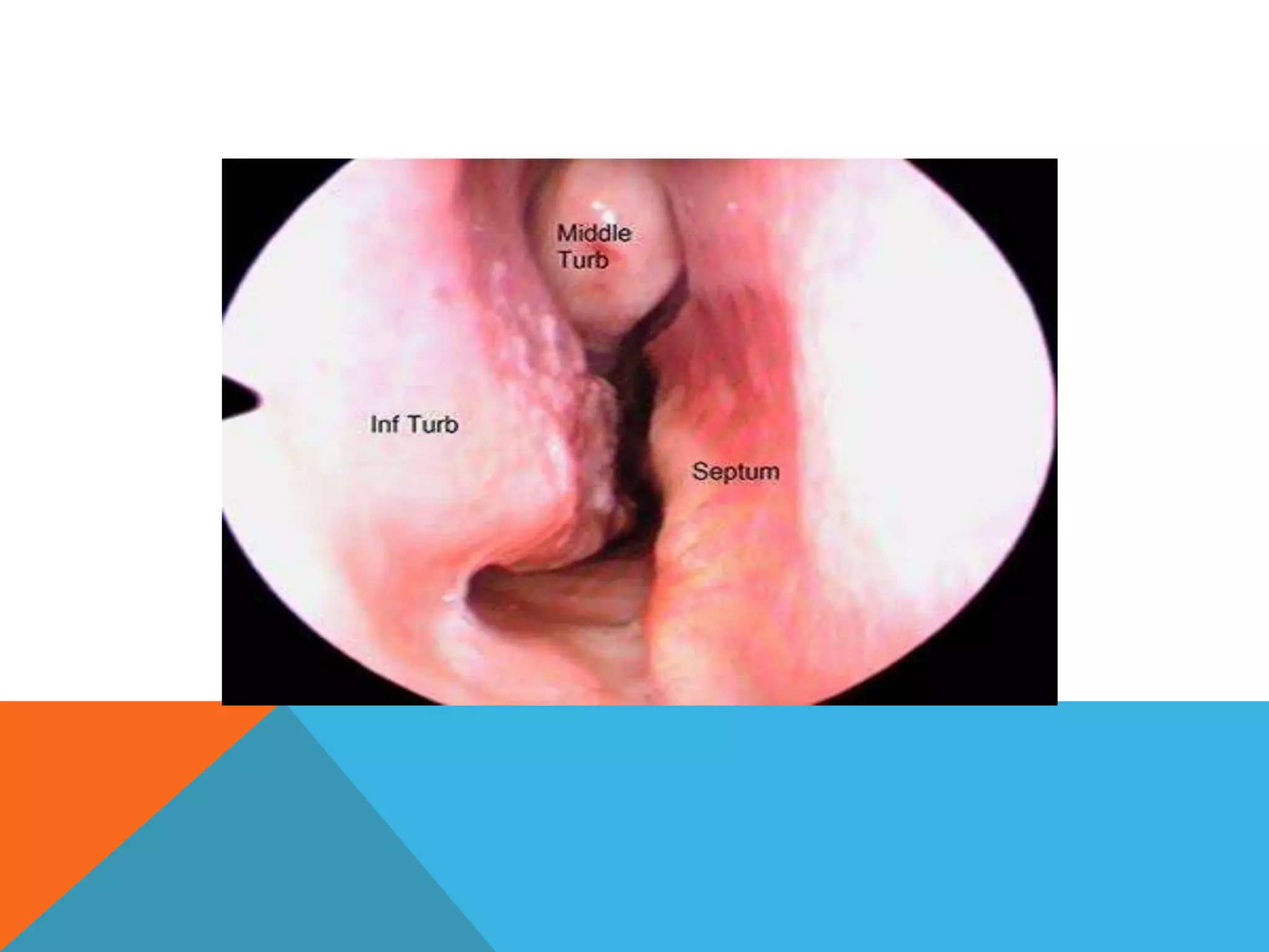
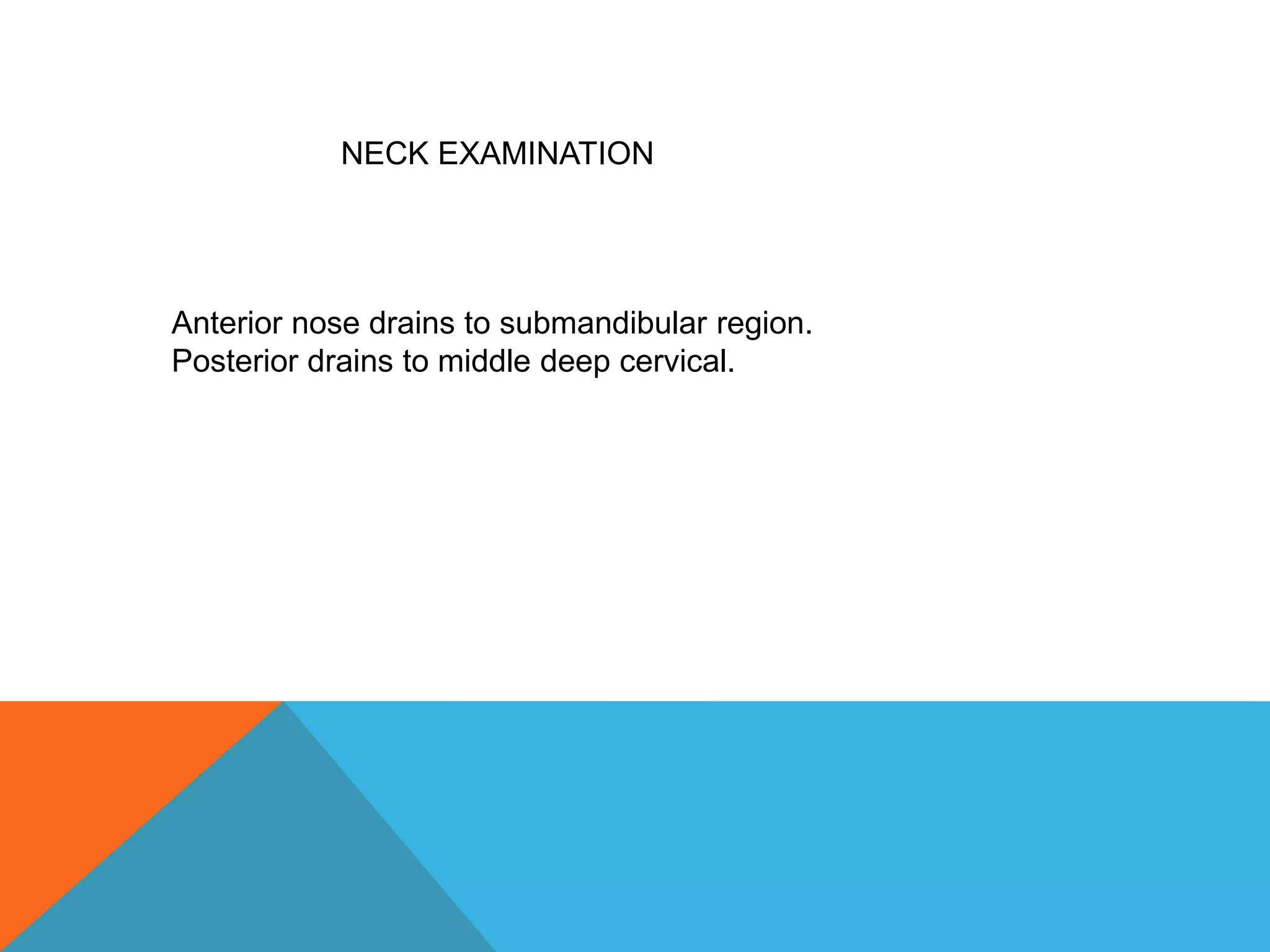
Examination of
Nose
Presented by –
Dr.Varsha Dhage
Assistant Professor,Shalakyatantra Dept.
SNKDCT’s Nallasopara Ayurved Medical
College
INSPECTION
:
First look at the external nose. Ask the patient to remove any
glasses. Look at the nose from the front and side for any signs.
• Nasal bridge deformity – Saddle Nose/ Hump
deformity
• Swelling
• Ulcers
• Sinuses
• Growths on skin
• Scars
• Broadening of nose
• Inflammation/ cellulites
PALPATION:
• Crepitation
• Tenderness
Anterior
Rhinoscopy :
The nose is both the sense organ and a respiratory organ.
Anterior rhinoscopy is an examination of the nasal cavities
enabling to note
• Nasal Secretions,
• Swelling of the turbinates,
• Properties of the mucosal surface,
• Position of the nasal septum
• Ulcerations or presence of foreign bodies.
MATERIAL :
Thundicum nasal speculum.
Light is focused at different sites in nose to examine nasal
septum, roof, floor and lateral wall by tilting patient’s head in
different directions.
• Nasal passage
- Narrow (septal deviation or hypertrophy of turbinates)
- Wide (atrophic rhinitis)
• Septum
- Position, Spurs, deviation, colour of mucosa, ulcers, crusting
and preforations, swellings (haematoma or abscess)
• Floor of the nose
- Defects (cleft palate or fistula)
- Swelling (dental cyst)
- Neoplasm (haemangioma)
- Granulations (foreign body or osteitis)
• Roof : Only seen in case of atrophic rhinitis
• Lateral wall :
- Colour of mucosa (congested in inflammation and pale in allergy)
- Size of turbinate’s (during rhinitis)
- Discharge (infection of maxillary, frontal or anterior ethmoidal
sinuses)
- Mass (polyp, rhinosporidiosis and carcinoma)
If any growth or polyp is suspected confirm by the probe test
Probe Test : it is carried out by spraying the nose with 4%
Lignocaine with 1:100000 adrenaline or 10% cocaine. The
lesion or area is palpated to determine its character and
mobility.
Posterior
Rhinoscopy :
Technique :
Patient sits facing examiner opens his mouth breaths quietly from
mouth
Examiner depresses tongue with tongue depressor and introduces
posterior rhinoscopic mirror warmed and tested on back of hand
Mirror is held like a pen and carried behind soft palate. Without
touching posterior third of tongue to avoid gag reflex.
Light from head mirror is focused on rhinoscopic mirror which further
illuminates part to be examined.
Functional Examination of
Nose:
• Patency of nose :
- Spatula test: By placing a cold tongue depressor below the nostril
to look for the area of mist formation. Compare the two sides always.
- Cotton wool wisp test: Fluff of cotton is held against each nostril
and its movements are noticed when patient inhales and exhales.
Sense of smell:
Ask the patient to identify the smell of the solutions held
before the nostril while keeping the eyes closed. Each nostril
is tested separately.
Common substances used : Clove oil, peppermint, coffee
and essence of rose.
EXAMINATION OF PARANASAL
SINUSES:
It includes both inspection and palpation of anterior group of
PNS, i.e.,
Maxillary
Frontal,
Anterior ethmoidal.
Look for any redness, swelling, mass, sinus in the area of
PNS.
NECK EXAMINATION
Anterior nose drains to submandibular region.
Posterior drains to middle deep cervical.
Thank you.
Nose examination ppt

Nose examination ppt

  • 1.
    Examination of Nose Presented by– Dr.Varsha Dhage Assistant Professor,Shalakyatantra Dept. SNKDCT’s Nallasopara Ayurved Medical College
  • 3.
    INSPECTION : First look atthe external nose. Ask the patient to remove any glasses. Look at the nose from the front and side for any signs. • Nasal bridge deformity – Saddle Nose/ Hump deformity • Swelling • Ulcers • Sinuses • Growths on skin • Scars • Broadening of nose • Inflammation/ cellulites
  • 4.
  • 5.
    Anterior Rhinoscopy : The noseis both the sense organ and a respiratory organ. Anterior rhinoscopy is an examination of the nasal cavities enabling to note • Nasal Secretions, • Swelling of the turbinates, • Properties of the mucosal surface, • Position of the nasal septum • Ulcerations or presence of foreign bodies.
  • 6.
    MATERIAL : Thundicum nasalspeculum. Light is focused at different sites in nose to examine nasal septum, roof, floor and lateral wall by tilting patient’s head in different directions.
  • 7.
    • Nasal passage -Narrow (septal deviation or hypertrophy of turbinates) - Wide (atrophic rhinitis) • Septum - Position, Spurs, deviation, colour of mucosa, ulcers, crusting and preforations, swellings (haematoma or abscess)
  • 9.
    • Floor ofthe nose - Defects (cleft palate or fistula) - Swelling (dental cyst) - Neoplasm (haemangioma) - Granulations (foreign body or osteitis) • Roof : Only seen in case of atrophic rhinitis
  • 10.
    • Lateral wall: - Colour of mucosa (congested in inflammation and pale in allergy) - Size of turbinate’s (during rhinitis) - Discharge (infection of maxillary, frontal or anterior ethmoidal sinuses) - Mass (polyp, rhinosporidiosis and carcinoma) If any growth or polyp is suspected confirm by the probe test Probe Test : it is carried out by spraying the nose with 4% Lignocaine with 1:100000 adrenaline or 10% cocaine. The lesion or area is palpated to determine its character and mobility.
  • 11.
    Posterior Rhinoscopy : Technique : Patientsits facing examiner opens his mouth breaths quietly from mouth Examiner depresses tongue with tongue depressor and introduces posterior rhinoscopic mirror warmed and tested on back of hand Mirror is held like a pen and carried behind soft palate. Without touching posterior third of tongue to avoid gag reflex. Light from head mirror is focused on rhinoscopic mirror which further illuminates part to be examined.
  • 13.
    Functional Examination of Nose: •Patency of nose : - Spatula test: By placing a cold tongue depressor below the nostril to look for the area of mist formation. Compare the two sides always. - Cotton wool wisp test: Fluff of cotton is held against each nostril and its movements are noticed when patient inhales and exhales.
  • 14.
    Sense of smell: Askthe patient to identify the smell of the solutions held before the nostril while keeping the eyes closed. Each nostril is tested separately. Common substances used : Clove oil, peppermint, coffee and essence of rose.
  • 15.
    EXAMINATION OF PARANASAL SINUSES: Itincludes both inspection and palpation of anterior group of PNS, i.e., Maxillary Frontal, Anterior ethmoidal. Look for any redness, swelling, mass, sinus in the area of PNS.
  • 16.
    NECK EXAMINATION Anterior nosedrains to submandibular region. Posterior drains to middle deep cervical.
  • 17.