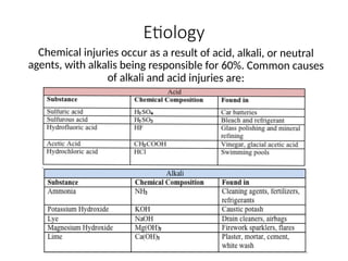
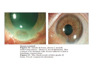
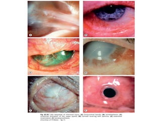
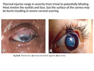
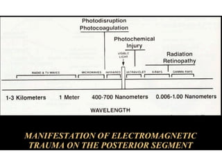
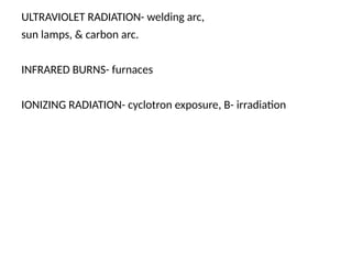
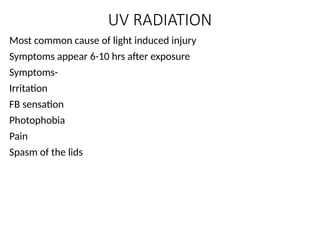
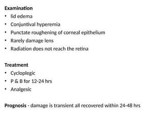
NON MECHANICAL
OCULAR INJURIES
Dr. SAURAV
JR1 OPHTHALMOLOGY
MUZAFFARNAGAR MEDICAL COLLEGE
Non mechanical ocular injuries
include:
Chemical
Thermal
Radiational
Electrical
CHEMICAL INJURIES
Etiology
Chemical injuries occur as a result of acid, alkali, or neutral
agents, with alkalis being responsible for 60%. Common causes
of alkali and acid injuries are:
PATHOPHYSIOLOGY
Alkali agents are lipophilic and therefore penetrate tissues more
rapidly than acids. They saponify the fatty acids of cell cell
membranes, penetrate the corneal stroma and destroy
proteoglycan ground substance and collagen bundles. The damaged
tissues then secrete proteolytic enzymes, which lead to further
damage.
Acids are generally less harmful than alkali substances. They
cause damage by denaturing and precipitating proteins in the
tissues they contact. The coagulated proteins act as a barrier
to prevent further penetration unlike alkali injuries. The one
exception to this is hydrofluoric acid, where the fluoride ion
rapidly penetrates the thickness of the cornea and causes
significant anterior segment destruction.
DIAGNOSIS
HISTORY
The severity of ocular injury depends on four factors:
• Toxicity of the chemical
• Duration of exposure (including retenion of particulate matter
on surface of globe or under the upper lid)
• The depth of penetration
• The area of involvement
The patient should be asked when the injury occurred, whether
they rinsed their eyes afterwards and for how long, the
mechanism of injury, the type of chemical that splashed in the
eye, and whether or not they were wearing eye protection.
PHYSICAL EXAMINATION
Prior to a full ophthalmic exam, the pH of both eyes should be checked. If
the pH is not in physiologic range, then the eye must be irrigated to bring
the pH to an appropriate range (between 7 and 7.2). It is recommended to
wait at least five minutes after irrigation before checking the pH to ensure
that the pH does not rise or fall secondary to retained particulate matter.
The physical exam should be used to assess the extent and depth of injury.
Specifically, the degree of corneal, conjunctival and limbal involvement
should be documented, as it can be used to predict ultimate visual
outcome.
The palpebral fissures should be checked and the fornices should be swept
during the initial exam. Both the palpebral and bulbar conjunctiva should
be examined with fluorescein under a cobalt blue light. As above, retained
particulate matter can cause persistent damage, despite irrigation. The
intraocular pressure should also be documented, as alkali injuries have
been found to both acutely and chronically cause an elevation of IOP.
Two major classification schemes for corneal burns are the Roper-Hall
(modified Hughes) classification and the Dua classification.
The Roper-Hall classification is based on the degree of corneal
involvement and limbal ischemia.
The Dua classification is based on an estimate of limbal involvement
(in clock hours) and the percentage of conjunctival involvement.
SYMPTOMS
• Severe pain
• Epiphora
• Blepharospasm
• Reduced visual acuity
MANAGEMENT
Irrigation
Irrigation is the cornerstone of managing chemical burns and should
be initiated by by bystanders and continued as transfer of care takes
place between EMS, ED physicians, and the ophthalmologist. Early
irrigation is critical in limiting the duration of chemical exposure. The
goal of irrigation is to remove the offending substance and restore
the physiologic pH.
Performing Irrigation:
• To position the patient for irrigation, the patient should ideally be
seated upright with their head supported and tilted toward the
affected side.
• Irrigation may require manual opening of the eyelid of the
affected eye to combat blepharospasm, while the irrigating
solution is delivered through IV tubing.
• The irrigating fluid should be administered nasal to lateral, poured
• Covering the noninjured eye with a shield may help prevent
additional chemical injury.
• During irrigation, the patient should be asked to blink frequently.
• Additionally, during irrigation, the patient should be asked to look
in all directions to ensure that the conjunctival sacs are irrigated.
Prior to beginning irrigation, disinfecting hands with an alcohol based
hand sanitizer is not recommended because it could cause further
irritation if gets into eye. Rather, hand washing is preferable.
Topical anesthetic such as Lidocaine can be applied prior to irrigation
to increase patient comfort.
The patient’s eyelids may be held open manually or with a speculum.
While there is no widespread consensus on how long the eye
should be irrigated, suggested guidelines recommend that
irrigation should be continued for a minimum of 30 minutes using
1-3 L of fluid or continued until a physiological pH is reached.
The pH can be monitored using pH paper and should be measured
at least 5 minutes after stopping irrigation.
Although irrigation is a crucial component of initial management,
over irrigation can result in corneal edema.
It is also crucial to identify the presence of and remove caustic
foreign bodies through visual inspection or by everting the eyelid.
Particles lodged under lids or in the conjunctival fornices cause
continued chemical exposure.
Sterile cotton tipped applicators wet with the irrigating fluid can
be used to remove visible foreign particles as well as used to
Types of Irrigating fluid:
Regardless of the type of irrigating fluid used, not delaying irrigation
is paramount to limiting the duration of chemical exposure and thus
minimizing ocular damage and restoring visual function.
Tap water is not sterile and is hypotonic to the corneal stroma. Use
of hypotonic solutions increases water influx into the cornea,
leading to further diffusion of corrosive material into the eye and
increased corneal edema.
Normal saline, Lactated Ringer’s, phosphate buffer solution, and
balanced saline solution, which are isotonic to the corneal stroma
and may be superior to tap water since they are sterile.
Use of hypertonic solutions is preferable, because they increase the
osmotic pressure to mobilize water and dissolved corrosives out of
corneal tissue. Thus, they prevent further uptake of the corrosive
chemical into the cornea, leading to a reduction in corneal swelling.
• Diphoterine is a hypertonic amphoteric solution that can be used
in both alkali and acid burns.
• Since Diphoterine is amphoteric, it is able to quickly neutralize
the corneal stroma to physiologic pH
• It has a buffering capacity similar to phosphate buffer.
• Diphoterine has been shown to lead to faster re-epithelialization
of the stroma and limit tissue damage, pain, and inflammation.
While Diphoterine may be the irrigating fluid of choice, immediate
irrigation should not be delayed if Diphoterine not readily available.
MEDICAL THERAPY
Patients with mild to moderate injury (Grade I and II) have a good
prognosis and can often be treated successfully with medical
treatment alone.
The aims of medical treatment are to enhance recovery of the
corneal epithelium and augment collagen synthesis, while also
minimizing collagen breakdown and controlling inflammation
Standard treatments:
Antibiotics - A topical antibiotic ointment like erythromycin ointment
four times daily can be used to provide ocular lubrication and prevent
superinfection. Stronger antibiotics (e.g. a topical fluoroquinolone)
are employed for more severe injuries (e.g. Grade II and above).
Cycloplegic agents such as atropine or cyclopentolate can help with
comfort.
Artificial tears- and other lubricating eye drops, preferably
preservative free, should be used generously for comfort.
Steroid drops- In the first week following injury, topical steroids can
help calm inflammation and prevent further corneal breakdown.
In mild injuries, topical prednisolone (Predforte) can be employed
four times daily. In more severe injuries, prednisolone can be used
every hour.
After about one week of intensive steroid use, the steroids should
be tapered because the balance of collagen synthesis vs. collagen
breakdown may tip unfavorably toward collagen breakdown.
Other treatments:
Ascorbic acid- is a cofactor in collagen synthesis and may be
depleted following chemical injury. Ascorbic acid can be used as a
topical drop (10% every hour) or orally (two grams, four times daily
in adults).
Doxycycline - acts independently of its antimicrobial properties to
reduce the effects of matrix metalloproteinases (MMPs), which can
degrade type I collagen.
Citrate drops: Deficiency in calcium inhibits the PMNs from
granulating and releasing proteolytic enzymes. Citrate is a potent
chelator and can therefore decrease proteolytic activity. Citrate also
inhibit collagenases.
1% Medroxyprogesterone- is a progestational steroid and has less
anti-inflammatory potency than corticosteroids, but has a minimum
effect on stromal repair. Medroxyprogesterone can therefore be
substituted for cortical steroids after 10-14 days of steroid
treatment.
Platelet rich plasma eye drops- have been found to be rich in growth
SURGICAL TREATMENTS
Debridement of necrotic epithelium- should be performed as early as
possible because necrotic tissue serves as a source of inflammation
and can inhibit epithelialization.
Conjunctival/Tenon’s transposition (Tenonplasty)- in Grade IV burns,
anterior segment necrosis can result from loss of limbal vascular
blood supply. In severe limbal ischemia, a sterile corneal ulceration
can ensue. After removal of necrotic tissue, a tenonplasty
(advancement of the conjunctiva and Tenon’s to the limbus) can be
employed to reestablish limbal vascularity and facilitate re-
epithelialization.
Amniotic membrane transplantation (AMT)- the purpose of AMT is to
rapidly restore the conjunctival surface and to reduce limbal and
stromal inflammation.
Limbal stem cell transplant- Much of the damage following chemical
Cultivated oral mucosal epithelial transplantation (COMET)- can also
be used to promote re-epithelialization and reduce inflammation in
corneal burns. The cells are harvested from the patient’s own buccal
mucosa so that systemic immunosuppression is not necessary.
Boston Keratoprosthesis
RECOMMENDED TREATMENT
Grade I
• Topical antibiotic ointment (erythromycin ointment or similar) four
times a day
• Prednisolone acetate 1% four times a day
• Preservative free artificial tears as needed
• If there is pain, consider a short acting cycloplegic like
cyclopentolate three times a day
Grade II
• Topical antibiotic drop like fluoroquinolone four times daily
• Prednisolone acetate 1% hourly while awake for the first 7-10
days. Consider tapering the steroid if the epithelium has not
healed by day 10-14. If an epithelial defect persists after day 10,
consider progestational steroids (1% medroxyprogesterone four
times daily)
• Long acting cycloplegic like atropine
• Oral Vitamin C, 2 grams four times a day
• Doxycycline, 100 mg twice a day (avoid in children)
• Sodium ascorbate drops (10%) hourly while awake
• Preservative free artificial tears as needed
• Debridement of necrotic epithelium and application of tissue
adhesive as needed
Grade III
• As for Grade II
• Consider amniotic membrane transplant/Prokera placement. This
should ideally be performed in the first week of injury.
Experienced surgeons have emphasized placement of the amniotic
membrane to cover the palpebral conjunctiva by suturing to the
lids in the operating room, not just covering the cornea and bulbar
conjunctiva.
Grade IV
• As for Grade II/III
• Early surgery is usually necessary. For significant necrosis, a
Tenonplasty can help reestablish limbal vascularity. An amniotic
membrane transplant is often necessary due to the severity of the
ocular surface damage.
STAGES OF OCULAR RECOVERY
FOLLOW UP
With severe chemical burns, patients should initially be followed
daily.
If there is concern for compliance with medication or if the patient is
a child, one should consider inpatient admission.
Once the health of the ocular surface has been restored, follow up
can be spread apart.
However, even in the healthiest appearing eyes, patients need long
term monitoring for glaucoma and dry eye.
Direct chemical damage to the conjunctiva can lead to scarring,
forniceal shortening, symblepharon formation and ciccatricial
entropion or ectropion.
These entities are encountered weeks to months after injury and can
be treated by suppressing inflammation and with early amniotic
THERMAL INJURIES
Thermal injuries range in severity from trivial to potentially blinding.
Most involve the eyelids and face, but the surface of the cornea may
be burnt resulting in severe corneal scarring
HYPERTHERMAL INJURIES:
◦ Flame burns, contact burns
Clinical Presentations:
• Conj hyperemia, chemosis
• Corneal superficial /deep burns- corneal opacification, sloughing
• Healing-leucoma formation
• Bullous keratitis, ectasia, staphyloma,
• symblepharon
• Scleral involvement- uveal prolapse, uveitis,
• panophthalmitis
Treatment
◦ Clean with saline
◦ Antibiotic cream
◦ Full thickness burns of lid- grafting
◦ Topical-atropine, antibiotics, lubricating e/d, steroids
◦ Glass rod passed in fornices
◦ Conj transposition flap, amniotic membrane graft,
limbal cell transplant
◦ PK or LK for leucomatous corneal opacity later stage
RADIATIONAL INJURIES
ULTRAVIOLET RADIATION- welding arc,
sun lamps, & carbon arc.
INFRARED BURNS- furnaces
IONIZING RADIATION- cyclotron exposure, B- irradiation
UV RADIATION
Most common cause of light induced injury
Symptoms appear 6-10 hrs after exposure
Symptoms-
Irritation
FB sensation
Photophobia
Pain
Spasm of the lids
Examination
• lid edema
• Conjuntival hyperemia
• Punctate roughening of corneal epithelium
• Rarely damage lens
• Radiation does not reach the retina
Treatment
• Cycloplegic
• P & B for 12-24 hrs
• Analgesic
Prognosis - damage is transient all recovered within 24-48 hrs
INFRARED BURNS
Acute exposure produces temporary lid
edema & erythema
No damage to globe
Chronic exposure seen in glass blower &
metal furnace stocker
Develop cataract
No other anterior & posterior segment
changes are found
BIOLOGICAL EFFECT OF IONIZING RAD.
• Produces ds break in DNA to kill a cell
• Interact with water to generate free radical eg. Hydroxyl ion
• Free radicals are unstable compound
• Biological effect expressed when damaged cell attempt to divide
• SIGN & SYMPTOMS
• Erythema of skin
• Conjuntival hyperemia
• Circumcorneal injection
• Watery/ mucopurulent discharge
• Stromal edema, interstitial keratitis, aseptic corneal necrosis
• Cataract- (500-700 rad), younger more vulnerable
• For cataract latency period- 6M-12 years
• Uveal tract - vascular dilatation & boggy edema
• Intraretinal Hg.
• Retinopathy
• Papilledema
• CRV thrombosis
TEATMIENT
• Cycloplegic
• Topical antibiotic
• BCL
• Cataract extraction
Infrared radiation (glassblower's cataract)
Ionbizing radition

nON MECHANICAL OCULAR INJURIES .pptx

  • 1.
    NON MECHANICAL OCULAR INJURIES Dr.SAURAV JR1 OPHTHALMOLOGY MUZAFFARNAGAR MEDICAL COLLEGE
  • 2.
    Non mechanical ocularinjuries include: Chemical Thermal Radiational Electrical
  • 3.
  • 4.
    Etiology Chemical injuries occuras a result of acid, alkali, or neutral agents, with alkalis being responsible for 60%. Common causes of alkali and acid injuries are:
  • 5.
    PATHOPHYSIOLOGY Alkali agents arelipophilic and therefore penetrate tissues more rapidly than acids. They saponify the fatty acids of cell cell membranes, penetrate the corneal stroma and destroy proteoglycan ground substance and collagen bundles. The damaged tissues then secrete proteolytic enzymes, which lead to further damage. Acids are generally less harmful than alkali substances. They cause damage by denaturing and precipitating proteins in the tissues they contact. The coagulated proteins act as a barrier to prevent further penetration unlike alkali injuries. The one exception to this is hydrofluoric acid, where the fluoride ion rapidly penetrates the thickness of the cornea and causes significant anterior segment destruction.
  • 6.
    DIAGNOSIS HISTORY The severity ofocular injury depends on four factors: • Toxicity of the chemical • Duration of exposure (including retenion of particulate matter on surface of globe or under the upper lid) • The depth of penetration • The area of involvement The patient should be asked when the injury occurred, whether they rinsed their eyes afterwards and for how long, the mechanism of injury, the type of chemical that splashed in the eye, and whether or not they were wearing eye protection.
  • 7.
    PHYSICAL EXAMINATION Prior toa full ophthalmic exam, the pH of both eyes should be checked. If the pH is not in physiologic range, then the eye must be irrigated to bring the pH to an appropriate range (between 7 and 7.2). It is recommended to wait at least five minutes after irrigation before checking the pH to ensure that the pH does not rise or fall secondary to retained particulate matter. The physical exam should be used to assess the extent and depth of injury. Specifically, the degree of corneal, conjunctival and limbal involvement should be documented, as it can be used to predict ultimate visual outcome. The palpebral fissures should be checked and the fornices should be swept during the initial exam. Both the palpebral and bulbar conjunctiva should be examined with fluorescein under a cobalt blue light. As above, retained particulate matter can cause persistent damage, despite irrigation. The intraocular pressure should also be documented, as alkali injuries have been found to both acutely and chronically cause an elevation of IOP.
  • 8.
    Two major classificationschemes for corneal burns are the Roper-Hall (modified Hughes) classification and the Dua classification. The Roper-Hall classification is based on the degree of corneal involvement and limbal ischemia. The Dua classification is based on an estimate of limbal involvement (in clock hours) and the percentage of conjunctival involvement.
  • 11.
    SYMPTOMS • Severe pain •Epiphora • Blepharospasm • Reduced visual acuity
  • 12.
    MANAGEMENT Irrigation Irrigation is thecornerstone of managing chemical burns and should be initiated by by bystanders and continued as transfer of care takes place between EMS, ED physicians, and the ophthalmologist. Early irrigation is critical in limiting the duration of chemical exposure. The goal of irrigation is to remove the offending substance and restore the physiologic pH. Performing Irrigation: • To position the patient for irrigation, the patient should ideally be seated upright with their head supported and tilted toward the affected side. • Irrigation may require manual opening of the eyelid of the affected eye to combat blepharospasm, while the irrigating solution is delivered through IV tubing. • The irrigating fluid should be administered nasal to lateral, poured
  • 13.
    • Covering thenoninjured eye with a shield may help prevent additional chemical injury. • During irrigation, the patient should be asked to blink frequently. • Additionally, during irrigation, the patient should be asked to look in all directions to ensure that the conjunctival sacs are irrigated. Prior to beginning irrigation, disinfecting hands with an alcohol based hand sanitizer is not recommended because it could cause further irritation if gets into eye. Rather, hand washing is preferable. Topical anesthetic such as Lidocaine can be applied prior to irrigation to increase patient comfort. The patient’s eyelids may be held open manually or with a speculum.
  • 14.
    While there isno widespread consensus on how long the eye should be irrigated, suggested guidelines recommend that irrigation should be continued for a minimum of 30 minutes using 1-3 L of fluid or continued until a physiological pH is reached. The pH can be monitored using pH paper and should be measured at least 5 minutes after stopping irrigation. Although irrigation is a crucial component of initial management, over irrigation can result in corneal edema. It is also crucial to identify the presence of and remove caustic foreign bodies through visual inspection or by everting the eyelid. Particles lodged under lids or in the conjunctival fornices cause continued chemical exposure. Sterile cotton tipped applicators wet with the irrigating fluid can be used to remove visible foreign particles as well as used to
  • 15.
    Types of Irrigatingfluid: Regardless of the type of irrigating fluid used, not delaying irrigation is paramount to limiting the duration of chemical exposure and thus minimizing ocular damage and restoring visual function. Tap water is not sterile and is hypotonic to the corneal stroma. Use of hypotonic solutions increases water influx into the cornea, leading to further diffusion of corrosive material into the eye and increased corneal edema. Normal saline, Lactated Ringer’s, phosphate buffer solution, and balanced saline solution, which are isotonic to the corneal stroma and may be superior to tap water since they are sterile.
  • 16.
    Use of hypertonicsolutions is preferable, because they increase the osmotic pressure to mobilize water and dissolved corrosives out of corneal tissue. Thus, they prevent further uptake of the corrosive chemical into the cornea, leading to a reduction in corneal swelling. • Diphoterine is a hypertonic amphoteric solution that can be used in both alkali and acid burns. • Since Diphoterine is amphoteric, it is able to quickly neutralize the corneal stroma to physiologic pH • It has a buffering capacity similar to phosphate buffer. • Diphoterine has been shown to lead to faster re-epithelialization of the stroma and limit tissue damage, pain, and inflammation. While Diphoterine may be the irrigating fluid of choice, immediate irrigation should not be delayed if Diphoterine not readily available.
  • 17.
    MEDICAL THERAPY Patients withmild to moderate injury (Grade I and II) have a good prognosis and can often be treated successfully with medical treatment alone. The aims of medical treatment are to enhance recovery of the corneal epithelium and augment collagen synthesis, while also minimizing collagen breakdown and controlling inflammation Standard treatments: Antibiotics - A topical antibiotic ointment like erythromycin ointment four times daily can be used to provide ocular lubrication and prevent superinfection. Stronger antibiotics (e.g. a topical fluoroquinolone) are employed for more severe injuries (e.g. Grade II and above). Cycloplegic agents such as atropine or cyclopentolate can help with comfort.
  • 18.
    Artificial tears- andother lubricating eye drops, preferably preservative free, should be used generously for comfort. Steroid drops- In the first week following injury, topical steroids can help calm inflammation and prevent further corneal breakdown. In mild injuries, topical prednisolone (Predforte) can be employed four times daily. In more severe injuries, prednisolone can be used every hour. After about one week of intensive steroid use, the steroids should be tapered because the balance of collagen synthesis vs. collagen breakdown may tip unfavorably toward collagen breakdown.
  • 19.
    Other treatments: Ascorbic acid-is a cofactor in collagen synthesis and may be depleted following chemical injury. Ascorbic acid can be used as a topical drop (10% every hour) or orally (two grams, four times daily in adults). Doxycycline - acts independently of its antimicrobial properties to reduce the effects of matrix metalloproteinases (MMPs), which can degrade type I collagen. Citrate drops: Deficiency in calcium inhibits the PMNs from granulating and releasing proteolytic enzymes. Citrate is a potent chelator and can therefore decrease proteolytic activity. Citrate also inhibit collagenases. 1% Medroxyprogesterone- is a progestational steroid and has less anti-inflammatory potency than corticosteroids, but has a minimum effect on stromal repair. Medroxyprogesterone can therefore be substituted for cortical steroids after 10-14 days of steroid treatment. Platelet rich plasma eye drops- have been found to be rich in growth
  • 20.
    SURGICAL TREATMENTS Debridement ofnecrotic epithelium- should be performed as early as possible because necrotic tissue serves as a source of inflammation and can inhibit epithelialization. Conjunctival/Tenon’s transposition (Tenonplasty)- in Grade IV burns, anterior segment necrosis can result from loss of limbal vascular blood supply. In severe limbal ischemia, a sterile corneal ulceration can ensue. After removal of necrotic tissue, a tenonplasty (advancement of the conjunctiva and Tenon’s to the limbus) can be employed to reestablish limbal vascularity and facilitate re- epithelialization. Amniotic membrane transplantation (AMT)- the purpose of AMT is to rapidly restore the conjunctival surface and to reduce limbal and stromal inflammation. Limbal stem cell transplant- Much of the damage following chemical
  • 21.
    Cultivated oral mucosalepithelial transplantation (COMET)- can also be used to promote re-epithelialization and reduce inflammation in corneal burns. The cells are harvested from the patient’s own buccal mucosa so that systemic immunosuppression is not necessary. Boston Keratoprosthesis
  • 22.
    RECOMMENDED TREATMENT Grade I •Topical antibiotic ointment (erythromycin ointment or similar) four times a day • Prednisolone acetate 1% four times a day • Preservative free artificial tears as needed • If there is pain, consider a short acting cycloplegic like cyclopentolate three times a day
  • 23.
    Grade II • Topicalantibiotic drop like fluoroquinolone four times daily • Prednisolone acetate 1% hourly while awake for the first 7-10 days. Consider tapering the steroid if the epithelium has not healed by day 10-14. If an epithelial defect persists after day 10, consider progestational steroids (1% medroxyprogesterone four times daily) • Long acting cycloplegic like atropine • Oral Vitamin C, 2 grams four times a day • Doxycycline, 100 mg twice a day (avoid in children) • Sodium ascorbate drops (10%) hourly while awake • Preservative free artificial tears as needed • Debridement of necrotic epithelium and application of tissue adhesive as needed
  • 24.
    Grade III • Asfor Grade II • Consider amniotic membrane transplant/Prokera placement. This should ideally be performed in the first week of injury. Experienced surgeons have emphasized placement of the amniotic membrane to cover the palpebral conjunctiva by suturing to the lids in the operating room, not just covering the cornea and bulbar conjunctiva. Grade IV • As for Grade II/III • Early surgery is usually necessary. For significant necrosis, a Tenonplasty can help reestablish limbal vascularity. An amniotic membrane transplant is often necessary due to the severity of the ocular surface damage.
  • 25.
  • 27.
    FOLLOW UP With severechemical burns, patients should initially be followed daily. If there is concern for compliance with medication or if the patient is a child, one should consider inpatient admission. Once the health of the ocular surface has been restored, follow up can be spread apart. However, even in the healthiest appearing eyes, patients need long term monitoring for glaucoma and dry eye. Direct chemical damage to the conjunctiva can lead to scarring, forniceal shortening, symblepharon formation and ciccatricial entropion or ectropion. These entities are encountered weeks to months after injury and can be treated by suppressing inflammation and with early amniotic
  • 29.
  • 30.
    Thermal injuries rangein severity from trivial to potentially blinding. Most involve the eyelids and face, but the surface of the cornea may be burnt resulting in severe corneal scarring
  • 31.
    HYPERTHERMAL INJURIES: ◦ Flameburns, contact burns Clinical Presentations: • Conj hyperemia, chemosis • Corneal superficial /deep burns- corneal opacification, sloughing • Healing-leucoma formation • Bullous keratitis, ectasia, staphyloma, • symblepharon • Scleral involvement- uveal prolapse, uveitis, • panophthalmitis
  • 32.
    Treatment ◦ Clean withsaline ◦ Antibiotic cream ◦ Full thickness burns of lid- grafting ◦ Topical-atropine, antibiotics, lubricating e/d, steroids ◦ Glass rod passed in fornices ◦ Conj transposition flap, amniotic membrane graft, limbal cell transplant ◦ PK or LK for leucomatous corneal opacity later stage
  • 33.
  • 35.
    ULTRAVIOLET RADIATION- weldingarc, sun lamps, & carbon arc. INFRARED BURNS- furnaces IONIZING RADIATION- cyclotron exposure, B- irradiation
  • 36.
    UV RADIATION Most commoncause of light induced injury Symptoms appear 6-10 hrs after exposure Symptoms- Irritation FB sensation Photophobia Pain Spasm of the lids
  • 37.
    Examination • lid edema •Conjuntival hyperemia • Punctate roughening of corneal epithelium • Rarely damage lens • Radiation does not reach the retina Treatment • Cycloplegic • P & B for 12-24 hrs • Analgesic Prognosis - damage is transient all recovered within 24-48 hrs
  • 38.
    INFRARED BURNS Acute exposureproduces temporary lid edema & erythema No damage to globe Chronic exposure seen in glass blower & metal furnace stocker Develop cataract No other anterior & posterior segment changes are found
  • 39.
    BIOLOGICAL EFFECT OFIONIZING RAD. • Produces ds break in DNA to kill a cell • Interact with water to generate free radical eg. Hydroxyl ion • Free radicals are unstable compound • Biological effect expressed when damaged cell attempt to divide • SIGN & SYMPTOMS • Erythema of skin • Conjuntival hyperemia • Circumcorneal injection • Watery/ mucopurulent discharge • Stromal edema, interstitial keratitis, aseptic corneal necrosis • Cataract- (500-700 rad), younger more vulnerable
  • 40.
    • For cataractlatency period- 6M-12 years • Uveal tract - vascular dilatation & boggy edema • Intraretinal Hg. • Retinopathy • Papilledema • CRV thrombosis TEATMIENT • Cycloplegic • Topical antibiotic • BCL • Cataract extraction
  • 41.
    Infrared radiation (glassblower'scataract) Ionbizing radition