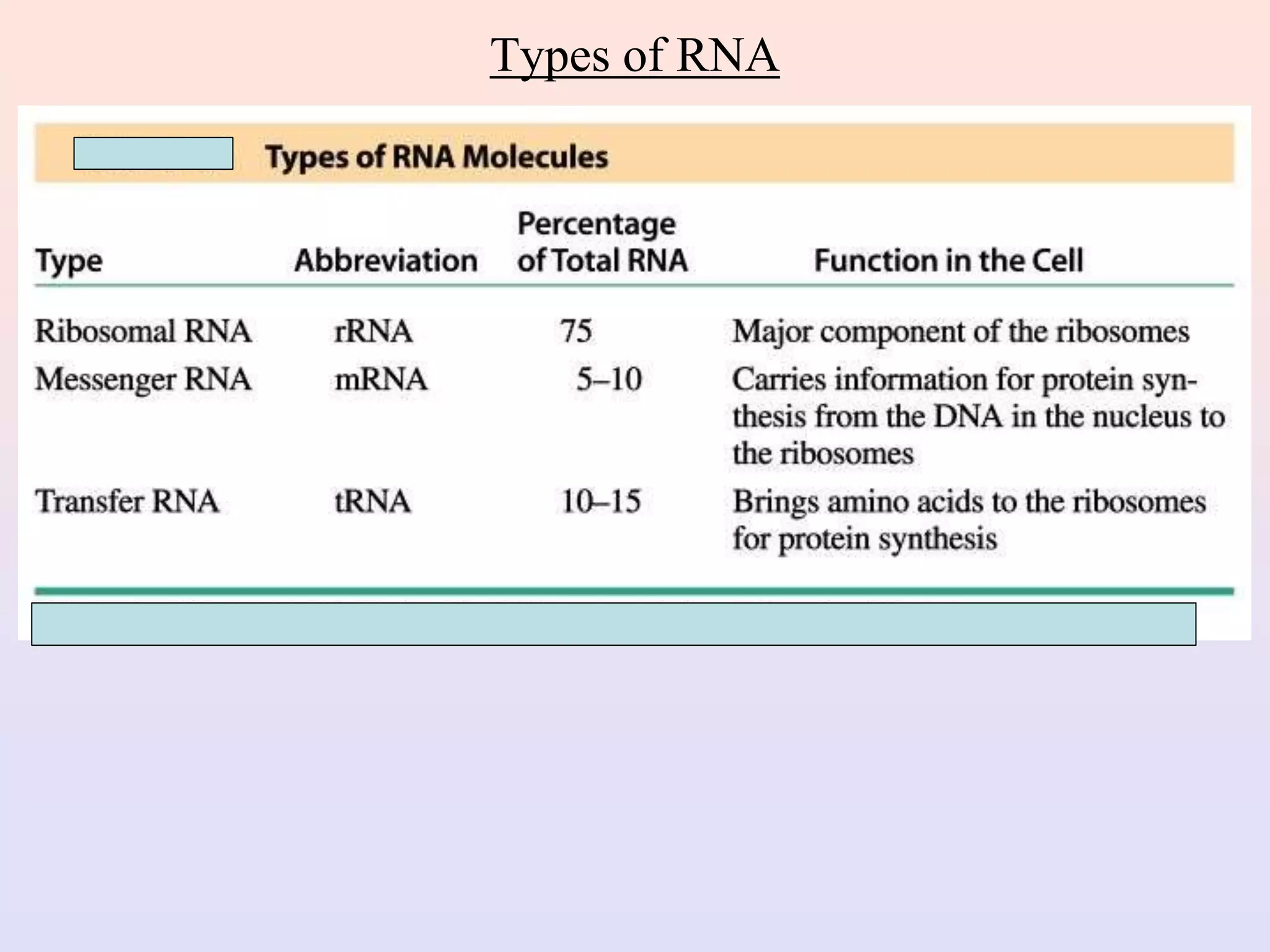
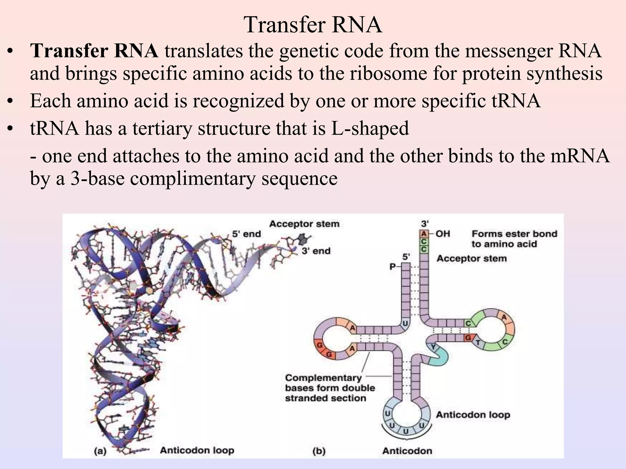
Call Girls in Munirka Delhi 💯Call Us 🔝9953322196🔝 💯Escort.
Unit III (Nucleic acid and genetic code).ppt
1. Nucleic Acids
• Nucleic acids are molecules that store information for cellular
growth and reproduction
• There are two types of nucleic acids:
- deoxyribonucleic acid (DNA) and ribonucleic acid (RNA)
• These are polymers consisting of long chains of monomers
called nucleotides
• A nucleotide consists of a nitrogenous base, a pentose sugar
and a phosphate group:
2. Nitrogen Bases
• The nitrogen bases in nucleotides consist of two general types:
- purines: adenine (A) and guanine (G)
- pyrimidines: cytosine (C), thymine (T) and Uracil (U)
3. Pentose Sugars
• There are two related pentose sugars:
- RNA contains ribose
- DNA contains deoxyribose
• The sugars have their carbon atoms numbered with primes
to distinguish them from the nitrogen bases
4. Nucleosides and Nucleotides
• A nucleoside consists of a nitrogen base linked by a glycosidic bond to C1’ of a ribose or
deoxyribose
• Nucleosides are named by changing the nitrogen base ending to -osine for purines and –idine
for pyrimidines
• A nucleotide is a nucleoside that forms a phosphate ester with the C5’ OH group of ribose or
deoxyribose
• Nucleotides are named using the name of the nucleoside followed by 5’-monophosphate
6. AMP, ADP and ATP
• Additional phosphate groups can be added to the nucleoside 5’-
monophosphates to form diphosphates and triphosphates
• ATP is the major energy source for cellular activity
7. Primary Structure of Nucleic Acids
• The primary structure of a nucleic acid is the nucleotide sequence
• The nucleotides in nucleic acids are joined by phosphodiester bonds
• The 3’-OH group of the sugar in one nucleotide forms an ester bond
to the phosphate group on the 5’-carbon of the sugar of the next
nucleotide
8. Reading Primary Structure
• A nucleic acid polymer has a free
5’-phosphate group at one end and a
free 3’-OH group at the other end
• The sequence is read from the free
5’-end using the letters of the bases
• This example reads
5’—A—C—G—T—3’
9. Example of RNA Primary Structure
• In RNA, A, C, G, and U are linked by 3’-5’ ester bonds
between ribose and phosphate
10. Example of DNA Primary Structure
• In DNA, A, C, G, and T are linked by 3’-5’ ester bonds
between deoxyribose and phosphate
11. Secondary Structure: DNA Double Helix
• In DNA there are two strands of nucleotides that wind together
in a double helix
- the strands run in opposite directions
- the bases are arranged in step-like pairs
- the base pairs are held together by hydrogen bonding
• The pairing of the bases from the two strands is very specific
• The complimentary base pairs are A-T and G-C
- two hydrogen bonds form between A and T
- three hydrogen bonds form between G and C
• Each pair consists of a purine and a pyrimidine, so they are the
same width, keeping the two strands at equal distances from
each other
12. Chargaff's rule (1949):
In a DNA double helix, the amount of purines
always equals to that of pyrimidines.
The number of A always equals to T, and the
number of C always equals to G.
14. Storage of DNA
• In eukaryotic cells (animals, plants, fungi) DNA is stored in
the nucleus, which is separated from the rest of the cell by a
semipermeable membrane
• The DNA is only organized into chromosomes during cell
replication
• Between replications, the DNA is stored in a compact ball
called chromatin, and is wrapped around proteins called
histones to form nucleosomes
15. DNA Replication
• When a eukaryotic cell divides, the process is called mitosis
- the cell splits into two identical daughter cells
- the DNA must be replicated so that each daughter cell has a copy
• DNA replication involves several processes:
- first, the DNA must be unwound, separating the two strands
- the single strands then act as templates for synthesis of the new
strands, which are complimentary in sequence
- bases are added one at a time until two new DNA strands that
exactly duplicate the original DNA are produced
• The process is called semi-conservative replication because one
strand of each daughter DNA comes from the parent DNA and one
strand is new
• The energy for the synthesis comes from hydrolysis of phosphate
groups as the phosphodiester bonds form between the bases
17. Direction of Replication
• The enzyme helicase unwinds several sections of parent DNA
• At each open DNA section, called a replication fork, DNA
polymerase catalyzes the formation of 5’-3’ester bonds of the
leading strand
• The lagging strand, which grows in the 3’-5’ direction, is
synthesized in short sections called Okazaki fragments
• The Okazaki fragments are joined by DNA ligase to give a
single 3’-5’ DNA strand
18. Ribonucleic Acid (RNA)
• RNA is much more abundant than DNA
• There are several important differences between RNA and DNA:
- the pentose sugar in RNA is ribose, in DNA it’s deoxyribose
- in RNA, uracil replaces the base thymine (U pairs with T)
- RNA is single stranded while DNA is double stranded
- RNA molecules are much smaller than DNA molecules
• There are three main types of RNA:
- ribosomal (rRNA), messenger (mRNA) and transfer (tRNA)
20. Ribosomal RNA and Messenger RNA
• Ribosomes are the sites of protein synthesis
- they consist of ribosomal DNA (65%) and proteins (35%)
- they have two subunits, a large one and a small one
• Messenger RNA carries the genetic code to the ribosomes
- they are strands of RNA that are complementary to the
DNA of the gene for the protein to be synthesized
21. Transfer RNA
• Transfer RNA translates the genetic code from the messenger RNA
and brings specific amino acids to the ribosome for protein synthesis
• Each amino acid is recognized by one or more specific tRNA
• tRNA has a tertiary structure that is L-shaped
- one end attaches to the amino acid and the other binds to the mRNA
by a 3-base complimentary sequence
22. Protein Synthesis
• The two main processes involved in protein synthesis are
- the formation of mRNA from DNA (transcription)
- the conversion by tRNA to protein at the ribosome (translation)
• Transcription takes place in the nucleus, while translation takes
place in the cytoplasm
• Genetic information is transcribed to form mRNA much the same
way it is replicated during cell division
23. The Genetic Code
• The genetic code is found in the sequence of nucleotides in
mRNA that is translated from the DNA
• A codon is a triplet of bases along the mRNA that codes for a
particular amino acid
• Each of the 20 amino acids needed to build a protein has at least 2
codons
• There are also codons that signal the “start” and “end” of a
polypeptide chain
• The amino acid sequence of a protein can be determined by
reading the triplets in the DNA sequence that are complementary
to the codons of the mRNA, or directly from the mRNA sequence
• The entire DNA sequence of several organisms, including
humans, have been determined, however,
- only primary structure can be determined this way
- doesn’t give tertiary structure or protein function
25. Reading the Genetic Code
• Suppose we want to determine the amino acids coded for
in the following section of a mRNA
5’—CCU —AGC—GGA—CUU—3’
• According to the genetic code, the amino acids for these
codons are:
CCU = Proline AGC = Serine
GGA = Glycine CUU = Leucine
• The mRNA section codes for the amino acid sequence of
Pro—Ser—Gly—Leu
26. Translation and tRNA Activation
• Once the DNA has been
transcribed to mRNA, the
codons must be tranlated to the
amino acid sequence of the
protein
• The first step in translation is
activation of the tRNA
• Each tRNA has a triplet called
an anticodon that complements
a codon on mRNA
• A synthetase uses ATP
hydrolysis to attach an amino
acid to a specific tRNA
27. Initiation and Translocation
• Initiation of protein synthesis occurs when a mRNA attaches to
a ribosome
• On the mRNA, the start codon (AUG) binds to a tRNA with
methionine
• The second codon attaches to a tRNA with the next amino acid
• A peptide bond forms between the adjacent amino acids at the
first and second codons
• The first tRNA detaches from the ribosome and the ribosome
shifts to the adjacent codon on the mRNA (this process is called
translocation)
• A third codon can now attach where the second one was before
translocation
28. Termination
• After a polypeptide with all the amino acids for a protein is
synthesized, the ribosome reaches the the “stop” codon:
UGA, UAA, or UAG
• There is no tRNA with an anticodon for the “stop” codons
• Therefore, protein synthesis ends (termination)
• The polypeptide is released from the ribosome and the
protein can take on it’s 3-D structure
(some proteins begin folding while still being synthesized,
while others do not fold up until after being released from
the ribosome)