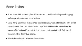
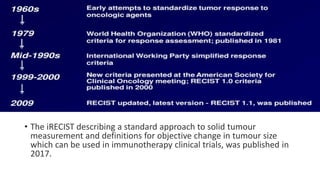
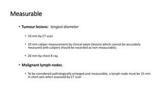
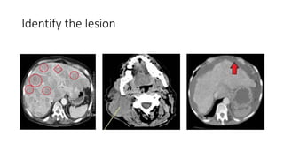
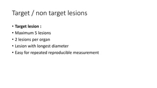
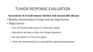
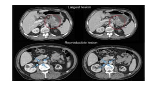
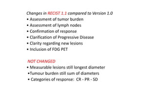
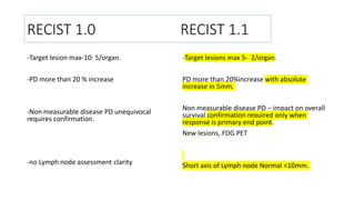
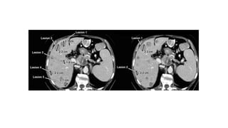
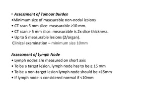
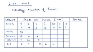
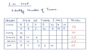
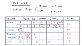
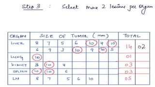
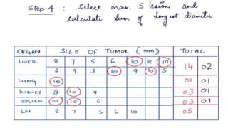
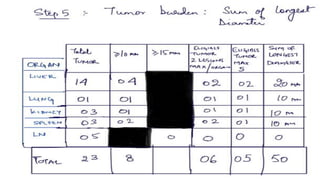
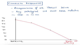
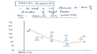
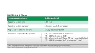
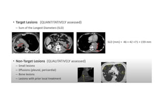
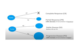
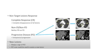
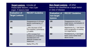
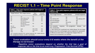
SlideShare a Scribd company logo
RECIST 1.1
RESPONSE EVALUATION CRITERIA IN SOLID TUMORS
NEED OF ANY CRITERIA
• For standardization of assessment and reporting of the results
• To develop a common language to describe the results of cancer Rx
• Working group formed in 1995
• RECIST 1st version published in 2000
• 2nd update in 2009 as RECIST 1.1
• The iRECIST describing a standard approach to solid tumour
measurement and definitions for objective change in tumour size
which can be used in immunotherapy clinical trials, was published in
2017.
RECIST (Response Evaluation Criteria in
Solid Tumours)
• It provides a simple and pragmatic methodology to evaluate the
activity and efficacy of new cancer therapeutics in solid tumors,
using validated and consistent criteria to assess changes in
tumor burden.
• Mission of the working group : To ensures that RECIST
undergoes continued testing, validation and updating.
RECIST 1.0
Development of RECIST
In 1994 International task force
1. European Organization for Research and Treatment of
Cancer (EORTC)
2. National Cancer Institute (NCI) of the U.S.
3. National Cancer Institute of Canada Clinical Trials Group
Review of 4000 patients for tumour response
Recommendation to simplify response evaluation
 1999: Criteria was publicly presented/accepted the
American Society for Clinical Oncology meeting.
 2000: Published in Journal of the National Cancer
Institute in 2000.
Intended for solid tumor response assessment in
Phase II clinical trials but is actually being used for
response assessment in all Phases
PURPOSE OF DEVELOPMENT
• Tumor response as a prospective end point in clinical trials
• Objective tumour response is used as a surrogate end point for other
measures of clinical benefit, including time to event (death or disease
progression) and symptom control
• Tumour response as a guide for the clinician and patient or study subject
in decisions about continuation of current therapy. This purpose is
applicable both to clinical trials and to routine practice
WHY UPDATE RECIST : ISSUES WITH ORIGINAL
RECIST
• How to apply RECIST in RCT ph III trials, where progression , not
response is the primary endpoint
• Integration of newer modalities like PET/MRI
• LN assessment
• Applicability in targeted non cytotoxic agents
Measurability of tumor at baseline
• At baseline , all tumor lesions and LN will be categorized measurable
or non measurable
Measurable
• Tumour lesions: longest diameter
• 10 mm by CT scan
• 10 mm caliper measurement by clinical exam (lesions which cannot be accurately
measured with calipers should be recorded as non-measurable).
• 20 mm by chest X-ray.
• Malignant lymph nodes:
• To be considered pathologically enlarged and measurable, a lymph node must be 15 mm
in short axis when assessed by CT scan
Identify the lesion
Non measurable
• Small lesions
• Leptomeningeal disease,
• Ascites, pleural or pericardial effusion,
• Inflammatory breast disease,
• Lymphangitic involvement of skin or lung,
• Abdominal masses/abdominal organomegaly identified by physical exam that is not
measurable by reproducible imaging techniques
• All baseline evaluations should be performed as closely as possible to
the beginning of treatment and never more than 4 weeks before the
beginning of treatment
Bone lesions
• Bone scan, PET scan or plain films are not considered adequate imaging
techniques to measure bone lesions.
• Lytic bone lesions or mixed lytic-blastic lesions, with identifiable soft tissue
components, that can be evaluated by CT or MRI can be considered as
measurable lesions if the soft tissue component meets the definition of
measurability described above.
• Blastic bone lesions are non-measurable.
Cystic lesions
• Lesions that meet the criteria for radiographically defined simple cysts should
not be considered as malignant lesions (neither measurable nor non-
measurable) since they are, by definition, simple cysts.
• ‘Cystic lesions’ thought to represent cystic metastases can be considered as
measurable lesions, if they meet the definition of measurability described above.
• However, if noncystic lesions are present in the same patient, these are preferred
for selection as target lesions.
Lesions with prior local treatment
• Tumour lesions situated in a previously irradiated area, or in an area
subjected to other loco-regional therapy, are usually not considered
measurable unless there has been demonstrated progression in the
lesion.
• Study protocols should detail the conditions under which such lesions
would be considered measurable.
Identify the lesion
Target / non target lesions
• Target lesion :
• Maximum 5 lesions
• 2 lesions per organ
• Lesion with longest diameter
• Easy for repeated reproducible measurement
Specifications by methods of measurement
• Clinical examination methods, radiological ,Histological, and by
using tumour markers
• The same method of assessment and the same technique should be
used to characterize each identified and reported lesion at baseline
and during follow-up.
• Imaging-based evaluation is preferred to evaluation by clinical
examination when both methods have been used to assess the
antitumor effect of a treatment.
• Clinical examination may be used to follow superficial lesions over time.
• Recommended that skin lesion assessment include taking a lesion
colour picture with a ruler to document the size of the lesion.
Chest x ray
• Measurable if lesion borders are clearly defined and
surrounded by aerated lung.
• Lesions bordering the thoracic wall are not suitable for
measurements by chest x-ray, since a slight change in
position of the patients can cause considerable differences in
the plane in which the lesion is projected and may appear to
cause a change that is actually an artifact.
Ultrasound examinations
• May be used to perform superficial target lesions measurements, such
as subcutaneous lesions and thyroid.
• Should not be used to follow deeper lesions.
• Cannot be reproduced in their entirety for independent review at a
later date and, because they are operator dependent.
• If new lesions are identified by ultrasound in the course of the study,
confirmation by CT or MRI is advised.
• If there is concern about radiation exposure at CT, MRI may be used
instead of CT in selected instances.
CT SCAN
• Preferred imaging modality
• CT scans of the thorax, abdomen, and pelvis should be contiguous
throughout the anatomic region of interest.
• The longest diameter of each target lesion should be selected in the
axial plane only.
• All images in a series should be reviewed for new disease rather than
reviewing selected target lesion images only.
• Lesions smaller than 10 mm may be difficult to accurately and
reproducibly measure.
• While this rule is applicable to baseline scans, as lesions potentially
decrease in size at follow-up CT studies, they should still be
measured.
• Lesions which are reported as ‘too small to measure’ should be
assigned a default measurement of 5 mm if they are still visible
• Optimal visualisation and measurement of metastases in solid tumours requires
contrast administration.
• Typically, most abdominal imaging is performed during the portal venous phase
and (optimally) about the same time frame after injection on each examination
• Most solid tumours may be scanned with a single phase after administration of
contrast.
• Triphasic CT scans are required in vascular tumours (Hepatocellular and
neuroendocrine tumours)
• Oral contrast to help differentiate the bowel from other soft tissue in the
abdomen.
• If any contraindication to Contrast CT, the decision as to whether a
non-contrast CT or MRI (with or without IV contrast) should be
guided by the tumour type and the anatomic location of the disease
after discussing with the radiologist
• Non contrast CT of the chest will be preferred over MRI of chest and
MRI of abdomen will be preferred over non contrast CT of abdomen.
MRI
• MRI scans may be used to identify target lesions, perform lesion
measurements, and follow the lesions over time
• Excellent contrast, spatial and temporal resolution
• There are many image acquisition variables involved in MRI, which
greatly impact image quality, lesion conspicuity and measurement.
• Availability of MRI is variable globally.
• As with CT, if an MRI is performed, the technical specifications of
the scanning sequences used should be optimised for the evaluation
of the type and site of disease.
• Furthermore, as with CT, the modality used at follow-up should be
the same as was used at baseline and the lesions should be
measured/assessed on the same pulse sequence.
• Generally, axial imaging of the abdomen and pelvis with T1 and T2
weighted imaging along with gadolinium enhanced imaging should
be performed.
• The field of view, matrix, number of excitations, phase encode steps,
use of fat suppression and fast sequences should be optimised
• Recommended that ideally the same MRI scanner be used to obtain
repeat images and the same anatomic place when following lesions
over time using MRI images.
• Endoscopy and laparoscopy –
• The utilization of these techniques for objective tumour evaluation has not yet
been fully or widely validated
• Used mainly to confirm complete pathological response when biopsies are
obtained or to determine relapse in trials where recurrence following complete
response or surgical resection.
• Tumour markers.
• Tumour markers alone cannot be used to assess response. However, if markers
are initially above the upper normal limit, they must return to normal levels for
a patient to be considered in complete clinical response when all tumour lesions
have disappeared.
TUMOR RESPONSE EVALUATION
Assessment of overall tumour burden and measurable disease
• Baseline documentation of target and non target lesions
• Target lesions :
• max of 5 lesions total ( max of 2 lesions per organ)
• Selected on the basis of their size (longest diameter)
• Be representative of involved organs
• Those that lend themselves to reproducible repeated measurements
Tumor burden measurement
• A sum of the longest diameter for all target lesions will be calculated
and reported as the baseline sum longest diameter.
• The baseline sum longest diameter will be used as the reference by
which to characterize the objective tumour response.
Non-Target Lesions
• Any lesion or site of disease not classified as a target lesion (e.g.:
pleural effusion, 5 mm lung nodule).
• Measurement of the lesions is not required
• Required to be identified and recorded at baseline
• For follow-up, non-target lesions are noted as either present or absent.
Response Assessment
--Response assessment using RECIST 1.0 involves determining:
• Target lesion response
• Non-Target lesion response
• Appearance of New lesions.
Changes in RECIST 1.1 compared to Version 1.0
• Assessment of tumor burden
• Assessment of lymph nodes
• Confirmation of response
• Clarification of Progressive Disease
• Clarity regarding new lesions
• Inclusion of FDG PET
NOT CHANGED
• Measurable lesions still longest diameter
•Tumour burden still sum of diameters
• Categories of response: CR - PR - SD
RECIST 1.0 RECIST 1.1
-Target lesion max-10: 5/organ.
-PD more than 20 % increase
-Non measurable disease PD unequivocal
requires confirmation.
-no Lymph node assessment clarity
-Target lesions max 5- 2/organ.
PD more than 20%increase with absolute
increase in 5mm.
Non measurable disease PD – impact on overall
survival confirmation required only when
response is primary end point.
New lesions, FDG PET
Short axis of Lymph node Normal <10mm.
• Assessment of Tumour Burden
•Minimum size of measurable non-nodal lesions
• CT scan 5 mm slice: measurable ≥10 mm.
• CT scan > 5 mm slice: measurable is 2x slice thickness.
• Up to 5 measurable lesions (2/organ).
Clinical examination – minimum size 10mm
Assessment of Lymph Node
• Lymph nodes are measured on short axis
• To be a target lesion, lymph node has to be ≥ 15 mm
• To be a non-target lesion lymph node should be <15mm
• If lymph node is considered normal if <10mm
Clarification of New Lesions
• Must be unequivocal and not attributed to different scanning
technique or non tumour.
• When in doubt, continue to treat and repeat.
• If scan showing new lesions is of anatomical region which wasn’t
included in BL, it is still PD.
Clarification of Unequivocal
Progression of Nontarget Lesions
• When measurable disease or a target lesion is present, to call it as
progression based on nontarget lesion there should be substantial
worsening in nontarget disease, which leads to an increase of overall
tumor burden even with SD or PR in target disease
• such that, even in presence of SD or PR in target disease, the overall
tumour burden has increased sufficiently to merit discontinuation of
therapy
• Modest increase in the size of one or more nontarget lesions is usually
not sufficient.
If no measurable disease is present…
• An increase of tumor burden that would be required to declare PD
for measurable disease should be present.
• Examples include an increase in pleural effusion from trace to large
or an increase in lymphangitic disease from localized to widespread.
Unequivocal progression of non target lesions
FDG-PET
• Valuable tool for detecting, staging and restaging.
• Criteria for incorporating (or substituting) FDG-PET into anatomical
assessment of tumour response in phase II trials are not yet available.
• If FDG-PET scans are included in a protocol an FDG uptake period of
60 min prior to imaging is best.
• Whole-body acquisition to detect all lesions - Images from the base
of the skull to the level of the mid-thigh should be obtained 60 min
post injection.
• PET camera - same scanner or the same model scanner, for serial
scans on the same patient.
PET/CT SCANS
• Combined modality scanning such as with PET–CT is increasingly used in
clinical care.
• Low dose or attenuation correction CT portions of a combined PET–CT are of
limited use – should not be substituted for contrast CT for anatomically based
RECIST measurements.
• If CT performed as part of a PET–CT is of identical diagnostic quality to a
diagnostic CT (with IV and oral contrast) then the CT portion of the PET–CT can
be used for RECIST measurements.
• “−” FDG-PET at BL and “+” at follow-up = PD
• No FDG-PET at BL and “+” at follow-up:
• PD: corresponds to new site in CT
• Equivocal: no new site on CT. Repeat CT and if new site, PD date is
that of initial “+” FDG-PET.
• Not PD: corresponds to pre-existing site on CT that is not progressing
Response Assessment
•
Splitting Lesions
• If a target lesion separates, measure the longest
diameter of each lesion separately.
• The individual longest diameters of all the resulting
lesions shall contribute to the sum of diameters
(e.g., a target lesion splits into 3 lesions)
Merging Lesions
• If target lesions become confluent, calculate the
longest diameter of the resulting lesion.
• The resulting longest diameter accounts for the
contribution of ALL involved target lesions to the sum
of diameters (SOD)
Merging Lesions
RECIST NOT USED FOR
• HCC
• GIST
• LYMPHOMA
• MESOTHELIOMA
Thank-you

More Related Content

What's hot

Rectal cancer chemo and radiotherapy trials
Rectal cancer chemo and radiotherapy trialsRectal cancer chemo and radiotherapy trials
Rectal cancer chemo and radiotherapy trials
Cancer surgery By Royapettah Oncology Group
 
RADIOTHERAPY FOR ENDOMETRIUM AND CERVICAL CANCERS
RADIOTHERAPY FOR ENDOMETRIUM AND CERVICAL CANCERSRADIOTHERAPY FOR ENDOMETRIUM AND CERVICAL CANCERS
RADIOTHERAPY FOR ENDOMETRIUM AND CERVICAL CANCERS
Kanhu Charan
 
Radiotherapy In Carcinoma Of The Breast
Radiotherapy In Carcinoma Of The BreastRadiotherapy In Carcinoma Of The Breast
Radiotherapy In Carcinoma Of The Breastfondas vakalis
 
Cross trial esophagus updated result
Cross trial esophagus updated resultCross trial esophagus updated result
Cross trial esophagus updated result
Bharti Devnani
 
APBI Accelerated Partial Breast Irradiation in Early Breast Cancer
APBI Accelerated Partial Breast Irradiation in Early Breast CancerAPBI Accelerated Partial Breast Irradiation in Early Breast Cancer
APBI Accelerated Partial Breast Irradiation in Early Breast Cancer
Ajay Sasidharan
 
Neoadjuvant therapy of rectal cancer
Neoadjuvant therapy of rectal cancerNeoadjuvant therapy of rectal cancer
Neoadjuvant therapy of rectal cancer
Mohamed Abdulla
 
Landmark trials in carcinoma breast
Landmark trials in carcinoma breastLandmark trials in carcinoma breast
Landmark trials in carcinoma breast
Stalinsurgeon Joseph Antonymuthu
 
Accelerated partial breast irradiation
Accelerated partial breast irradiationAccelerated partial breast irradiation
Accelerated partial breast irradiation
Bharti Devnani
 
Esophageal cancer-role of RT
Esophageal cancer-role of RTEsophageal cancer-role of RT
Esophageal cancer-role of RTBharti Devnani
 
Contouring guidelines Cervix IMRT
Contouring guidelines Cervix IMRTContouring guidelines Cervix IMRT
Contouring guidelines Cervix IMRT
Debarshi Lahiri
 
Radiosurgery for lung cancer short version
Radiosurgery for lung cancer short versionRadiosurgery for lung cancer short version
Radiosurgery for lung cancer short version
Robert J Miller MD
 
Role of Post-op Radiotherapy in Head and Neck Cancers
Role of Post-op Radiotherapy in Head and Neck CancersRole of Post-op Radiotherapy in Head and Neck Cancers
Role of Post-op Radiotherapy in Head and Neck Cancers
Ashutosh Mukherji
 
Radiotherapy in Breast Cancer: Current Issues
Radiotherapy in Breast Cancer: Current IssuesRadiotherapy in Breast Cancer: Current Issues
Radiotherapy in Breast Cancer: Current Issues
Jyotirup Goswami
 
SBRT Contouring Guidelines
SBRT  Contouring  GuidelinesSBRT  Contouring  Guidelines
SBRT Contouring Guidelines
Dr Rushi Panchal
 
EWINGS SARCOMA & RADIOTHERAPY
EWINGS SARCOMA & RADIOTHERAPYEWINGS SARCOMA & RADIOTHERAPY
EWINGS SARCOMA & RADIOTHERAPY
Paul George
 
HIPPOCAMPUS TARGET DELINEATION
HIPPOCAMPUS TARGET DELINEATIONHIPPOCAMPUS TARGET DELINEATION
HIPPOCAMPUS TARGET DELINEATION
Kanhu Charan
 
Rectal cancer surgery trials
Rectal cancer  surgery trialsRectal cancer  surgery trials
Rectal cancer surgery trials
Cancer surgery By Royapettah Oncology Group
 
MANAGEMENT OF EARLY STAGE NON SMALL CELL LUNG CARCINOMA
MANAGEMENT OF EARLY STAGE NON SMALL CELL LUNG CARCINOMAMANAGEMENT OF EARLY STAGE NON SMALL CELL LUNG CARCINOMA
MANAGEMENT OF EARLY STAGE NON SMALL CELL LUNG CARCINOMA
Isha Jaiswal
 
Osteosarcoma and Radiation therapy
Osteosarcoma and Radiation therapyOsteosarcoma and Radiation therapy
Osteosarcoma and Radiation therapy
Dr. Aaditya Prakash
 

What's hot (20)

Rectal cancer chemo and radiotherapy trials
Rectal cancer chemo and radiotherapy trialsRectal cancer chemo and radiotherapy trials
Rectal cancer chemo and radiotherapy trials
 
Radiation for Lung Cancer
Radiation for Lung CancerRadiation for Lung Cancer
Radiation for Lung Cancer
 
RADIOTHERAPY FOR ENDOMETRIUM AND CERVICAL CANCERS
RADIOTHERAPY FOR ENDOMETRIUM AND CERVICAL CANCERSRADIOTHERAPY FOR ENDOMETRIUM AND CERVICAL CANCERS
RADIOTHERAPY FOR ENDOMETRIUM AND CERVICAL CANCERS
 
Radiotherapy In Carcinoma Of The Breast
Radiotherapy In Carcinoma Of The BreastRadiotherapy In Carcinoma Of The Breast
Radiotherapy In Carcinoma Of The Breast
 
Cross trial esophagus updated result
Cross trial esophagus updated resultCross trial esophagus updated result
Cross trial esophagus updated result
 
APBI Accelerated Partial Breast Irradiation in Early Breast Cancer
APBI Accelerated Partial Breast Irradiation in Early Breast CancerAPBI Accelerated Partial Breast Irradiation in Early Breast Cancer
APBI Accelerated Partial Breast Irradiation in Early Breast Cancer
 
Neoadjuvant therapy of rectal cancer
Neoadjuvant therapy of rectal cancerNeoadjuvant therapy of rectal cancer
Neoadjuvant therapy of rectal cancer
 
Landmark trials in carcinoma breast
Landmark trials in carcinoma breastLandmark trials in carcinoma breast
Landmark trials in carcinoma breast
 
Accelerated partial breast irradiation
Accelerated partial breast irradiationAccelerated partial breast irradiation
Accelerated partial breast irradiation
 
Esophageal cancer-role of RT
Esophageal cancer-role of RTEsophageal cancer-role of RT
Esophageal cancer-role of RT
 
Contouring guidelines Cervix IMRT
Contouring guidelines Cervix IMRTContouring guidelines Cervix IMRT
Contouring guidelines Cervix IMRT
 
Radiosurgery for lung cancer short version
Radiosurgery for lung cancer short versionRadiosurgery for lung cancer short version
Radiosurgery for lung cancer short version
 
Role of Post-op Radiotherapy in Head and Neck Cancers
Role of Post-op Radiotherapy in Head and Neck CancersRole of Post-op Radiotherapy in Head and Neck Cancers
Role of Post-op Radiotherapy in Head and Neck Cancers
 
Radiotherapy in Breast Cancer: Current Issues
Radiotherapy in Breast Cancer: Current IssuesRadiotherapy in Breast Cancer: Current Issues
Radiotherapy in Breast Cancer: Current Issues
 
SBRT Contouring Guidelines
SBRT  Contouring  GuidelinesSBRT  Contouring  Guidelines
SBRT Contouring Guidelines
 
EWINGS SARCOMA & RADIOTHERAPY
EWINGS SARCOMA & RADIOTHERAPYEWINGS SARCOMA & RADIOTHERAPY
EWINGS SARCOMA & RADIOTHERAPY
 
HIPPOCAMPUS TARGET DELINEATION
HIPPOCAMPUS TARGET DELINEATIONHIPPOCAMPUS TARGET DELINEATION
HIPPOCAMPUS TARGET DELINEATION
 
Rectal cancer surgery trials
Rectal cancer  surgery trialsRectal cancer  surgery trials
Rectal cancer surgery trials
 
MANAGEMENT OF EARLY STAGE NON SMALL CELL LUNG CARCINOMA
MANAGEMENT OF EARLY STAGE NON SMALL CELL LUNG CARCINOMAMANAGEMENT OF EARLY STAGE NON SMALL CELL LUNG CARCINOMA
MANAGEMENT OF EARLY STAGE NON SMALL CELL LUNG CARCINOMA
 
Osteosarcoma and Radiation therapy
Osteosarcoma and Radiation therapyOsteosarcoma and Radiation therapy
Osteosarcoma and Radiation therapy
 

Similar to RECIST

surgical oncology.pptx
surgical oncology.pptxsurgical oncology.pptx
surgical oncology.pptx
cinsonandrews321
 
Radiotherapy in ca esophagus
Radiotherapy in ca esophagusRadiotherapy in ca esophagus
Radiotherapy in ca esophagus
Isha Jaiswal
 
cervical cancer conformal radiotherapy planning (3D CRT)
cervical cancer conformal radiotherapy planning (3D CRT)cervical cancer conformal radiotherapy planning (3D CRT)
cervical cancer conformal radiotherapy planning (3D CRT)
Gebrekirstos Hagos Gebrekirstos, MD
 
Monitoring after therapies for hcc
Monitoring after therapies for hccMonitoring after therapies for hcc
Monitoring after therapies for hcc
Dhaval Mangukiya
 
Breast cancer- Current Concepts in Staging and Management
Breast cancer- Current Concepts in Staging and ManagementBreast cancer- Current Concepts in Staging and Management
Breast cancer- Current Concepts in Staging and Management
Sudeep Singh
 
stockholm trial
stockholm trialstockholm trial
stockholm trial
Dr. Pallavi Jain
 
RT in Ca esophagus
RT in Ca esophagusRT in Ca esophagus
RT in Ca esophagus
Dr.Rashmi Yadav
 
Treatment of breast cancer
Treatment of breast cancerTreatment of breast cancer
Treatment of breast cancer
Animesh Agrawal
 
Trauma audit presentation
Trauma audit presentationTrauma audit presentation
Trauma audit presentation
fathima Hasan Mohamed
 
Diagnosis of cancer
Diagnosis of cancerDiagnosis of cancer
Diagnosis of cancer
Jaineel Dharod
 
TNMR Vs TNM and new Classification of Rectal Cancer
TNMR Vs TNM and new Classification of Rectal CancerTNMR Vs TNM and new Classification of Rectal Cancer
TNMR Vs TNM and new Classification of Rectal Cancer
Aliaa Farag
 
Modalities of treatment for cancer
Modalities of treatment for cancerModalities of treatment for cancer
Modalities of treatment for cancer
ANILKUMAR BR
 
Colorectal liver metastases multidisciplinary approach 2 (2)
Colorectal liver metastases multidisciplinary approach 2 (2)Colorectal liver metastases multidisciplinary approach 2 (2)
Colorectal liver metastases multidisciplinary approach 2 (2)
mostafa hegazy
 
Colorectal liver metastases multidisciplinary approach 2 (2)
Colorectal liver metastases multidisciplinary approach 2 (2)Colorectal liver metastases multidisciplinary approach 2 (2)
Colorectal liver metastases multidisciplinary approach 2 (2)
mostafa hegazy
 
Radiotherapy in Early stage invasive breast carcinoma
Radiotherapy in Early stage invasive breast carcinomaRadiotherapy in Early stage invasive breast carcinoma
Radiotherapy in Early stage invasive breast carcinoma
astha17srivastava
 
GROSSING OF BREAST.pptx
GROSSING OF BREAST.pptxGROSSING OF BREAST.pptx
GROSSING OF BREAST.pptx
aditisikarwar2
 
Fusion prostatic biopsy
Fusion prostatic biopsyFusion prostatic biopsy
Fusion prostatic biopsy
Mohammad Ihmeidan
 
RADIOTHERAPY IN CARCINOMA BREAST (EARLY AND LOCALLY ADVANCED)
RADIOTHERAPY IN CARCINOMA BREAST (EARLY AND LOCALLY ADVANCED)RADIOTHERAPY IN CARCINOMA BREAST (EARLY AND LOCALLY ADVANCED)
RADIOTHERAPY IN CARCINOMA BREAST (EARLY AND LOCALLY ADVANCED)
DrAnkitaPatel
 
spinal metastasis
spinal metastasisspinal metastasis
spinal metastasis
DheerajJonnalagadda1
 

Similar to RECIST (20)

surgical oncology.pptx
surgical oncology.pptxsurgical oncology.pptx
surgical oncology.pptx
 
Radiotherapy in ca esophagus
Radiotherapy in ca esophagusRadiotherapy in ca esophagus
Radiotherapy in ca esophagus
 
Breast cancer staging
Breast cancer stagingBreast cancer staging
Breast cancer staging
 
cervical cancer conformal radiotherapy planning (3D CRT)
cervical cancer conformal radiotherapy planning (3D CRT)cervical cancer conformal radiotherapy planning (3D CRT)
cervical cancer conformal radiotherapy planning (3D CRT)
 
Monitoring after therapies for hcc
Monitoring after therapies for hccMonitoring after therapies for hcc
Monitoring after therapies for hcc
 
Breast cancer- Current Concepts in Staging and Management
Breast cancer- Current Concepts in Staging and ManagementBreast cancer- Current Concepts in Staging and Management
Breast cancer- Current Concepts in Staging and Management
 
stockholm trial
stockholm trialstockholm trial
stockholm trial
 
RT in Ca esophagus
RT in Ca esophagusRT in Ca esophagus
RT in Ca esophagus
 
Treatment of breast cancer
Treatment of breast cancerTreatment of breast cancer
Treatment of breast cancer
 
Trauma audit presentation
Trauma audit presentationTrauma audit presentation
Trauma audit presentation
 
Diagnosis of cancer
Diagnosis of cancerDiagnosis of cancer
Diagnosis of cancer
 
TNMR Vs TNM and new Classification of Rectal Cancer
TNMR Vs TNM and new Classification of Rectal CancerTNMR Vs TNM and new Classification of Rectal Cancer
TNMR Vs TNM and new Classification of Rectal Cancer
 
Modalities of treatment for cancer
Modalities of treatment for cancerModalities of treatment for cancer
Modalities of treatment for cancer
 
Colorectal liver metastases multidisciplinary approach 2 (2)
Colorectal liver metastases multidisciplinary approach 2 (2)Colorectal liver metastases multidisciplinary approach 2 (2)
Colorectal liver metastases multidisciplinary approach 2 (2)
 
Colorectal liver metastases multidisciplinary approach 2 (2)
Colorectal liver metastases multidisciplinary approach 2 (2)Colorectal liver metastases multidisciplinary approach 2 (2)
Colorectal liver metastases multidisciplinary approach 2 (2)
 
Radiotherapy in Early stage invasive breast carcinoma
Radiotherapy in Early stage invasive breast carcinomaRadiotherapy in Early stage invasive breast carcinoma
Radiotherapy in Early stage invasive breast carcinoma
 
GROSSING OF BREAST.pptx
GROSSING OF BREAST.pptxGROSSING OF BREAST.pptx
GROSSING OF BREAST.pptx
 
Fusion prostatic biopsy
Fusion prostatic biopsyFusion prostatic biopsy
Fusion prostatic biopsy
 
RADIOTHERAPY IN CARCINOMA BREAST (EARLY AND LOCALLY ADVANCED)
RADIOTHERAPY IN CARCINOMA BREAST (EARLY AND LOCALLY ADVANCED)RADIOTHERAPY IN CARCINOMA BREAST (EARLY AND LOCALLY ADVANCED)
RADIOTHERAPY IN CARCINOMA BREAST (EARLY AND LOCALLY ADVANCED)
 
spinal metastasis
spinal metastasisspinal metastasis
spinal metastasis
 

More from Sabeena Choudhary

Drug dev approval
Drug dev approvalDrug dev approval
Drug dev approval
Sabeena Choudhary
 
Role of olaparib in breast and ovarian cancers
Role of olaparib in breast and ovarian cancersRole of olaparib in breast and ovarian cancers
Role of olaparib in breast and ovarian cancers
Sabeena Choudhary
 
Response assessment lymphomas
Response assessment lymphomasResponse assessment lymphomas
Response assessment lymphomas
Sabeena Choudhary
 
Lymphoma staging and response evolution with current recommendations (2)
Lymphoma staging and response evolution with current recommendations (2)Lymphoma staging and response evolution with current recommendations (2)
Lymphoma staging and response evolution with current recommendations (2)
Sabeena Choudhary
 
Part 2 response in lymphomas
Part 2 response in lymphomasPart 2 response in lymphomas
Part 2 response in lymphomas
Sabeena Choudhary
 
lymphoma response
 lymphoma response lymphoma response
lymphoma response
Sabeena Choudhary
 
Detection, reporting and monitoring of ad rs final ppt
Detection, reporting and monitoring of ad rs final pptDetection, reporting and monitoring of ad rs final ppt
Detection, reporting and monitoring of ad rs final ppt
Sabeena Choudhary
 
phases of a clinical trial and accelerated drug
 phases of a clinical trial and accelerated drug phases of a clinical trial and accelerated drug
phases of a clinical trial and accelerated drug
Sabeena Choudhary
 

More from Sabeena Choudhary (9)

HIDAC.pptx
HIDAC.pptxHIDAC.pptx
HIDAC.pptx
 
Drug dev approval
Drug dev approvalDrug dev approval
Drug dev approval
 
Role of olaparib in breast and ovarian cancers
Role of olaparib in breast and ovarian cancersRole of olaparib in breast and ovarian cancers
Role of olaparib in breast and ovarian cancers
 
Response assessment lymphomas
Response assessment lymphomasResponse assessment lymphomas
Response assessment lymphomas
 
Lymphoma staging and response evolution with current recommendations (2)
Lymphoma staging and response evolution with current recommendations (2)Lymphoma staging and response evolution with current recommendations (2)
Lymphoma staging and response evolution with current recommendations (2)
 
Part 2 response in lymphomas
Part 2 response in lymphomasPart 2 response in lymphomas
Part 2 response in lymphomas
 
lymphoma response
 lymphoma response lymphoma response
lymphoma response
 
Detection, reporting and monitoring of ad rs final ppt
Detection, reporting and monitoring of ad rs final pptDetection, reporting and monitoring of ad rs final ppt
Detection, reporting and monitoring of ad rs final ppt
 
phases of a clinical trial and accelerated drug
 phases of a clinical trial and accelerated drug phases of a clinical trial and accelerated drug
phases of a clinical trial and accelerated drug
 

Recently uploaded

Adversarial Attention Modeling for Multi-dimensional Emotion Regression.pdf
Adversarial Attention Modeling for Multi-dimensional Emotion Regression.pdfAdversarial Attention Modeling for Multi-dimensional Emotion Regression.pdf
Adversarial Attention Modeling for Multi-dimensional Emotion Regression.pdf
Po-Chuan Chen
 
The French Revolution Class 9 Study Material pdf free download
The French Revolution Class 9 Study Material pdf free downloadThe French Revolution Class 9 Study Material pdf free download
The French Revolution Class 9 Study Material pdf free download
Vivekanand Anglo Vedic Academy
 
Biological Screening of Herbal Drugs in detailed.
Biological Screening of Herbal Drugs in detailed.Biological Screening of Herbal Drugs in detailed.
Biological Screening of Herbal Drugs in detailed.
Ashokrao Mane college of Pharmacy Peth-Vadgaon
 
Welcome to TechSoup New Member Orientation and Q&A (May 2024).pdf
Welcome to TechSoup   New Member Orientation and Q&A (May 2024).pdfWelcome to TechSoup   New Member Orientation and Q&A (May 2024).pdf
Welcome to TechSoup New Member Orientation and Q&A (May 2024).pdf
TechSoup
 
Unit 8 - Information and Communication Technology (Paper I).pdf
Unit 8 - Information and Communication Technology (Paper I).pdfUnit 8 - Information and Communication Technology (Paper I).pdf
Unit 8 - Information and Communication Technology (Paper I).pdf
Thiyagu K
 
TESDA TM1 REVIEWER FOR NATIONAL ASSESSMENT WRITTEN AND ORAL QUESTIONS WITH A...
TESDA TM1 REVIEWER  FOR NATIONAL ASSESSMENT WRITTEN AND ORAL QUESTIONS WITH A...TESDA TM1 REVIEWER  FOR NATIONAL ASSESSMENT WRITTEN AND ORAL QUESTIONS WITH A...
TESDA TM1 REVIEWER FOR NATIONAL ASSESSMENT WRITTEN AND ORAL QUESTIONS WITH A...
EugeneSaldivar
 
How to Make a Field invisible in Odoo 17
How to Make a Field invisible in Odoo 17How to Make a Field invisible in Odoo 17
How to Make a Field invisible in Odoo 17
Celine George
 
special B.ed 2nd year old paper_20240531.pdf
special B.ed 2nd year old paper_20240531.pdfspecial B.ed 2nd year old paper_20240531.pdf
special B.ed 2nd year old paper_20240531.pdf
Special education needs
 
June 3, 2024 Anti-Semitism Letter Sent to MIT President Kornbluth and MIT Cor...
June 3, 2024 Anti-Semitism Letter Sent to MIT President Kornbluth and MIT Cor...June 3, 2024 Anti-Semitism Letter Sent to MIT President Kornbluth and MIT Cor...
June 3, 2024 Anti-Semitism Letter Sent to MIT President Kornbluth and MIT Cor...
Levi Shapiro
 
Language Across the Curriculm LAC B.Ed.
Language Across the  Curriculm LAC B.Ed.Language Across the  Curriculm LAC B.Ed.
Language Across the Curriculm LAC B.Ed.
Atul Kumar Singh
 
Chapter 3 - Islamic Banking Products and Services.pptx
Chapter 3 - Islamic Banking Products and Services.pptxChapter 3 - Islamic Banking Products and Services.pptx
Chapter 3 - Islamic Banking Products and Services.pptx
Mohd Adib Abd Muin, Senior Lecturer at Universiti Utara Malaysia
 
Digital Tools and AI for Teaching Learning and Research
Digital Tools and AI for Teaching Learning and ResearchDigital Tools and AI for Teaching Learning and Research
Digital Tools and AI for Teaching Learning and Research
Vikramjit Singh
 
"Protectable subject matters, Protection in biotechnology, Protection of othe...
"Protectable subject matters, Protection in biotechnology, Protection of othe..."Protectable subject matters, Protection in biotechnology, Protection of othe...
"Protectable subject matters, Protection in biotechnology, Protection of othe...
SACHIN R KONDAGURI
 
Home assignment II on Spectroscopy 2024 Answers.pdf
Home assignment II on Spectroscopy 2024 Answers.pdfHome assignment II on Spectroscopy 2024 Answers.pdf
Home assignment II on Spectroscopy 2024 Answers.pdf
Tamralipta Mahavidyalaya
 
Additional Benefits for Employee Website.pdf
Additional Benefits for Employee Website.pdfAdditional Benefits for Employee Website.pdf
Additional Benefits for Employee Website.pdf
joachimlavalley1
 
The geography of Taylor Swift - some ideas
The geography of Taylor Swift - some ideasThe geography of Taylor Swift - some ideas
The geography of Taylor Swift - some ideas
GeoBlogs
 
Honest Reviews of Tim Han LMA Course Program.pptx
Honest Reviews of Tim Han LMA Course Program.pptxHonest Reviews of Tim Han LMA Course Program.pptx
Honest Reviews of Tim Han LMA Course Program.pptx
timhan337
 
Acetabularia Information For Class 9 .docx
Acetabularia Information For Class 9  .docxAcetabularia Information For Class 9  .docx
Acetabularia Information For Class 9 .docx
vaibhavrinwa19
 
The Roman Empire A Historical Colossus.pdf
The Roman Empire A Historical Colossus.pdfThe Roman Empire A Historical Colossus.pdf
The Roman Empire A Historical Colossus.pdf
kaushalkr1407
 
2024.06.01 Introducing a competency framework for languag learning materials ...
2024.06.01 Introducing a competency framework for languag learning materials ...2024.06.01 Introducing a competency framework for languag learning materials ...
2024.06.01 Introducing a competency framework for languag learning materials ...
Sandy Millin
 

Recently uploaded (20)

Adversarial Attention Modeling for Multi-dimensional Emotion Regression.pdf
Adversarial Attention Modeling for Multi-dimensional Emotion Regression.pdfAdversarial Attention Modeling for Multi-dimensional Emotion Regression.pdf
Adversarial Attention Modeling for Multi-dimensional Emotion Regression.pdf
 
The French Revolution Class 9 Study Material pdf free download
The French Revolution Class 9 Study Material pdf free downloadThe French Revolution Class 9 Study Material pdf free download
The French Revolution Class 9 Study Material pdf free download
 
Biological Screening of Herbal Drugs in detailed.
Biological Screening of Herbal Drugs in detailed.Biological Screening of Herbal Drugs in detailed.
Biological Screening of Herbal Drugs in detailed.
 
Welcome to TechSoup New Member Orientation and Q&A (May 2024).pdf
Welcome to TechSoup   New Member Orientation and Q&A (May 2024).pdfWelcome to TechSoup   New Member Orientation and Q&A (May 2024).pdf
Welcome to TechSoup New Member Orientation and Q&A (May 2024).pdf
 
Unit 8 - Information and Communication Technology (Paper I).pdf
Unit 8 - Information and Communication Technology (Paper I).pdfUnit 8 - Information and Communication Technology (Paper I).pdf
Unit 8 - Information and Communication Technology (Paper I).pdf
 
TESDA TM1 REVIEWER FOR NATIONAL ASSESSMENT WRITTEN AND ORAL QUESTIONS WITH A...
TESDA TM1 REVIEWER  FOR NATIONAL ASSESSMENT WRITTEN AND ORAL QUESTIONS WITH A...TESDA TM1 REVIEWER  FOR NATIONAL ASSESSMENT WRITTEN AND ORAL QUESTIONS WITH A...
TESDA TM1 REVIEWER FOR NATIONAL ASSESSMENT WRITTEN AND ORAL QUESTIONS WITH A...
 
How to Make a Field invisible in Odoo 17
How to Make a Field invisible in Odoo 17How to Make a Field invisible in Odoo 17
How to Make a Field invisible in Odoo 17
 
special B.ed 2nd year old paper_20240531.pdf
special B.ed 2nd year old paper_20240531.pdfspecial B.ed 2nd year old paper_20240531.pdf
special B.ed 2nd year old paper_20240531.pdf
 
June 3, 2024 Anti-Semitism Letter Sent to MIT President Kornbluth and MIT Cor...
June 3, 2024 Anti-Semitism Letter Sent to MIT President Kornbluth and MIT Cor...June 3, 2024 Anti-Semitism Letter Sent to MIT President Kornbluth and MIT Cor...
June 3, 2024 Anti-Semitism Letter Sent to MIT President Kornbluth and MIT Cor...
 
Language Across the Curriculm LAC B.Ed.
Language Across the  Curriculm LAC B.Ed.Language Across the  Curriculm LAC B.Ed.
Language Across the Curriculm LAC B.Ed.
 
Chapter 3 - Islamic Banking Products and Services.pptx
Chapter 3 - Islamic Banking Products and Services.pptxChapter 3 - Islamic Banking Products and Services.pptx
Chapter 3 - Islamic Banking Products and Services.pptx
 
Digital Tools and AI for Teaching Learning and Research
Digital Tools and AI for Teaching Learning and ResearchDigital Tools and AI for Teaching Learning and Research
Digital Tools and AI for Teaching Learning and Research
 
"Protectable subject matters, Protection in biotechnology, Protection of othe...
"Protectable subject matters, Protection in biotechnology, Protection of othe..."Protectable subject matters, Protection in biotechnology, Protection of othe...
"Protectable subject matters, Protection in biotechnology, Protection of othe...
 
Home assignment II on Spectroscopy 2024 Answers.pdf
Home assignment II on Spectroscopy 2024 Answers.pdfHome assignment II on Spectroscopy 2024 Answers.pdf
Home assignment II on Spectroscopy 2024 Answers.pdf
 
Additional Benefits for Employee Website.pdf
Additional Benefits for Employee Website.pdfAdditional Benefits for Employee Website.pdf
Additional Benefits for Employee Website.pdf
 
The geography of Taylor Swift - some ideas
The geography of Taylor Swift - some ideasThe geography of Taylor Swift - some ideas
The geography of Taylor Swift - some ideas
 
Honest Reviews of Tim Han LMA Course Program.pptx
Honest Reviews of Tim Han LMA Course Program.pptxHonest Reviews of Tim Han LMA Course Program.pptx
Honest Reviews of Tim Han LMA Course Program.pptx
 
Acetabularia Information For Class 9 .docx
Acetabularia Information For Class 9  .docxAcetabularia Information For Class 9  .docx
Acetabularia Information For Class 9 .docx
 
The Roman Empire A Historical Colossus.pdf
The Roman Empire A Historical Colossus.pdfThe Roman Empire A Historical Colossus.pdf
The Roman Empire A Historical Colossus.pdf
 
2024.06.01 Introducing a competency framework for languag learning materials ...
2024.06.01 Introducing a competency framework for languag learning materials ...2024.06.01 Introducing a competency framework for languag learning materials ...
2024.06.01 Introducing a competency framework for languag learning materials ...
 

RECIST

  • 1. RECIST 1.1 RESPONSE EVALUATION CRITERIA IN SOLID TUMORS
  • 2. NEED OF ANY CRITERIA • For standardization of assessment and reporting of the results • To develop a common language to describe the results of cancer Rx
  • 3.
  • 4. • Working group formed in 1995 • RECIST 1st version published in 2000 • 2nd update in 2009 as RECIST 1.1 • The iRECIST describing a standard approach to solid tumour measurement and definitions for objective change in tumour size which can be used in immunotherapy clinical trials, was published in 2017.
  • 5. RECIST (Response Evaluation Criteria in Solid Tumours) • It provides a simple and pragmatic methodology to evaluate the activity and efficacy of new cancer therapeutics in solid tumors, using validated and consistent criteria to assess changes in tumor burden. • Mission of the working group : To ensures that RECIST undergoes continued testing, validation and updating.
  • 6. RECIST 1.0 Development of RECIST In 1994 International task force 1. European Organization for Research and Treatment of Cancer (EORTC) 2. National Cancer Institute (NCI) of the U.S. 3. National Cancer Institute of Canada Clinical Trials Group
  • 7. Review of 4000 patients for tumour response Recommendation to simplify response evaluation  1999: Criteria was publicly presented/accepted the American Society for Clinical Oncology meeting.  2000: Published in Journal of the National Cancer Institute in 2000. Intended for solid tumor response assessment in Phase II clinical trials but is actually being used for response assessment in all Phases
  • 8. PURPOSE OF DEVELOPMENT • Tumor response as a prospective end point in clinical trials • Objective tumour response is used as a surrogate end point for other measures of clinical benefit, including time to event (death or disease progression) and symptom control • Tumour response as a guide for the clinician and patient or study subject in decisions about continuation of current therapy. This purpose is applicable both to clinical trials and to routine practice
  • 9.
  • 10. WHY UPDATE RECIST : ISSUES WITH ORIGINAL RECIST • How to apply RECIST in RCT ph III trials, where progression , not response is the primary endpoint • Integration of newer modalities like PET/MRI • LN assessment • Applicability in targeted non cytotoxic agents
  • 11. Measurability of tumor at baseline • At baseline , all tumor lesions and LN will be categorized measurable or non measurable
  • 12. Measurable • Tumour lesions: longest diameter • 10 mm by CT scan • 10 mm caliper measurement by clinical exam (lesions which cannot be accurately measured with calipers should be recorded as non-measurable). • 20 mm by chest X-ray. • Malignant lymph nodes: • To be considered pathologically enlarged and measurable, a lymph node must be 15 mm in short axis when assessed by CT scan
  • 14.
  • 15. Non measurable • Small lesions • Leptomeningeal disease, • Ascites, pleural or pericardial effusion, • Inflammatory breast disease, • Lymphangitic involvement of skin or lung, • Abdominal masses/abdominal organomegaly identified by physical exam that is not measurable by reproducible imaging techniques
  • 16. • All baseline evaluations should be performed as closely as possible to the beginning of treatment and never more than 4 weeks before the beginning of treatment
  • 17. Bone lesions • Bone scan, PET scan or plain films are not considered adequate imaging techniques to measure bone lesions. • Lytic bone lesions or mixed lytic-blastic lesions, with identifiable soft tissue components, that can be evaluated by CT or MRI can be considered as measurable lesions if the soft tissue component meets the definition of measurability described above. • Blastic bone lesions are non-measurable.
  • 18.
  • 19. Cystic lesions • Lesions that meet the criteria for radiographically defined simple cysts should not be considered as malignant lesions (neither measurable nor non- measurable) since they are, by definition, simple cysts. • ‘Cystic lesions’ thought to represent cystic metastases can be considered as measurable lesions, if they meet the definition of measurability described above. • However, if noncystic lesions are present in the same patient, these are preferred for selection as target lesions.
  • 20. Lesions with prior local treatment • Tumour lesions situated in a previously irradiated area, or in an area subjected to other loco-regional therapy, are usually not considered measurable unless there has been demonstrated progression in the lesion. • Study protocols should detail the conditions under which such lesions would be considered measurable.
  • 22. Target / non target lesions • Target lesion : • Maximum 5 lesions • 2 lesions per organ • Lesion with longest diameter • Easy for repeated reproducible measurement
  • 23. Specifications by methods of measurement • Clinical examination methods, radiological ,Histological, and by using tumour markers • The same method of assessment and the same technique should be used to characterize each identified and reported lesion at baseline and during follow-up. • Imaging-based evaluation is preferred to evaluation by clinical examination when both methods have been used to assess the antitumor effect of a treatment.
  • 24. • Clinical examination may be used to follow superficial lesions over time. • Recommended that skin lesion assessment include taking a lesion colour picture with a ruler to document the size of the lesion.
  • 25. Chest x ray • Measurable if lesion borders are clearly defined and surrounded by aerated lung. • Lesions bordering the thoracic wall are not suitable for measurements by chest x-ray, since a slight change in position of the patients can cause considerable differences in the plane in which the lesion is projected and may appear to cause a change that is actually an artifact.
  • 26. Ultrasound examinations • May be used to perform superficial target lesions measurements, such as subcutaneous lesions and thyroid. • Should not be used to follow deeper lesions. • Cannot be reproduced in their entirety for independent review at a later date and, because they are operator dependent. • If new lesions are identified by ultrasound in the course of the study, confirmation by CT or MRI is advised. • If there is concern about radiation exposure at CT, MRI may be used instead of CT in selected instances.
  • 27. CT SCAN • Preferred imaging modality • CT scans of the thorax, abdomen, and pelvis should be contiguous throughout the anatomic region of interest. • The longest diameter of each target lesion should be selected in the axial plane only.
  • 28. • All images in a series should be reviewed for new disease rather than reviewing selected target lesion images only. • Lesions smaller than 10 mm may be difficult to accurately and reproducibly measure. • While this rule is applicable to baseline scans, as lesions potentially decrease in size at follow-up CT studies, they should still be measured. • Lesions which are reported as ‘too small to measure’ should be assigned a default measurement of 5 mm if they are still visible
  • 29. • Optimal visualisation and measurement of metastases in solid tumours requires contrast administration. • Typically, most abdominal imaging is performed during the portal venous phase and (optimally) about the same time frame after injection on each examination • Most solid tumours may be scanned with a single phase after administration of contrast. • Triphasic CT scans are required in vascular tumours (Hepatocellular and neuroendocrine tumours) • Oral contrast to help differentiate the bowel from other soft tissue in the abdomen.
  • 30. • If any contraindication to Contrast CT, the decision as to whether a non-contrast CT or MRI (with or without IV contrast) should be guided by the tumour type and the anatomic location of the disease after discussing with the radiologist • Non contrast CT of the chest will be preferred over MRI of chest and MRI of abdomen will be preferred over non contrast CT of abdomen.
  • 31. MRI • MRI scans may be used to identify target lesions, perform lesion measurements, and follow the lesions over time • Excellent contrast, spatial and temporal resolution • There are many image acquisition variables involved in MRI, which greatly impact image quality, lesion conspicuity and measurement. • Availability of MRI is variable globally. • As with CT, if an MRI is performed, the technical specifications of the scanning sequences used should be optimised for the evaluation of the type and site of disease.
  • 32. • Furthermore, as with CT, the modality used at follow-up should be the same as was used at baseline and the lesions should be measured/assessed on the same pulse sequence. • Generally, axial imaging of the abdomen and pelvis with T1 and T2 weighted imaging along with gadolinium enhanced imaging should be performed. • The field of view, matrix, number of excitations, phase encode steps, use of fat suppression and fast sequences should be optimised • Recommended that ideally the same MRI scanner be used to obtain repeat images and the same anatomic place when following lesions over time using MRI images.
  • 33. • Endoscopy and laparoscopy – • The utilization of these techniques for objective tumour evaluation has not yet been fully or widely validated • Used mainly to confirm complete pathological response when biopsies are obtained or to determine relapse in trials where recurrence following complete response or surgical resection. • Tumour markers. • Tumour markers alone cannot be used to assess response. However, if markers are initially above the upper normal limit, they must return to normal levels for a patient to be considered in complete clinical response when all tumour lesions have disappeared.
  • 34. TUMOR RESPONSE EVALUATION Assessment of overall tumour burden and measurable disease • Baseline documentation of target and non target lesions • Target lesions : • max of 5 lesions total ( max of 2 lesions per organ) • Selected on the basis of their size (longest diameter) • Be representative of involved organs • Those that lend themselves to reproducible repeated measurements
  • 35.
  • 36. Tumor burden measurement • A sum of the longest diameter for all target lesions will be calculated and reported as the baseline sum longest diameter. • The baseline sum longest diameter will be used as the reference by which to characterize the objective tumour response.
  • 37. Non-Target Lesions • Any lesion or site of disease not classified as a target lesion (e.g.: pleural effusion, 5 mm lung nodule). • Measurement of the lesions is not required • Required to be identified and recorded at baseline • For follow-up, non-target lesions are noted as either present or absent.
  • 38. Response Assessment --Response assessment using RECIST 1.0 involves determining: • Target lesion response • Non-Target lesion response • Appearance of New lesions.
  • 39. Changes in RECIST 1.1 compared to Version 1.0 • Assessment of tumor burden • Assessment of lymph nodes • Confirmation of response • Clarification of Progressive Disease • Clarity regarding new lesions • Inclusion of FDG PET NOT CHANGED • Measurable lesions still longest diameter •Tumour burden still sum of diameters • Categories of response: CR - PR - SD
  • 40. RECIST 1.0 RECIST 1.1 -Target lesion max-10: 5/organ. -PD more than 20 % increase -Non measurable disease PD unequivocal requires confirmation. -no Lymph node assessment clarity -Target lesions max 5- 2/organ. PD more than 20%increase with absolute increase in 5mm. Non measurable disease PD – impact on overall survival confirmation required only when response is primary end point. New lesions, FDG PET Short axis of Lymph node Normal <10mm.
  • 41.
  • 42. • Assessment of Tumour Burden •Minimum size of measurable non-nodal lesions • CT scan 5 mm slice: measurable ≥10 mm. • CT scan > 5 mm slice: measurable is 2x slice thickness. • Up to 5 measurable lesions (2/organ). Clinical examination – minimum size 10mm Assessment of Lymph Node • Lymph nodes are measured on short axis • To be a target lesion, lymph node has to be ≥ 15 mm • To be a non-target lesion lymph node should be <15mm • If lymph node is considered normal if <10mm
  • 43.
  • 44. Clarification of New Lesions • Must be unequivocal and not attributed to different scanning technique or non tumour. • When in doubt, continue to treat and repeat. • If scan showing new lesions is of anatomical region which wasn’t included in BL, it is still PD.
  • 45. Clarification of Unequivocal Progression of Nontarget Lesions • When measurable disease or a target lesion is present, to call it as progression based on nontarget lesion there should be substantial worsening in nontarget disease, which leads to an increase of overall tumor burden even with SD or PR in target disease • such that, even in presence of SD or PR in target disease, the overall tumour burden has increased sufficiently to merit discontinuation of therapy • Modest increase in the size of one or more nontarget lesions is usually not sufficient.
  • 46. If no measurable disease is present… • An increase of tumor burden that would be required to declare PD for measurable disease should be present. • Examples include an increase in pleural effusion from trace to large or an increase in lymphangitic disease from localized to widespread.
  • 47. Unequivocal progression of non target lesions
  • 48.
  • 49. FDG-PET • Valuable tool for detecting, staging and restaging. • Criteria for incorporating (or substituting) FDG-PET into anatomical assessment of tumour response in phase II trials are not yet available. • If FDG-PET scans are included in a protocol an FDG uptake period of 60 min prior to imaging is best. • Whole-body acquisition to detect all lesions - Images from the base of the skull to the level of the mid-thigh should be obtained 60 min post injection. • PET camera - same scanner or the same model scanner, for serial scans on the same patient.
  • 50. PET/CT SCANS • Combined modality scanning such as with PET–CT is increasingly used in clinical care. • Low dose or attenuation correction CT portions of a combined PET–CT are of limited use – should not be substituted for contrast CT for anatomically based RECIST measurements. • If CT performed as part of a PET–CT is of identical diagnostic quality to a diagnostic CT (with IV and oral contrast) then the CT portion of the PET–CT can be used for RECIST measurements.
  • 51.
  • 52. • “−” FDG-PET at BL and “+” at follow-up = PD • No FDG-PET at BL and “+” at follow-up: • PD: corresponds to new site in CT • Equivocal: no new site on CT. Repeat CT and if new site, PD date is that of initial “+” FDG-PET. • Not PD: corresponds to pre-existing site on CT that is not progressing
  • 54.
  • 55.
  • 56.
  • 57.
  • 58.
  • 59.
  • 60.
  • 61.
  • 62.
  • 63.
  • 64.
  • 65.
  • 66.
  • 67.
  • 68.
  • 69.
  • 70. Splitting Lesions • If a target lesion separates, measure the longest diameter of each lesion separately. • The individual longest diameters of all the resulting lesions shall contribute to the sum of diameters (e.g., a target lesion splits into 3 lesions)
  • 71. Merging Lesions • If target lesions become confluent, calculate the longest diameter of the resulting lesion. • The resulting longest diameter accounts for the contribution of ALL involved target lesions to the sum of diameters (SOD)
  • 73.
  • 74.
  • 75. RECIST NOT USED FOR • HCC • GIST • LYMPHOMA • MESOTHELIOMA

Editor's Notes

  1. Initiative was taken by WHO, meetings were held by rep of eortc, uicc , ecog, mrc, swog etc on standardizastion of reporting results of cancer rx
  2. Cr : disappearance of ALL KNOWN DS PR : 50% OR MORE DECREASE IN TOTAL TM LOAD PD : 25% OR MORE INC
  3. Assessing tumour growth and cancer cell proliferation in patients is important both for judging the effectiveness of individual treatment and for the evaluation of therapies in clinical trials. EORTC was among the international organisations that developed RECIST (Response Evaluation Criteria in Solid Tumours), a method of determining whether tumour measurement data can allow the conclusion that a patient’s disease has improved, stayed about the same, or worsened
  4. Must be accurately meaaured in atleast one dimension (longest diameter in the plane of measurement is to b recorded )
  5. These are for cconfirming presence or disapp of bone lesions
  6. Largest lesion may not be most reproducible: most reproducible should be selected as target. In this example, the primary gastric lesion (circled at baseline and at follow-up in the top two images) may be able to be measured with thin section volumetric CT with the same degree of gastric distention at baseline and follow-up. However, this is potentially challenging to reproduce in a multicentre trial and if attempted should be done with careful imaging input and analysis. The most reproducible lesion is a lymph node (circled at baseline and at follow-up in the bottom two images).