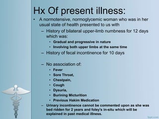
Case Presentation
DR. TAHMINA ZAFAR
How we will proceed with this
presentation:
Learning objectives
Introduction and
Case Scenario
Activity Related To
The Case
• Introduction
• Pathophysiology
• Presentation
• Investigations
• Diagnosis
Neuromyelitis
Optica
Closing Activity
Take Home
Message
• To be able to identify signs
and symptoms
• To be able to make a
differential diagnosis
• To reach a final diagnosis
Case Scenario:
• A 36 y/o Female, resident of Chungi,
Lahore presented to us with symptoms
of:
– Bi-lateral upperlimb numbnes 12 Days
– Fecal incontinence 10 Days
Hx Of present illness:
• A normotensive, normoglycemic woman who was in her
usual state of health presented to us with
– History of bilateral upper-limb numbness for 12 days
which was:
• Gradual and progressive in nature
• Involving both upper limbs at the same time
– History of fecal incontinence for 10 days
– No association of:
• Fever
• Sore Throat,
• Chestpain,
• Cough
• Dysuria,
• Burining Micturition
• Previous Hakim Medication
– Urinary incontinence cannot be commented upon as she was
bed ridden for 2 years and foley's in-situ which will be
explained in past medical illness.
Past Medical Hx
• In 2015 she presented with hx of bi-lateral
lower limb weakness and was diagnosed as
Multiple Sclerosis
• She was treated but not fully recovered, and
discharged on medication
• In 2017 she again presented with worsening of
previous symptoms and urinary incontinence
but was discharged again without complete
recovery
• According to the patient in 2020 her condition
got worsened but she did not consult any
doctor because of the Corona epidemic
• She has hx of laparoscopic cholecystectomy in
2015.
• No hx of blood transfusion
• No hx of any trauma/accident
Family Hx:
• No Family hx of
– DM
– HTN
– IHD
• No hx of similar illness in any other family
member
Musculoskeletal hx:
• No previous hx of joint pains,
swellings or rheumatic disease.
Gynecological Hx
• Age of menarche was at 16 with regular
menstruation cycles, with no hx of
menorrhagia or oligomenorrhea
• She has 4 children, all alive and healthy, all by
normal SVD
• No hx of OCPs
Personal hx:
• She is a housewife with 4 children, all alive
and healthy
• She has no history of smoking or any drug
abuse
• She lives in a 10 Marla house, 4 bathrooms, in
a combined family with total 12 people living
there.
• Good Ventilation
General Physical Examination:
• An ill looking female with average height and
built lying comfortably on bed with iv line on
right hand and folleys insitu
– Vitally stable with
– Bp120/70
– Pulse 77/min
– Temp 98.6f
• There is no pallor jaundice cyanosis raised jvp
clubbing edema feet palpable lymphnodes
kilonychea
Systemic examination
• Central nervous system examination
• HMF
• Oriented with time place and person
• Memory intact recent and past
– Speeh normal
– Cranial nerves all intact
Motor system
lower limbs
• Bulk tone normal
• Bilateral power 3/5
• No fasciculations
»Upper limbs
Tone bulk normal
• Power 5/5 no fasciculations
• Reflexes
• Upper limbs
• Normal( bicep tricep and brachioradialus)
• +2
• Reflexes lower limbs
– All absent
• Planters
– Bilateral downgoing
• Sensory system
– Sensory level at t7 to t10
• Spine and skull
– Normal
• Gait
– Cant b assessed
Other systems
• Respiratory
• Cardiac
• Git
• Musculoskeletal
– No positive findings
Investigations
• CBC, RFTS, LFTS, BSR, PT/INR/APTT, TFTS,
VIT B12 levels, RA FACTOR, ANA, VDRL:
– WITHIN NORMAL RANGES
• HEPATITIS B and C
– NEGATIVE
• CT BRAIN PLAIN
– NO SIGNIFICANT FINDINGS
• MRI BRAIN WITH CONTRAST
– NO SIGNIFICANT FINDINGS
• MRI CERVICODORSAL SPINE
– SHOWING HYPERINTENSE
LESION(2017), And cord atrophy at the
level of C3-6 (2021).
2017
Cervico-
Dorsal spine
with and
without
contrast
Showing
hyperintense
lesion (Blue
arrow)(T2):
2017 MRI
brain and
dorso-
cervical
spine with
iv contrast
• MRI
Brain
done in
2017
• (T2 and
T1):
2017 MRI
images
(Sagittal):
2017 MRI
images
(Coronal
With and
without
contrast):
2017Cevi
c-dorsal
spine
(T2):
MRI
Cervico-
dorsal
spine
with
contrast.
(2021)
Showing
atrophic
changes:
MRI
Cervico-
dorsal
spine
with
contrast.
(2021)
Showing
atrophic
changes:
2021
2021
2021
Diffrential diagnosis
• Transverse myelitis
• MS
• NMO spectrum disorder or Devic’s
disease
Activity:
• Can it be multiple sclerosis?
– Give your answers
– Give at least two reasons supporting
your answer
• Can it be transverse myelitis?
– Give answer
– Supporting evidence or reason in two
points at least
Neuromyelitis Optica:
• Introduction:
• Neuromyelitis optica (NMO), also known as Devic’s disease, is an
immune-mediated demyelinative disorder of the central nervous
system (CNS).
• Originally, it was considered a variant of multiple sclerosis (MS).
• Only in 2004, with the discovery of specific IgG antibodies
directed against aquaporin 4 (AQP4), considered as pathognomic
for NMO, did it become possible to classify this disorder as a
separate entity.
• In 2015, Wingerchuk et al. published the international consensus
on diagnostic criteria for NMOSD
• . According to these, NMOSD includes:
– classical NMO (optic neuritis—ON + longitudinal extensive transverse
myelitis—LETM)
– isolated ON or LETM
– ON and/or LETM associated with autoimmune systemic diseases
– ON and LETM accompanied by symptoms of brainstem, diencephalon
or cerebral involvement
Pathophysiology:
• The main pathological mechanism
involved in the background to
NMO is associated with
autoreactive anti-AQP4 IgG
antibodies
• Possible mechanisms include
inappropriate recognition of this
protein by T-cells or impairment of
early B-cell tolerance checkpoints
Presentation:
• Patients usually present with hx of bi lateral
limb numbness and decreasing visual acuity.
• Although symptoms may vary between various
types of NMOSD variants.
• It follows a relapsing course with incomplete
recovery and each episode worsening the
previous symptoms.
• Differentiating NMO from MS is particularly
relevant, because disease-modifying therapies,
effectively used in MS, appear to have weak or
even adverse effects upon NMO course.
Investigations:
• MRI is usually the first investigation of
choice.
• The presence of AQP4 antibodies in
serum confirms the diagnosis.
• For other variants of NMOSD, different
diagnostic criteria exist that can be
used to distinguish them from other
auto-immune disorders given in the
next slide.
Diagnosis:
Ending activity:
Group A:
• 1.
• 2.
Group B:
• 1.
• 2.
• MRI findings
• Antiaquaporin-4 antibodies positive in serum
• MOG serum antibody status
– CSF analysis:
» Raised protein levels
» Absence of oligoclonal bands
Confirming the Diagnosis:
Treatment Options:
• Methylprednisolone
• Plasma exchange
• Intravenous immunoglobulin
• Mycophenolate mofetil
• Azathioprine
• Methotrexate
• Cyclophosphamide
• Rituximab
Take Home Message:
• NMOSD is a distinct group of auto immune
disorders.
• The presence of AQP4 is pathognomic for
diagnosis of NMO.
• It is a relapsing condition with incomplete
recovery and each episode worsening the
previous injury.
• NMOSD Should be differentiated from MS
because the treatment modalities may worsen
the condition.
• Early diagnosis and treatment leads to better
outcomes and decrease in morbidity.
NMO Presentattion.ppt

NMO Presentattion.ppt

  • 1.
  • 2.
    How we willproceed with this presentation: Learning objectives Introduction and Case Scenario Activity Related To The Case • Introduction • Pathophysiology • Presentation • Investigations • Diagnosis Neuromyelitis Optica Closing Activity Take Home Message • To be able to identify signs and symptoms • To be able to make a differential diagnosis • To reach a final diagnosis
  • 3.
    Case Scenario: • A36 y/o Female, resident of Chungi, Lahore presented to us with symptoms of: – Bi-lateral upperlimb numbnes 12 Days – Fecal incontinence 10 Days
  • 4.
    Hx Of presentillness: • A normotensive, normoglycemic woman who was in her usual state of health presented to us with – History of bilateral upper-limb numbness for 12 days which was: • Gradual and progressive in nature • Involving both upper limbs at the same time – History of fecal incontinence for 10 days – No association of: • Fever • Sore Throat, • Chestpain, • Cough • Dysuria, • Burining Micturition • Previous Hakim Medication – Urinary incontinence cannot be commented upon as she was bed ridden for 2 years and foley's in-situ which will be explained in past medical illness.
  • 5.
    Past Medical Hx •In 2015 she presented with hx of bi-lateral lower limb weakness and was diagnosed as Multiple Sclerosis • She was treated but not fully recovered, and discharged on medication • In 2017 she again presented with worsening of previous symptoms and urinary incontinence but was discharged again without complete recovery • According to the patient in 2020 her condition got worsened but she did not consult any doctor because of the Corona epidemic
  • 6.
    • She hashx of laparoscopic cholecystectomy in 2015. • No hx of blood transfusion • No hx of any trauma/accident
  • 7.
    Family Hx: • NoFamily hx of – DM – HTN – IHD • No hx of similar illness in any other family member
  • 8.
    Musculoskeletal hx: • Noprevious hx of joint pains, swellings or rheumatic disease.
  • 9.
    Gynecological Hx • Ageof menarche was at 16 with regular menstruation cycles, with no hx of menorrhagia or oligomenorrhea • She has 4 children, all alive and healthy, all by normal SVD • No hx of OCPs
  • 10.
    Personal hx: • Sheis a housewife with 4 children, all alive and healthy • She has no history of smoking or any drug abuse • She lives in a 10 Marla house, 4 bathrooms, in a combined family with total 12 people living there. • Good Ventilation
  • 11.
    General Physical Examination: •An ill looking female with average height and built lying comfortably on bed with iv line on right hand and folleys insitu – Vitally stable with – Bp120/70 – Pulse 77/min – Temp 98.6f • There is no pallor jaundice cyanosis raised jvp clubbing edema feet palpable lymphnodes kilonychea
  • 12.
    Systemic examination • Centralnervous system examination • HMF • Oriented with time place and person • Memory intact recent and past – Speeh normal – Cranial nerves all intact
  • 13.
    Motor system lower limbs •Bulk tone normal • Bilateral power 3/5 • No fasciculations »Upper limbs Tone bulk normal • Power 5/5 no fasciculations • Reflexes • Upper limbs • Normal( bicep tricep and brachioradialus) • +2
  • 14.
    • Reflexes lowerlimbs – All absent • Planters – Bilateral downgoing • Sensory system – Sensory level at t7 to t10 • Spine and skull – Normal • Gait – Cant b assessed
  • 15.
    Other systems • Respiratory •Cardiac • Git • Musculoskeletal – No positive findings
  • 16.
    Investigations • CBC, RFTS,LFTS, BSR, PT/INR/APTT, TFTS, VIT B12 levels, RA FACTOR, ANA, VDRL: – WITHIN NORMAL RANGES • HEPATITIS B and C – NEGATIVE • CT BRAIN PLAIN – NO SIGNIFICANT FINDINGS • MRI BRAIN WITH CONTRAST – NO SIGNIFICANT FINDINGS • MRI CERVICODORSAL SPINE – SHOWING HYPERINTENSE LESION(2017), And cord atrophy at the level of C3-6 (2021).
  • 17.
  • 18.
  • 19.
  • 20.
  • 21.
  • 22.
  • 23.
  • 24.
  • 25.
  • 26.
  • 27.
  • 28.
    Diffrential diagnosis • Transversemyelitis • MS • NMO spectrum disorder or Devic’s disease
  • 29.
    Activity: • Can itbe multiple sclerosis? – Give your answers – Give at least two reasons supporting your answer • Can it be transverse myelitis? – Give answer – Supporting evidence or reason in two points at least
  • 30.
    Neuromyelitis Optica: • Introduction: •Neuromyelitis optica (NMO), also known as Devic’s disease, is an immune-mediated demyelinative disorder of the central nervous system (CNS). • Originally, it was considered a variant of multiple sclerosis (MS). • Only in 2004, with the discovery of specific IgG antibodies directed against aquaporin 4 (AQP4), considered as pathognomic for NMO, did it become possible to classify this disorder as a separate entity. • In 2015, Wingerchuk et al. published the international consensus on diagnostic criteria for NMOSD • . According to these, NMOSD includes: – classical NMO (optic neuritis—ON + longitudinal extensive transverse myelitis—LETM) – isolated ON or LETM – ON and/or LETM associated with autoimmune systemic diseases – ON and LETM accompanied by symptoms of brainstem, diencephalon or cerebral involvement
  • 31.
    Pathophysiology: • The mainpathological mechanism involved in the background to NMO is associated with autoreactive anti-AQP4 IgG antibodies • Possible mechanisms include inappropriate recognition of this protein by T-cells or impairment of early B-cell tolerance checkpoints
  • 32.
    Presentation: • Patients usuallypresent with hx of bi lateral limb numbness and decreasing visual acuity. • Although symptoms may vary between various types of NMOSD variants. • It follows a relapsing course with incomplete recovery and each episode worsening the previous symptoms. • Differentiating NMO from MS is particularly relevant, because disease-modifying therapies, effectively used in MS, appear to have weak or even adverse effects upon NMO course.
  • 33.
    Investigations: • MRI isusually the first investigation of choice. • The presence of AQP4 antibodies in serum confirms the diagnosis. • For other variants of NMOSD, different diagnostic criteria exist that can be used to distinguish them from other auto-immune disorders given in the next slide.
  • 34.
  • 35.
    Ending activity: Group A: •1. • 2. Group B: • 1. • 2.
  • 36.
    • MRI findings •Antiaquaporin-4 antibodies positive in serum • MOG serum antibody status – CSF analysis: » Raised protein levels » Absence of oligoclonal bands Confirming the Diagnosis:
  • 37.
    Treatment Options: • Methylprednisolone •Plasma exchange • Intravenous immunoglobulin • Mycophenolate mofetil • Azathioprine • Methotrexate • Cyclophosphamide • Rituximab
  • 38.
    Take Home Message: •NMOSD is a distinct group of auto immune disorders. • The presence of AQP4 is pathognomic for diagnosis of NMO. • It is a relapsing condition with incomplete recovery and each episode worsening the previous injury. • NMOSD Should be differentiated from MS because the treatment modalities may worsen the condition. • Early diagnosis and treatment leads to better outcomes and decrease in morbidity.