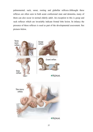
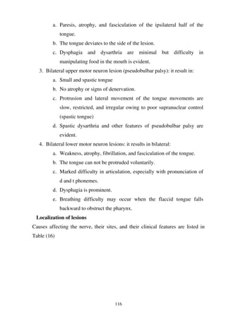
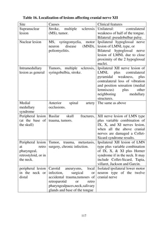
SlideShare a Scribd company logo
1 of 206
Download to read offline
BM-Publisher, ISBN 978-0-9864331-1-5
http://www.bmpublisher.net
All rights reserved. No part of this publication may be reproduced or transmitted in any
form or by any means, electronically or mechanically, including photocopying,
recording or any information storage or retrieval system, without the prior permission
from the BM-publisher
Whilst the information in this book is believed to be true and accurate at the date of
going to press, the BMpublisher cannot accept any legal responsibility or liability for
any errors or omissions that may be made
Neurological Examination Made Practical® is a registered trademark of the
BM-Publisher, Inc
ISBN 978-0-9864331-1-5
BM-Publisher
Dearborn, Michigan 48126
USA
Email: bmpublisher@bmpublisher.net
-----------------------------------------------------------------------------------------------
1
CHAPTER ONE
EXAMINATION OF HIGHER CEREBRAL FUNCTIONS
It includes examination of the following functions:
1-Level of consciousness
2-Memory
3-Orientation
4-Judjment &reasoning
5-Language
6-Integrative sensory functions
7-Integrative motor functions
8-Primitive reflexes
1-1: Level of Consciousness
Method of examination
1-Verbal stimulation
If the patient is a wake, talk to him, test his orientation for time, place and
person (to exclude confusion) and ask him to obey simple commands like lift
your hand, open your mouth, open your eye (to exclude wernicke’s aphasia),
look up, look down, and look at the tip of your nose (to exclude lock in
syndrome).
I. If the patient is in a sleepy state try to wake him by tactile stimulation or by
calling his name. If no response to tactile or verbal stimulation proceed to
the next stage of stimulation which is painful stimulation.
2. Painful (noxious) stimulation
Its done by supraorbital or sternal pressure (rubbing the sternum
vigorously by the knuckle of the hands), pinching the side of the neck or inner
parts of the arm or thighs or pressure on the nail bed, and by noticing the patient
responses (eye, verbal and motor) to these stimuli (both verbal and noxious) we
can roughly estimate the level of consciousness and it’s changes from hour to
hour.
2
Interpretation of the results
1. Normal consciousness: The patient is aware of himself and environment, he
is a wake, alert, with eyes open at rest and unless there is deafness or
language disorder, verbal stimulation results in appropriate verbal responses.
2. Impaired consciousness: Characterized by defect of arousal. There is infinite
gradation of altered consciousness but the terms drowsiness, stupor, and
coma usefully describe the three major stages which can be clinically
recognized.
Drowsiness
It is a sleepy state from which the patient can be aroused to a state of
complete wakefulness and cooperation by verbal or tactile stimuli, but he tends
to sink into sleep again if stimulation ceases. This condition is common in high
brainstem disturbances directly or indirectly and in drug toxicity.
Stupor
It is a sleeplike state from which the patient can be aroused by vigorous or
repeated stimulation (Verbal, tactile, or painful), he answer questions
appropriately or inappropriately, just to fall a sleep again. No satisfactory co-
operation is obtained. This condition is caused by disease or compression of the
upper brain stem, or by bilateral cerebral dysfunction (anoxic, toxic, traumatic or
infective).
Coma
It is a sleep like state from which the patient cannot be aroused. The
patient is not aware of his internal or external needs. The eyes are typically
closed and do not open spontaneously. The patient doesn’t speak and there is no
purposeful movement of the face or limbs. Verbal stimulation produce no
response, painful stimulation may also produce no response or may elicit non-
purposeful reflex movement mediated through the spinal cord or brainstem
3
pathways. Coma results from a disturbance in the function of either the
brainstem reticular activating system above the midpons or both cerebral
hemispheres. Coma is best evaluated by Glasgow coma scale, Table (1).
3. Turbidity of consciousness
Confusion
It is inability to think with customary speed and clarity, with incoherent
speech associated with disorientation and impairment of the memory. Confusion
most often results from a process that influences the brain globally such as toxic
or metabolic disturbances or dementia. Confusion also may results from any
condition that leads to drowsiness or stupor. Important differential diagnosis
includes:
I. Wernicke’s aphasia.
II. Psychosis.
III.Non-dominant hemisphere lesions.
IV. Subdural hematoma.
V. Disorders of memory or visuospatial orientation.
Delirium
It is a confusional state plus agitation and aggressive behavior, commonly
associated with hallucination and delusion plus autonomic over activity and
motor abnormalities like (tremor, myoclonus and asterexis). A delirious like
state is commonly caused by infective and toxic state and in delirium tremens of
alcoholism.
4. Dissociated consciousness
It means dissociation between consciousness and motor performance, where
the patient performs programmed action that you can not resist and after
which the patient can not remember it. This is called automatism. It is
commonly occur in partial complex epilepsy and fugue state of hysteria.
4
Important related syndromes
1. Persistent vegetative state:
This condition means preserved autonomic and brain stem function without
cognition, some patients who sustained severe coma due to cerebral hypoxia or
ischemia or structural brain lesion may regain wakefulness but not awareness.
After one month, this condition is called persistent vegetative state. Such
patients have spontaneous eye opening and sleep-wake cycle which differentiate
them from comatose patients.
2. Locked-in syndrome (Akinetic mutism)
Such patient appears comatose but is awake and alert, although they are mute
and quadriplegic. The diagnosis is made by noting that voluntary eye opening,
vertical eye movements, ocular convergence, or some combination of these
midbrain mediated movements is preserved. During the examination of any
apparently comatose patient, the patient should be told to open your eyes, look
up, look down and look at the tip of your nose, to elicit such movements. The
cause of this syndrome is due to functional transection of the brain stem below
the midpons level. The usual causes are pontine infarction, hemorrhage, central
pontine myelinolysis, tumor or encephalitis.
3. Psychogenic unresponsiveness:
We must always think of this possibility in any seemingly comatose patient,
in whom general, physical and neurological examination is normal. This
condition may occur in schizophrenia (catatonic type), conversion disorders,
somatization disorders and malingering.
4. Brain death:
We must always think of this possibility in any patient who is deeply
comatose and maintained on a ventilator because spontaneous breathing had
been inadequate or had ceased. Preconditions for considering a diagnosis of
brain death and tests for confirming it is beyond the scope of this book.
Pictures at page 5 give the anatomical site of the above lesions.
5
Table 1. The Glasgow Coma Scale.
Eye opening Verbal response Motor response
Spontaneous 4 Oriented 5 Obeying 5
To speech 3 Confused 4 Localizing 4
To pain 2 Inappropriate 3 Flexing 3
None 1 Incomprehensible 2 Extension 2
None 1 None 1
6
1-2: Memory
Tests of memory
Memory is assessed clinically by testing immediate recall, recent memory and
remote memory, which correspond roughly to registration, storage and retrieval,
respectively.
1. Immediate recall (registration)
a. Digit number: ask the patient to repeat a random series of numbers; most
normal adult can repeat a series of seven numbers forward and five
numbers backward without difficulty.
b. Babcock’s sentence: “there is one thing a nation must have to be rich and
great, and that is a large, secure supply of wood”, or any other sentence
and ask the patient to repeat this sentence directly after the examiner.
2. Recent memory (short term memory)
a. Ask the patient about events that occurred before minutes to hours, like
what was your breakfast.
b. Ask the patient to recall the names of three objects repeated to the patient
before three minutes.
c. Non-verbal testing: ask the patient to select an object previously shown
to him from a group of objects. This test is especially suitable for patients
with expressive aphasia.
3. Remote memory (long term memory)
a. Ask the patient about historical, personal or geographical events.
b. Ask the patient to give association or connection between these events.
The questions must be appropriate for the patient, that some one of
comparable cultural and educational background can be assumed to
know, for example (when was your marriage? And what was the color of
your neck tie? )
7
Introduction
Memory is the ability to register, store, and finally retrieve information.
Short term memory can be impaired by diseases which affect the medial
temporal lobe bilaterally while long term memory is usually a manifestation of
diffuse cortical disease. The functional components of memory are:
1. Registration: which is the ability to receive information through the
appropriate primary sensory areas as well as the corresponding association
cortex, ablation of the primary auditory and the adjacent association auditory
cortices will abolish the immediate memory of auditory type. The duration
of recall is a matter of seconds.
2. Storage: it is the process by which new information and tasks are learned. It
is the function of the limbic structures including the hippocampus and
related structures like the mamillary bodies and the dorsomedial nuclei of
the thalamus. Stored memory is reinforced by repetitions and emotional
significance. Short term memory may be recalled for minutes to hours.
3. Retrieval: this is the ability to project previously learned information. It can
be recalled months or years after it has been stored. It reflects the integrity of
the whole cerebral cortex and the subcortical structures in addition.
Amnesia
It is a memory disorder which can be classified into
1. Isolated amnesic syndromes.
2. Non-isolated that is part or one feature of global cognitive disorder which
can occur in two conditions.
I. Acute confusional state
II. Dementia
3. Psychogenic amnesia
1- Isolated amnesic syndromes
These syndromes are usually characterized by the following features:
I. In acute amnestic syndromes there is usually impairment of short term
memory and registration.
8
II. Confabulation: Some patients may attempt to fill in gaps in memory with
false recollections.
III.Retrograde amnesia for a variable period preceding the event may be
present, but this period shrinks as the condition improves.
IV. In chronic amnestic syndromes long term memory is also affected but
to a lesser extent than short term. Registration is intact.
V. Site of lesion is bilateral damage to hippocampus, and related structures
such as the dorsomedial nucleous of the thalamus or mamllary body.
VI. Other features of amnestic syndrome are usually related to the site of
lesions other than that of memory.
Causes of isolated amnestic syndrome
Acute
I. Head trauma.
II. Hypoxia or ischemia.
III. Bilateral posterior cerebral artery occlusion.
IV. Transient global amnesia (TGA).
V. Alcoholic blackouts.
Chronic
I. Alcoholic korsakoff amnestic syndrome.
II. Post encephalitic amnesia.
III.Brain tumor.
IV. Paraneoplastic limbic encephalitis.
2-Amnesia accompanying acute confusional state
In this condition, attention is impaired, resulting in impairment of
registration and inability to from new memories. Decrease in the level of
consciousness is the chief complaint. Those patients are usually in a sleepy state
and disoriented for time and place.
3-Amnesia accompany dementias
Dementias are usually chronic, steadily, progressive and the level of
consciousness is not impaired. Amnesia is usually associated with impairment of
9
other spheres of cognition like language, parietal lobe functions (construction,
left-right orientation), frontal lobe or diffuse cerebral cortical functions
(Judgment, abstraction and the ability to perform previously learned skills as
shown in pictures at page 11. The mini mental status examination provides a
useful bed side screening test when dementia is suspected, Table (2).
Causes of Dementia
For better approach and management of the patient the causes of
dementias are best divided into reversible and irreversible.
1. Often reversible:
Infectious: e.g. Neurosyphilis.
Deficiency states: e.g. .vitamin B1 or B12, folic acid.
Toxins: drugs, heavy metals.
Metabolic: e.g. hypothyroidism, hypoglycemia, hypocalcaemia,
Cushing syndrome, liver or renal failure.
Trauma: e.g. subdural haematoma.
Others: hydrocephalus, anoxia, tumors.
2. Irreversible
Hereditary: e.g. Huntington’s chorea
Prion disease: e.g. Greutzfeldt-Jakob disease
Non metastatic manifestation of malignancy: e.g. bronchial carcinoma
Others: Alzheimer’s or pick’s disease.
N.B The commonest causes of dementia in order of frequency include.
I. Alzheimer
II. Dementia with lewy bodies.
III. Vascular dementia (multi infarct Dementia).
3- Psychogenic amnesia
In this condition subjective and emotionally charged memories are more
affected than is recalling of objective facts and events. In organic amnesia the
reverse is true. Isolated loss of memory for personal identity in awake and alert
person is virtually diagnostic of a psychogenic problem.
10
Table 2. Minimental status examination
Item. Points
Orientation
Time(1 point each for year, season, month, date, and day of the week)
Place(1 point each for state, country, city, building and Floor or room)
Registration....repeat names of three objects (1point per object
Attention and calculation.
Serial 7s or spell “world” Backward (1 point per subtraction or letter
Recall names of three objects repeated previously(1point per object)
Language
Repeat “no islands or buts”
Read and follow “close your eyes)
Write a complete sentence.
Construction...Copy two interesting pentagons.
Total
5
5
3
5
3
1
1
1
1
30
A total of less than 24 should generally lead to more detailed investigation of the
possibility of dementia, although norms vary to some extent with age and
education.
11
Rohkamm, Color Atlas of Neurology @ 2004 Thieme.
12
1-3 Orientation
Orientation is usually tested in three dimensions.
1. Time – Ask the patient about (day, date, month, year), what time of the day
is it?
2. Place – Ask where he is now (room, building, town, country), what floor is
it on?
3. Person – Ask the patient about his (name, age, work, address) also ask him
about the relative’s names who are around his bed?
Interpretation of results
Orientation is usually impaired in acute confusional states, and it must be
tested early to assess the level of consciousness. If the level of consciousness is
normal disorientation is usually occur in amnestic syndromes and psychiatric
disorders. The latter condition is suggested when there is disorientation for
personal identity rather than place and time.
Common causes of acute confusional states are listed in table (3).and
partial list of drugs that can produce it is provided in table (4).
Table 3. Common Causes of acute Confusional States
Causes Examples
1-Metabolic disorders:
Drugs Ethanol intoxication or withdrawal, Sedative drug
intoxication or withdrawal, opioids, anticholinergic
Endocrine disorder Hypothyroidism, hyperthyroidism. hypoglycemia,
hyperglycemia
Nutritional disorders hyponatremia, hypocalcemia, hypercalcemia
Organ system failure hepatic encephalopathy, Reye’s syndrome, uremia,
dialysis disequilibrium, pulmonary encephalopathy
,organ transplantation
2-Infectious and
Noninfectious.
Bacterial meningitis, viral encephalitis, tuberculous
meningitis, leptomeningeal metastases, AIDS, others
3-Vascular disorders Hypertensive encephalopathy, subarachnoid
hemorrhage,vertebrobasilar ischemia, right (non
dominant) hemisphere infarction, SLE, DIC,TTP
4-Head trauma Concussion, Intracranial hemorrhage
5-Seizure Post ictal state, Complex partial seizure
13
Table 4. Therapeutic drugs associated with acute confusional states
List (A) List (B)
Acyclovir , Disulfiram Amantadine , Ergot alkaloids
Aminocaproic acid , Ethanol Amphetamines , Ganciclovir
Anticholinergic , Hallucinogens Anticonvulsants , Isoniazid
Antidepressants , Ketamine Antihistamines , Levodopa
Antipsychotics , Lidocaine Baclofen , Methylxanthines
Barbiturates Benzodiazepines , Opioids
B-blocker , Penicillin Cephalosporins , Quinacrine
Chloroquine , Quinidine Cocaine , Quinine
Corticosteroids , Salicylate Cyclosporine , Selegiline
Digoxin , Thyroid hormones Non steroidal anti-inflammatory
Pictures at page 14, from Rohkamm, color atlas of neurology, illustrate the
mechanism of normal consciousness, apallic syndrome, acute confusion,
disturbances of arousal and stupor state,
14
15
1-4 Judgment and Reasoning
This important category of cognitive abilities must be included in every attempt
to evaluate the mental status, it includes at least testing of Abstraction,
calculation and general behavior.
1. Abstraction and calculation
Method of examination
I. Proverb interpretation: Ask the patient to explain proverb or
fable.
II. Similarities and dissimilarities ask the patient to describe
similarities and differences between groups of object (orange
and a ball, child and dwarf, etc...)
III. 100-7 test: subtraction of serials 3’s or 7’s from l00 is a good
test of calculation and concentration.
2. General behavior: It includes mood, hallucinations, delusions and other
abnormalities of behavior like attitude, manner of dress…etc
Interpretation of the results
Abnormalities of these tests can help to distinguish between organic and
psychiatric disease. Abstraction and calculation are usually impaired in
Dementia. Visual hallucinations are common in acute confusional states, while
auditory hallucinations and fixed delusions are most common with psychiatric
disorders.
Demented patients may be apathetic, inappropriately elated or depressed,
and their moods can fluctuate. If the examination is other wise normal, early
dementia can easily be confused with depression.
16
1- 5: Language Examination
The essential elements of language are comprehension, expression, repetition,
naming, reading and writing. The important preliminary information needed for
correct assessment of Language are nationality, native Language, educational
level, mental state, handedness and the previous ability to read or write. The
different anatomical areas controlling language are well illustrated below at
pages 16 and 17.
17
18
Methods of Examination
* Language is examined in six aspects
1. Comprehension to spoken Language
Ask the patient to obey simple commands for example (Lift your hand, Close
your eye, Open your mouth, Point to certain objects around the room). If he did
the previous tests easily then ask him to obey more complex commands for e.g.
(lift your hand and close your eyes).
2. Expression
Ask the patient to talk about his illness without interruption from his family
and note the following important parameters of his speech.
I. Fluency: Is he fluent or non-fluent?
If the patient is severely non-fluent, communication is still feasible by asking
the patient to answer by yes or no. if he is totally mute and the comprehension is
relatively spared communication is done by eyes movement.
II. Awareness: Is he a ware of his deficit or his speech?
III. Errors: Is he doing mistakes likes, paraphasias, Neologism, perseverations
and circumlocutions
Paraphasia: these are of two types:-
Literal: which means substitution of a syllable within the word e.g. the grass is
greeI
Verbal: It means substitution of one word for another e.g. the grass is blue
• Neologism. It means appearance of phonemes, syllable or words that are
not part of the language e.g. the grass is grumps.
• Perseveration: The patient is involuntarily repeats the same words or acts
just after it has been employed.
• Circumlocution: The patient uses along sentence to overcome failure to find
particular word.
3. Repetition
Ask the patient to repeat a complex phrase, and if he fails ask him to
repeat simpler one or even a single word.
19
4. Naming
Ask the patient about the names of common objects, if he fails to give the
right names, ask him to choose the correct one form a list of alternatives. Use
about twenty objects before deciding that no abnormality exists.Palients with
true nominal aphasia usually fails to give the correct names of three objects out
of twenty.
5. Reading
I. Ask the patient to read words, then sentences, then longer passages.
II. Ask him to read and obey a written command, this will link the motor and
sensory side.
III. Patient with alexia is unable to understand a written speech.
6. Writing
I. Test the ability to write to dictation then ask him to construct a sentence or
to compose apiece about his job, town or weather.
II. Patient with agraphia may be able to write his name or address but unable
to construct a sentence.
Aphasia: It means a disturbance of the ability to use language, whether in
speaking, writing or comprehension.
Clinical varieties of aphasia
The preceding systematic examination of language will usually enable one to
disclose the following types of dysphasia.
1. Broca’s aphasia.
2. Wernicke’s aphasia.
3. Total or global aphasia.
4. Dissociative or (Disconnection) syndromes such as:
I. Conduction aphasias.
II. Word deafness (Auditory verbal agnosia).
III. Word blindness (visual verbal agnosia or alexia).
1V. Pure word mutism.
V. Transcortical aphasias.
20
5. Anomic aphasia
6. Agraphias
Broca’s aphasia
• Speech is non-fluent even to the point of mutism in extreme examples.
• Relative preservation of comprehension.
• Poor, repetition, reading and writing.
• Phrases length is abbreviated and many of the small words (article,
prepositions and conjunctions) are omitted giving the speech a telegraphic
character (so-called agrammatism).
• Other important features of Broca’s aphasia includes: verbal stereotypy,
apraxia of speech, limbs and right facio- brachial palsy.
• Site of lesion is usually includes Broca’s area plus deep white matter
between Broca’s area and motor cortex, the anterior insula, frontal- parietal
operculum, and adjacent cerebrum..
• Causes: embolic infarction in the territory of upper main (rolandic) division
of the middle cerebral artery is probably the most frequent cause of
vascular lesion. Other causes include: thrombosis, tumor, sub cortical
hypertensive hemorrhage, traumatic hemorrhage and seizure.
Wernicke’s aphasia
• Speech is Fluent and paraphasic.
• Comprehension is impaired for both spoken and written language.
• Impairment of repetition, naming and writing.
• The patient is unaware of his deficit or his own errors. Fluent paraphasic
speech may be entirely incomprehensible (Jargon aphasia).
• Site of Lesion: Posterior perisylvian region.
• Causes: embolic (less often thrombotic) occlusion of the lower division of
the left middle cerebral artery. Other rare causes include Hemorrhage.
Tumor, abscess and extension of a small putaminal or thalamic
hemorrhage.
21
Total or global aphasia
• All aspects of language are affected, but they can utter few syllable such as,
ah, or cry, shout or moan. In other words, they are not mute.
• With the passage of time, some degree of comprehension of language may
return, and the patient is left with severe Broca’s aphasia.
• Varying degree of right hemiplegia, hemianesthesia, and homonymous
hemianopia almost invariably accompanying global aphasia of vascular
origin.
• Site of lesion: large part of the language area (sensory and motor parts).
• Causes: the lesion is usually due to occlusion of the left internal carotid or
middle cerebral artery. But it may be caused by tumor, hemorrhage or other
lesion and it may occur transiently as a post-ictal effect.
Dissociative syndromes
Conduction aphasia
• Speech is fluent and paraphasic but comprehension is relatively spared, thus
distinguishing it from Wernick’s aphasia.
• Site of the lesion: arcuate fasiculus which connect the sensory and motor
areas of speech.
• Causes: embolic occlusion of the ascending parietal or posterior temporal
branch of the middle cerebral artery, but other causes like neoplasm, or
trauma in the region may produce the same syndrome.
Pure word deafness
• Impairment of auditory comprehension and repetition and an inability to
write to dictation.
• Spontaneous speech, writing, and reading are normal, thus distinguishing
this disorder from the classic Wernick’s aphasia.
• Site of lesion: bilateral in the middle third of the superior temporal gyri, in a
position to interrupt the connection between primary auditory cortex and
the association areas. In rare cases unilateral lesions have been localized in
this part of the dominant temporal lobe.
22
• Causes: Embolic occlusion of small branch of the lower division of the
middle cerebral artery.
Alexia
The patient can not understand the written speech. It can be divided into
2 types:
1. Alexia without agraphia (pure word blindness): The patient is able to
speak, understand spoken language, he can write both spontaneously and
to dictation but can not understand or copy the written words.
Site of the lesion: left occipital cortex and splenium of the corpus-
callosum so both visual cortices are separated from the language area of
the left hemisphere.
2. Alexia with agraphia: The patient can not read or write spontaneously.
This is part of Gerstman syndrome i.e. right-left confusion, acalculia,
finger agnosia, anomia in addition to alexia and agraphia.
Site of the lesion: angular gyrus or the subjacent white mater.
In such cases, there is no right homonymous hemianopi. The entire
collection of symptoms is sometimes called the Syndrome of angular
gyrus.
Pure word mutism
• The patient loses all capacity to articulate or he has a sever slurring of
speech.
• Comprehension, reading and writing are normal from the onset.
• After recovery, the language show neither loss of vocabulary nor
agrammatism, or there may be mild dysarhtria (hence cortical dysarthria),
anomia, and paraphasic substitutions.
• The most notable feature of this type of speech disorder is its transience,
and within few weeks or months, language is returned to normal.
• Site of the lesion: The anatomic site has not been determined precisely. In a
few postmortem cases, reference is made to a lesion in Broca’s area.
• Causes: Commonly vascular.
23
Transcortical aphasias
These dysphasias characterized by normal repetition due to pathology
which isolate the intact motor and sensory language areas from the rest of the
cortex of the same hemisphere. They usually follow water-shed infarction
between middle, anterior and posterior cerebral arteries, as a result of prolonged
hypotension, carbon monoxide poisoning or other forms of anoxia. They are of
three types:
1. transcortical sensory aphasia
It is similar to Wernicke’s aphasia (fluent, paraphasic speech) but the
repetition is remarkably preserved.
Site of the lesion: posterior parietal-occipital region, posterior and inferior
to Wernick’s area. This location explains the frequent concurrence of
transient visual agnosia and hemianopia.
Causes: Water-shed infarction that disconnects the auditory language area
from the referent centers.
2. transcortical motor aphasia
The speech is non-fluent speech with relative preservation of
comprehension, but the repetition is strikingly intact.
Site of the lesion: anterior to Sylvain fissure but superior to Boca’s area
(anterior isolation syndrome).
Causes: it occur in 2 clinical situations:
• Mild, partially recovered Broca’s aphasia.
• In states of abulia and a kinetic mutism with frontal lobe damage, for the
same region.
3. Echolalia
The speech is non fluent, comprehension is poor but repetition is still
possible leading to persistent echolalia which means involuntary repetition of
words or phrases spoken by someone else without the intention of doing so,
and without understanding their meanings.
24
Site of the lesion: Infarction of the whole area surrounding the perisylvian
regions, and the latter becomes isolated from other cortical area. The lesion
lies in the temporo-parietal region.
Causes: Water-shed infarction due to sudden hemodynamic disturbances of
perfusion.
Anomic aphasia
• Disorder confined to naming of people and objects.
• Other language functions (reading, writing spelling…etc) being adequate.
• Recall of the names of letters, digits, and other printed verbal material is
almost invariably preserved, and immediate repetition of a spoken name is
intact. That the deficit is principally one of the naming is shown by the
patient correct use of the object and usually by an ability to point to the
correct object on hearing or seeing the name and to choose the correct name
from a list. The patient’s understanding of what is heard or read is normal.
• Site of the lesion: Lower temporal lobe in a place which interrupt
connections between sensory language areas and the hippocampal regions
concerned with learning and memory.
• Causes: Mass lesions e.g. tumors, herpes encephalitis or an abscess are the
most frequent causes; as they enlarge, a contra lateral upper quadrantic
visual field defect or a Wernick’s aphasia is added. Occasionally, anomia
appears with lesions due to occlusion of the temporal branches of the
posterior cerebral artery.
• N.B.: some degree of anomia is part of every type of language disorders,
but when the anomia is the most notable aspect of language difficulty, the
term anomic aphasia is employed, so anomia may part of the following
speech disorders:
I. Transcortical motor aphasia.
II. Gerstmann syndrome.
III.Alzheimer disease.
IV. Old age.
25
V. Finally anomic aphasia may be the only residual abnormality after
partial recovery from Wernick’s aphasia, conduction, transcortical
sensory, or rarely Broca’s aphasia.
5. Agraphia
Writing is disturbed in all types of aphasia except in 2 conditions (pure
word mutism and pure word blindness). Pure agraphia as the initial and sole
disturbance of language function is very rare, but such case can occur in lesion
between the angular gyrus and the motor area, but sometimes be anterior or
posterior to this.
The notion of a specific center for writing in the posterior part of second
frontal convulsion (extner’s writing area) has questioned by some aphasiologist ,
however, case of pure dysgraphia have been reported due to hematoma located
in the Centrum semiovale beneath the motor parts of frontal cortex.
Types of agraphia or dysgraphia
1. Aphasic agraphia-- Language formulation is defective, spelling and
grammatical errors are prominent.
2. Constructional agraphia-- In this circumstance, letter and words are formed
clearly enough but are wrongly arranged on the page. Words may be
superimposed, written diagonally, in haphazard arrangement, or from right
to left. With right parietal lobe lesions, only the right half of the page is
used.
3. Apraxic agraphia—here language formulation is correct and the spatial
arrangements of words are respected, but the hand had lost its skills in
forming letters and words. The patient is uncertain about how to held pen
and how to apply it on paper.
Schematic approach of aphasic patient
This can be done by listening to the patient fluency, testing
comprehension and lastly asking the patient to repeat certain phrases and
sentences (table-5- ). Other disorders of speech which include, agraphias,
26
anomias, and alexia, can be tested by examining writing, naming and reading
respectively.
Table (5) schematic approach of aphasia
Fluency
Non-fluent Fluent
1-Broca’s aphasia 1-Wernicke’s aphasia
2-Pure word mutism 2-Pure word deafness
3-Transcortical motor aphasia 3-Transcortical sensory aphasia
4-Echolalia 4-Conductive aphasia
Comprehension Comprehension
Impaired Normal Normal Impaired
(1) (4) (2) (3) (4) (1) (2) (3)
Repetition Repetition Repetition
Normal Impaired Normal Impaired Normal Impaired
(4) (1) (3) (2) (3) (1) (2)
Reading & Writing
Normal (2) Impaired (1)
27
1- 6: Integrative Sensory Functions
Sensory integrative disorders from parietal lobe lesions are manifested by
misperception of or inattention to sensory stimuli on the contra lateral side of
the body, when the primary sensory modalities are intact. Patients with parietal
lobe lesions may exhibit the following signs:
1. Asteriognosis.
2. Agraphesthesia.
3. Absence of two point’s discrimination.
4. Allesthesia (failure of localization of touch).
5. Disorders of body scheme or image.
6. Agnosia.
1-Astereognosis:
The patient can not identify, by touch, an object placed in his hand.
Methods of examination
Ask the patient to close his eyes; the object is placed first into the hand
suspected of abnormality. If he fails or take a long time to decide, it should then
be placed into the other hand, for comparison of the accuracy and speed of
response. Tested object must be familiar and large enough for a weak hand to
feel.
Result of examination
Patient with astereognosis can not identify any detail of the object at all.
He is unaware that anything has been placed in his hand. Its particular value in
localization arise when, though other form of sensation (touch, position) are
normal or only slightly affected, yet there is astereognosis, the lesion responsible
then lies in the parietal lobe.
2-Agraphesthesia
The patient is unable to recognize letters or numbers written on the skin
with a blunt point.
28
Method of examination
Ask the patient to close his eyes, and letters or numerals are written on the
palm, anterior aspect of the forearm, thigh or lower leg. Clear figures should be
used first, and if correct, the more difficult, can be used as a finer test.
Result of examination
If peripheral sensation is normal, agraphesthesia indicates parietal lobe
lesion.
3-Absence of two point discrimination
This is an inability to differentiate between a single and two
simultaneously applied adjacent, but separated, stimuli that can be distinguished
by a normal person.
Method of examination
Ask the patient to close his eyes, and touch the pulp of his finger firmly
with the two point’s discriminator, starting with them far apart, and
approximating them until the patient begins to make errors. The threshold is thus
determined and the other hand can be compared. The same test can be carried on
the dorsum of the foot.
Result of examination
The normal ability to distinguish the two points from one varies in
different parts of the body. On the fingers it should be possible at less than 5 mm
separation, but on the dorsum of the foot, separation of even 5 cm may be still
normal. Impairment of the two point discrimination test indicates parietal lobe
lesion when light touch only slightly affected or normal and no signs of sever
dorsal column disease is present.
4-Allesthesia (failure of localization of touch)
Method of examination
Ask the patient to close his eyes and then he is touched on some point of
the body with examiners finger or a pin. He is then asked to indicate the point
touched with his own forefinger.
29
Result: the significance of this test is the same as for agraphesthesia and two
point discrimination
5-Asomatognosia (Disorders of body scheme or images).
Methods of examination
1-Ask the patient the patient which is his right and left hand or leg.
2-Ask him to point to different fingers of his right and left hands, make the
questions more difficult gradually.
3-Try to know whether: the patient is aware about his disability or not. If he
seems unaware, draw his attention specifically to the weak limb and ask him
to explain his inability to move it.
4-Check for sensory inattention which means that a visual, tactile or auditory
stimulus is perceived when applied alone to the side contralateral to the
lesion, but not when stimuli are applied bilaterally and simultaneously.
Results of Examination
A-Unilateral asomatognosia, include the following types:
1-Amorphosynthesis.The patient in this condition ignore or neglect the opposite
side of the body and the extratrapersonal space of this side. This condition
represents the full extent of this disturbance, which in lesser degree consist
only of tactile and visual extinctions.
2-Anosognosia.The patient here is unaware, neglect or even deny the opposite
side of his body which is usually the left side in a right handed patient.
3-Sensory inattention. In this condition the neglect of one side may be present,
but not in the gross degree described above, it is then necessary to present to
the patient simultaneous and bilateral stimulation of one of the common
senses in order to detect the defect which includes visual, auditory, and
tactile inattention.
Site of Lesion
The lesion responsible for the various forms of unilateral asomatognosia
lies in the cortex and white matter of the superior parietal lobule, but may extend
into the post central gyrus,frontal motor areas,
30
Temporal and occipital lobes account for some of the associated
abnormalities. Rarely, a deep lesion of the ventrolateral thalamus and juxtaposed
white matter of the parietal lobe will produce contra lateral neglect.Unilatral
asomatognosia is seven times as frequent with right (non-dominant) parietal
lesion as with left sided ones.
31
B-Bilateral asomatognosia (gerstmann syndrome of finger agnosia, left-right
confusion, acalculia and agraphia) .This syndrome provides the most striking
example of bilateral asomatognosia and, it is due to a left or dominant parietal
lobe lesion. The characteristic features are
I.Inability to designate or name the different fingers of the two
hands.
II. Confusion of the right and the left sides of the body.
III.Inability to write
IV.Inability to calculate.
5-Agnosia
It means failure to recognize some object or sound when the sense by
which it is normally recognized remains intact.
Examination of Agnosias
1-Visual Agnosia
Methods of examination
1-Show the patient a number of common small objects and asks him about their
names and uses. If he fails to do so allow him to fell the object in a hand in
which the sensation is normal and ask him the same question.
2-Ask him about the names of different colors.
3-Ask him lastly to walk toward a particular point, having placed some chairs in
the way and observe whether he is able to find his way to the correct place
and around the obstruction.
Results of examination
1-Visual object agnosia--The patient is unable to name or describe the use of the
object shown ,but he is able to identify them by totch.Patient with nominal
aphasia will be able to find the exact name whichever sense is used ,also he is
usually able to describe the uses of the object.
2-Visual color agnosia --The patient is unable to identify colors, this is often
associated with object agnosia, the site of lesion for both condition lies in the
dominant occipital hemisphere.
32
3-Visual space agnosia -- The patient is unable to find his way around an
obstruction or to find his way to a given point .It is most marked in bilateral
posterior –inferior parietal lesions but a unilateral lesion can cause agnosia
for one –half of the space, so that the patient will turn always to the opposite
side and is liable to return to the point of starting .This is most clearly
demonstrated when the non dominant hemisphere is affected.
2-Tactile Agnosia
Methods of examination
1-Ask the patient to close his eyes and then place a number of common objects
in turn in one or other hand and ask him to name them ,to describe their shape,
size and texture and to indicate their use
2-If he fails to do so, allow him to look at them and see if he is able to know
them.
Results of examination
In tactile agnosia the patient is able to describe the object regarding their
size, shape and texture, but unable to give it’s name or it’s use, and particularly
if on seeing it he is then able to name it .The site of the lesion is in the contra
lateral parietal lobe (supramarginal gyrus)
3-Auditory Agnosia
Method of examination
Ask the patient to close his eyes and to identify sounds made by friction,
striking a match.etc.
Results of examination
In auditory agnosia, the patient is unable to recognize these sounds, but
can recognize the objects on sight or totch, and as there is often associated word
deafness, it may be necessary, to write out construction, the site of the lesion is
in the posterior part of the temporal convolution of the dominant hemisphere.
33
1-7: Integrative Motor Function (Apraxia)
Apraxia: It is the inability to perform previously learned skills, despite intact
motor, senory and co-ordination functions.
Methods of Examination
1-Ask the patient to put out his tongue, blow out match, close eyes, and show his
teeth, for (facial-oral apraxia)
2-Make fist, wave goodbye, point, imitates use of comb, tooth brush and scissor
for ideomotor apraxia.
3-Ask the patient to light cigarette for ideational apraxia.
4-Note how he manipulates buttons, or studs, how he takes off his coat and
jackets and puts them on again, for dressing apraxia.
5-Commands and copy cube, clock, house, flower, star, matches, etc.if this
appear difficult do it for him and ask him to copy it, for constructional
apraxia.
Results of Examination
Four types of apraxia are usually recognized
1-Ideomotor apraxia: the patient performs automatic acts normally, such as
shaking hands, blowing his nose, etc, and is able to formulate the idea of an
act and to describe how it should be done, but he is unable to do it correctly
on command .This is probably the commonest type of apraxia, the basic
disturbance is an inability to imitate an act involving the use of objects.
2-Ideational apraxia: the formulation of the method carrying out the whole of
complex act is defective, although the execution of different parts of the
complete act may be normal .The best example is lightening cigarrete.The
basic problem here is inability to use object properly, and there is no problem
with imitation .In clinical practice, ideomotor and ideational apraxia
frequently coexist
3-Constuctional apraxia: the patient is defeated by attempts to make design
with, for instance, matches, either spontaneously or by copying; often in
writing he cramps every thing into one small corner of the paper.
34
4-Dressing apraxia: the patient becomes hopeless when he try to dress and
undress, put his clothes on the wrong way round ,or may be quite unable to
start the necessary motions. This defect, which is also present if ideational
apraxia is present, may be found entirely by its self.
Localization of Lesions Causing Apraxia
Lipman who introduced the term of apraxia in 1990 suggested that a
planned or commanded action is normally developed in the parietal lobe of the
dominant hemisphere, where visual, auditory, and somasthetic information is
integrated.
*Lesions producing ideomotor apraxia
A-In the dominant supramarginal gyrus should produce bilateral apraxia.
b-Between this gyrus and the left motor cortex should produced right sided
apraxia in the right handed people.
c-In the corpus callosum should produced left sided apraxia, but this site is
questionable in so far as left limb apraxia has not been observed in patients
with lesions or surgical section confined to the anterior third of corpus
callosum.More commonly left limb apraxia is usually seen in left frontal lobe
lesion, and includes broca’s area, the left motor cortex and deep underlying
white matter. Clinically there is a motor speech disorder, a right hemi paresis
and apraxia of the non-paralyzed hand i.e. sympathetic apraxia.
* Lesions producing constructional apraxia.
It occurs in angular gyrus lesions of either hemisphere, but when isolated is
usually from non-dominant parietal lesion.
* Lesions producing dressing apraxia.
Usually occurs in non-dominant posterior parietal lobe lesion.
Causes of apraxia; vascular and degenerative lesions are more often responsible
than neoplasm. Lesions that cause different types of apraxias are shown below at
page 35.
35
36
1- 8: Primitive Reflexes
Methods of Examination
1-The palmer grasp reflex. It is elicited by stroking the skin of the patient’s
palm with the examiner’s fingers. If the reflex is present, the patient’s
fingers close around those of the examiner. The force of the patient’s grasp
increase when the examiner attempts to withdraw the fingers, and the patient
be unable to voluntarily release the grasp.
2-The plantar reflex. It consists of flexion and adduction of the toes in response
to stimulation of the sole of the foot.
3-The palmomental reflex. It is elicited by scratching along the length of the
palm of the hand and results in contraction of the ipsilateral chin (mentalis)
and perioral (orbicularis) muscle.
4-The suck reflex. It consists of involuntary sucking movements following the
stimulation of the lips.
5-The snout reflex. It is elicited by gently tapping the lips and results in their
protrusion
6-The rooting reflex. Stimulation of the lips causes them to deviate towards the
stimulus.
7-The glabellar reflex. It is elicited by repetitive tapping on the
forehead. Normal person blink only in response to the first several
taps.Persistant blinking is abnormal response (myerson’s sign)
8-The avoiding response. It consists of a tendency for the patients hand to move
away from palmer or dorsal stroking or touch. It is usually evoked by stimuli
on the ulnar surface of the hand. It is found in patients with contra lateral
parietal lobe disease, or lesions in its connections.
Interpretation
A number of reflexes that are present in infancy and subsequently
disappear may be released again by frontal lobe dysfunction in later life. It is
presumed that such release results from loss of cortical inhibition of these
primitive reflexes (frontal release signs) which include palmer& planter grasps,
37
palmomental, suck, snout, rooting and glabellar reflexes.Althought these
reflexes are often seen in both acute confusional state and dementia, many of
them can also occur in normal elderly adult .An exception to this is grasp and
suck reflexes which are invariably indicate frontal lobe lesion. In infancy the
presence of these reflexes is used as part of the developmental assessment. See
pictures below.
38
CHAPTER TWO
EXAMINATIONS OF THE CRANIAL NERVES
2-1: The First Cranial Nerve (The olfactory nerve)
Function
It carries sensation of smell from the nasal mucosa to the olfactory bulb.
The stimuli then pass through the olfactory tract and roots, especially the lateral
root, running to the peri-amygdaloid, and pre-piriform areas of the cortex and
probably not to the hippocampus as usually thought (Figure 1).
Purposes of the tests:
1- To determine whether any impairment of smell is unilateral or bilateral.
2- To determine whether this impairment is due to local nasal disorders or
due to olfactory (neural) lesion.
Methods of examination
1- Several tubes are prepared containing substances which are familiar and
non-irritant like coffee, peppermint, tobacco, almond, oil of lemon, …etc.
2- The patient must then compress each nostril in turn and by sniffing
through the other, show that the airway is open.
3- The test odor is then placed under one nostril while the other is
compressed, and the patient after closing his eyes is told to take two good
sniffs. Then he will be asked:
a. If he can smell any thing and
b. If he can identify the odor.
The test is then repeated using the second nostril and he is asked:
c. If the odor is the same in each nostril.
After an interval to allow the odor to disperse, the test is then
repeated with two other odors and the patient must be asked another
additional questions that:
d. If he can distinguish the different odors.
39
Figure 1.Schematic diagram of the olfactory pathways
Results of examination
1- Normal results. This include: those who recognize and name odors quickly
(usually women), those who recognize but they cannot name the odors (usually
men), and those who can detect a smell and easily distinguish differences, but
can neither recognize nor name odors.
2-Parosmia. Those for whom, each odor smells the same but is distorted and
unpleasant.
3-Anosmia. Those who can smell nothing in one or both nostrils, or those whose
sense of smell is much reduced on one side compared with the other, but careful
examination of the nasal passages is necessary to distinguish neural from local
disease.
40
Localization of lesions affecting the olfactory nerve
Lesions causing anosmia
1- Local nasal disease: e.g. allergic rhinitis, heavy smoking. These are usually
bilateral and a local examination solves the problem.
2- A neural lesion: After a local nasal disease had been ruled out, anosmia,
especially unilateral anosmia, should raise the suspicion that a lesion may
affect the olfactory nerve, filaments, bulb, tract, or stria and because the
cortical representation for smell in the piriform cortex is bilateral, a
unilateral lesion distal to the decussation of the olfactory fibers causes no
olfactory impairment. Sites of damage to the olfactory pathway and their
common causes are as follow:
a. Olfactory filaments: Head injury is probably the commonest cause
of disruption of the olfactory fibers prior to their decussation. The
olfactory nerve proper (olfactory filaments) may be torn by fractures
involving the cribriform plate of the ethmoid bone, but closed head
injury without fracture may also disrupt the olfactory pathways
unilaterally or bilaterally.
b. The olfactory bulb and tract: These are frequently affected by
tumors of the olfactory groove (especially meningiomas), which may
cause the Foster-Kennedy syndrome. Other causes include tumors of
the sphenoid or the frontal bones e.g. osteomas, pituitary tumors with
suprasellar extension, aneurysms of circle of Willis, diffuse meningeal
process e.g. meningitis, and mass lesions of the frontal lobe e.g.
glioma or abscess. Damage to the olfactory system may be bilateral
(trauma and infection) or unilateral (e.g. frontal lobe tumors). Table
(6) illustrates the important sites of damage to the olfactory pathway.
41
Lesions causing parosmia
This phenomenon is usually seen after head injury or with psychiatric diseases
like depression.
Table 6. The sites of damage to the olfactory pathway.
Site Example
Nasal mucosa Upper respiratory tract infection
Cribriform plate Head trauma
Olfactory bulb and tract Frontal meningioma
Temporal lobe Trauma or tumor
2-2 The Second Cranial Nerve (The optic nerve)
Functions
1- It carries visual impulses from the retina to the lateral geniculate
body.
2- It acts as the afferent pathway for the pupillary light reflex.
Purposes of the tests
1- To measure the visual acuity (VA) and to decide if any defect is
due to local ocular disease or in the visual pathway.
2- To examine the visual fields (VF).
3- To examine the fundus.
Visual acuity
Methods of testing
1-Test for distant vision: VA is measured with Snellen's test types*
. The patient
reads down the chart as far as he can. If only the top letter of the chart is
visible, the VA is 6/60. A normal person should read at least the seventh line
i.e. VA of 6/6. If the VA is less than 6/60, the patient is moved toward the
test chart until he can read the top letter. If the top letter is visible at 2 meters,
VA will be 2/60. VA of less than 1/60 is considered as counting fingers (CF),
hand movement (HM), perception of light (PL), or no perception of light
(NoPL).
42
*
The patient is normally placed at 6 meters from the chart and each eye is tested
separately.
2- Test for near vision: Jaegar type cards are used for testing, it must be held one
foot from the patient's eye. Average acuity lies between Jaegar 1 and Jaegar
4. Rough examination can be made by asking the patient to read a
newspaper.
Loss of visual acuity (localization)
1- Ocular disorders: They represent the commonest causes of visual failure,
they are too numerous to list, but include all refractive errors, cataract and
vitreous opacities.
2- Retina: VA is normal, if macula is spared, decreased if macula is affected.
3- Optic nerve: Virtually all compressive and most of the non-compressive
lesions of the optic nerve cause drop in VA, often before a visual field
defect can be detected.
4- Optic chiasm: VA is decreased in both eyes when the medial part of the
chiasm is affected. Decreased in the eye ipsilateral to a lateral chiasmatic
lesion.
5- Retrochiasmatic lesion: Unilateral lesions of the optic tract, lateral
geniculate body, visual radiation or the occipital cortex do not impair VA.
When the lesion is bilateral VA falls to the same degree in both eyes.
Visual fields (VF)
The shape and distribution of VF loss reflect the site of the lesion exactly, so
good examination of the VF is most helpful in localization of lesions of the
visual pathways when we examine a cooperative patient.
Purposes of the tests
1- To chart the periphery of the VF and their extent. This is about 160
degrees in the horizontal plane and about 135 degrees in the vertical plane
when we examine one eye.
2- To detect the position, size, and shape of the blind spot.
3- To detect any abnormal scotomas.
43
Methods of examination
Numerous methods exist for measuring the VF which like the VA must be
examined separately for each eye. The followings are the most practical and
valuable methods:
A- Confrontation
Do not underestimate the value of testing by confrontation. A surprising degree
of accuracy is possible with a cooperative patient. The following parameters can
be tested by confrontation.
1- The peripheral fields: The patient and the examiner face each other at a
distance of about one meter from each. The examiner close (say) the right
eye and the patient cover the left eye. Ask the patient to fix carefully on
the examiner's pupil while the examiner move test objects of varying size
(the whole hand, moving finger, a white head pin) inward in each of the
four quadrants from just outside the limits of his own fields, at first from
almost a full arm length to the side midway between the patient face and
your own. The patient must say the moment he sees the object and
whether it is of equal clarity in each quadrant, for graying of vision often
precedes a measurable defect. If he fails to see the moving object
(fingers), keep bringing the hand nearer until he does see them. The
boundaries are then defined by determining the farthest peripheral sites at
which the patient can detect slight movement of the fingertips.
2- The blind spot: The patient's blind spot can be located in the region of the
examiner's own blind spot, and the size of these spots can be compared
using a red head pin. The procedure is then repeated for the other eye. It
should always possible to identify this blind spot when testing the VF to
confrontation, and its size should be correspond with the examiner's own
blind spot when the red pin is held equidistant between the examiner's
eye and the patient's eye. The physiological blind spot is situated to the
temporal side of central point of fixation of the VF. It represents the optic
disc which is devoid of receptor cells. The blind spot sometimes appears
44
to be absent in an uncooperative patient, or if the patient is attempting to
mislead the examiner.
3- The central fields: The central area of vision can be tested in order to map
out any scotoms by using a red head pin and by confrontation. The
examiner moves the target from the central area of the field outwards in
every direction upward, downward, to the right and to the left, or he may
move the red head pin from outside inward in a cross manner. Central
scotomas can be recognized by this method either because the red head
can not be perceived in the scotoma, or in less severe examples, because
the intensity of the red color is reduced in this part of the field.
45
B-Perimetry
In cooperative patient, central VF testing with the tangent (B Jerrum) screen and
peripheral testing with a modern perimeter, using a stimuli of different colors
provide a detailed map of the VF.
Interpretation of the results: Peripheral fields defects (Figure 2).
1- Total unilateral loss of vision: This is caused by a lesion of the ipsilateral
optic nerve. It is commonly caused by retrobulbar neuritis, and optic
nerve compression.
2- Bitemporal hemianopia: This is caused by lesions at the optic chiasm. It
is commonly caused by pituitary tumors, craniopharyngioma, suprasellar
meningioma, midline aneurysms, hypothalamic neoplasm, gross 3rd
ventricle dilatation and optic chiasmal glioma.
3- Bitemporal upper quadrantic defect: This is caused by early stages of
chiasmal compression from below. It is caused commonly by pituitary
tumors.
4- Bitemporal lower quadrantic defect: This is caused by early stages of
chiasmal compression from above. It is commonly caused by intrinsic
tumors of the hypothalamus, suprasellar cysts or meningioma. This
defect is not a very common defect.
5- Homonymous hemianopia: This is the commonest major defect, caused
by a lesion anywhere from the optic tract to the occipital cortex. In the
tract it is usually complete, incongruous and without macular sparing. In
the radiations, it is usually incomplete, congruous with macular sparing.
In the calcarine cortex, it is usually complete, congruous, and with
macular sparing, but may show associated scotomas, congruity and
macular sparing are variable. It is caused commonly by vascular
disorders, cerebral tumors, vascular anomalies and injuries.
6- Upper quadrantic homonymous defect: It can be caused by temporal lobe
lesions involving the optic radiation, where they sweep round the
temporal horn of the lateral ventricle, less commonly in the lower
46
calcarine lesions, occasionally in partial tract lesions. It is caused
commonly by cerebral tumors, vascular diseases, cerebral abscesses, and
injuries.
7- Lower quadrantic homonymous defect: This could be caused by lesions
of the upper radiations in the parietal lobe or the calcarine area. It is
caused commonly by vascular diseases, injuries and tumors.
8- Binasal hemianopia: It is very rare but can be caused by bilateral lesions
confined to the uncrossed optic fibers on each side of the optic chiasm,
and it may also occur in open angle glaucoma.
9- Concentric constriction: Sometimes occur in long standing papilloedema,
in bilateral lesions of the striate (visual) cortex and in some retinal
disorders like retinitis pigmentosa. It is also found in hysteria.
Figure 2. Schematic diagram of the visual pathway showing clinical manifestations of lesions in various
sites
47
A- Central field defects
The most important factor in localizing the lesion is to note whether the field
defect is monocular, in which case the lesion usually affect the retina or the
optic nerve; or binocular, in which case the lesion is localized to or beyond the
optic chiasm. Obviously, multiple lesions in the visual pathway, which occur
frequently with multiple sclerosis and other conditions, may result in bilateral
loss even when the anterior optic pathways are involved.
I-monocular scotomas
1- Central and centrocaecal scotomas: Intrinsic lesions of the optic nerve
between the chiasm and the optic nerve head, central scotoma indicate
macular involvement, cemtrocaecal scotoma indicate involvement of the
macula and the papillomaucular bundle. Causes include multiple sclerosis
and optic nerve gliomas.
2- Arcute scotoma: It indicates involvement of the retina or the optic nerve
back to the chiasm. They are located in the nasal field. In retinal disease the
tip of the scotoma not reach the blind spot while in optic nerve lesions it
reaches the blind spot. They take this shape because of the arrangement of
the retinal fibers in the temporal part which like an arc. Causes include:
vascular lesions, toxins, optic nerve gliomas, demyelinating lesions.
3- Sector shape scotoma: Defect in the temporal field lateral to the blind spot
have an appearance of sector rather than an arc, the straight course of the
nasal retinal fibers toward the optic nerve head explain this shape. They also
occur in retinal and optic nerve lesions due to the same causes.
4- Crescent shape scotoma: Peripheral field defect due to involvement of retina
or optic nerve back to the chiasm, but also could occur in lesions located in
the most anterior extent of the calcarine cortex. Common causes include
vascular and demyelinating lesions.
5- Junctional scotoma: A central defect in one field with superior temporal
defect in the opposite field. It points to lesions in the anterior angle of the
48
chiasm, which damage the ipsilateral optic nerve and the loop made by
fibers from the inferonasal retina of the other eye called the Wilbrand's knee.
6- Monocular altitudinal defect: It is characteristic for diseases in the
distribution of the central retinal artery and usually accompanied by macular
sparing because it gets its blood supply from the cilioretinal arteries.
7- Enlarged blind spot: Commonly caused by papilledema from increased
intracranial pressure.
-Binocular scotomas
1- Bilateral altitudinal defect: It may result from bilateral ischemic disease of
the retinas or optic nerves, but bilateral occipital lesions are more
commonly responsible for this type of defect.
2- Bilateral ring scotomas: It may be the consequence of retinal disease, like
retinitis pigmentosa. These ring defects have characteristic horizontal step
between the 2 halves of the ring. Bilateral occipital lesions may cause a
similar defect. However in occipital lesions, vertical step can regularly be
identified between the 2 halves of the ring.
3- Bitemporal scotomas: In chiasmal lesions, the peripheral field may be
affected later. Commonly caused by the same causes of bitemporal
hemianopia, but with special reference to glioma of the optic chiasm if in
children.
4- Homonymous scotoma: unilateral lesion of the tip of the calcarine cortex.
Commonly caused by injuries or tumors.
Fundoscopy
The fundus must always being examined in all patients whether they have
neurological or medical complaints. Skills in detecting early abnormalities come
only from inspecting a large number of fundi. Finding unexpected abnormalities
may alter the whole problem.
49
Methods of examination
1- Instruct the patient to look at a distant point which had been clearly defined
for him, thus overcoming the accommodation reflex.
2- Diminish if necessary the illumination of the room in order to overcome the
light reflex.
3- Use the right eye and the right hand to examine the right eye of the patient
and then walk around the bed to examine the left eye of the patient with your
left eye holding the ophthalmoscope with your left hand. Leaning over the
patient is uncomfortable and prevents him from fixing on a distant object,
while using the wrong eye result in nasal collision.
4- The ophthalmoscope should then be brought as close as possible to the
patient eye and the light slightly directed nasally. In this way the optic disc
can be found and, in addition, the light will not shine directly on the macula.
If the patient's pupils are not dilated shining a light on the macula will lead
to miosis and may make the examination of the fundus difficult or
impossible.
5- If the optic disc is not in focus, the strength of the lenses of the
ophthalmoscope should be gradually adjusted until the disc is sharply
focused. The optic disc, the retinal blood vessels, the retina itself including
the macular region and the periphery of the fundus should be examined in
turn.
Features to be examined
For purpose of easy memorization these include 6 Cs
1- Color: The normal disc has a pale- pink color, distinctly paler than the
surrounding fundus. The temporal side of the disc is usually paler than the
nasal side. In atrophy of the optic nerve the disc becomes pale and may even
become white or grey-white in color. I edema the optic nerve head, the disc
is pinker than normal and may approach the color of the surrounding retina.
In psedopapilloedema, a congenital anomaly sometimes associated with
hypermetropia, the disc may appear swollen and pinker than normal, but the
50
retinal blood vessels are normal in appearance, corrected vision is normal
and the condition is stationary
2- Contour (edge of the disc): This is normally well defined a part from the
nasal edge which is normally somewhat blurred. In normal eyes there is
sometimes a white scleral ring, a dark-pigmented ring, or a stippled
choroidal ring surrounding the optic disc.
3- Cup (physiological cup): It is depression in the center of the disc which is
paler than the surrounding disc, and from it the retinal vessels enter and
leave the eye. In glaucoma, the cup may be greatly increased in size and the
retinal vessels will kink as they cross the edge of the disc, this may give a
wrong impression of optic atrophy. However in this condition there is
usually a rim of normal disc. In addition there is always increased
intraocular pressure which must be always be checked if you face this
problem.
4- Circumference: The normal disc is round or slightly oval. If astigmatism is
present, the disc may appear more oval than normal.
5- Circulation (the retinal vessels): These radiate from the disc dividing into
many branches as they pass toward the periphery of the retina. The
following points must be noted when you examine the retinal vessels.
a. The character of the vessels: The arteries are narrower and lighter than
veins and often have a central reflecting line, so that a "silver wire"
appearance can be quite normal.
b. Continuity of the vessels: See if the vessels are continuously visible or
if they appear and disappear as they reach the disc as in cases of
papilledema and glaucoma.
c. The curvature of the vessels: Note if the curves of the vessels are
gradual (normal) or acute and tortuous (Abnormal).
d. Crossing point: It is important to study the points where arteries and
veins cross. More frequently it is the artery that crosses the vein, and
51
in normal conditions neither the artery nor the vein show any change
in color, diameter and direction.
e. Spontaneous venous pulsation: It is frequently seen in normal fundus,
it never occurs in papilledema. If it is absent, it doesn’t necessarily
indicate increased intracranial pressure.
6- Curiosities (chorioretinal): Lastly we inspect the retina systematically,
inspecting each quadrant in a clock wise pattern. Each abnormality must
be described in details. Its position described in term of clock’s numbers.
The distance of the abnormality from the optic disc in term of disc
diameter from the border of the optic disc. For example, we say that dot or
blot hemorrhage is noticed at 9 o'clock about 2 disc diameter from the
optic disc border.
After we complete the examination of the retina and while the patient is fixing at
a distant object, now we ask him to look directly at the light, when the vessels
free macula can be seen. This area is situated 2 disc diameter from the optic disc
border. It is recognized by being darker in color than the surrounding fundus.
The following are the most important for neurologists:
Abnormalities of the fundus
1- Abnormalities of the optic disc:
a. Optic atrophy.
b. Optic nerve swelling.
c. Opaque or medullated nerve fibers.
d. Myopic crescent.
2- Retinal vessels abnormalities:
a. Retinal arteriosclerosis.
b. Hypertensive retinopathy.
c. Occlusion of the central retinal artery.
d. Occlusion of the central retinal vein.
3- Retinal abnormalities:
a. Retinal hemorrhages.
52
b. Retinal exudates.
c. Retinal tubercles.
d. Retinal phakomata.
e. Pigmentory abnormalities.
Optic atrophy
In this condition the optic disc appear paler than normal and may even be
white. Because of the wide variation in color of the optic disc, a useful sign of
optic atrophy is the decrease in the number of capillaries on the disc. In optic
atrophy, the number of capillaries that cross the disc margin is reduced from the
normal of (10-5) (kestenbaum's sign). There are three main varieties of marked
optic atrophy:
1- Primary optic atrophy: The disc is flat and quite white with clear cut
edges. It is due to local lesions of the optic nerve such as, ischemic
neuropathy, pressure on the nerve, optic neuritis, injuries, toxins e.g.
tobacco, alcohol, lead, …etc; tabes dorsalis and certain familial disorders
e.g. leber's disease.
2- Secondary (consecutive) optic atrophy: The disc is grayish-white in color
and its margins are indistinct (blurred). It follows swelling of the optic
disc due to papilledema.
3- Temporal pallor: The disc is apparently pale in quadrantic or crescentric
manner on the temporal half due to lesion principally of the
papillomacular bundle. This is often seen in cases of multiple sclerosis,
but it is neither constant in that disease, nor pathognomonic of it.
Differential diagnosis of optic atrophy
1- Myopic disc: Normally looks very pale and appears greatly
enlarged. Examine it through the patient's own spectacles will help
you to get the best view. If the patient is not known to be myopic,
adjustment of the ophthalmoscope lenses to get the best focus of the
disc will solve the problem.
2- Enlarged physiological cup.
53
Optic nerve swellings
This is of two types:
A- Papilledema: This is swelling of the optic nerve head due to increased
intracranial pressure. Fundoscopic appearance varies according to the
stage of the papilledema. The followings are the different stages of
evolution of papilledema:
1- Engorgement of the retinal vessels.
2- Blurring of the disc margins.
3- A redder disc with loss of the physiological cupping of the disc.
4- Advanced stage: In addition to the above findings, there are a lot of
retinal hemorrhages and exudates.
B- Papillitis: This is due to local disease that affects the optic nerve head.
There is hyperemia and some swelling of the optic disc. It must not be
confused with papilledema in spite of their ophthalmoscopic appearances.
(Table 7) list the important differences between the 2 conditions
Table 7. Clinical differences between papiledema and papillitis
Papilledema Papillitis
1- Usually bilateral 1- Usually unilateral
2- VA is normal 2- VA is markedly reduced.
3- VF shows enlargement of the
blind spot
3- VF discloses central or
centrocaecal scotomas.
4- The swelling of the optic disc is
usually severe with marked
engorgement of the vessels.
4- The swelling of the optic disc is
usually slight, retinal veins
engorgement is less marked.
5- No signs of inflammation. 5- There may be signs of
inflammation
6- There are usually exudates and
hemorrhages in the advanced
stages of papilledema.
6- There are usually no exudates or
hemorrhages. Although, it can
occur in adolescent with very
acute optic neuritis.
54
Opaque or medullated nerve fibers
These usually present as one or more white patches radiating form the optic disc
for a short distance. The patch has a characteristic feathered edge and retinal
vessels may disappear for a short distance within it. This condition is a harmless
and non-progressive congenital anomaly
A myopic crescent
This is a ring of exposed white sclera, usually on the temporal side of the optic
disc, but sometimes extending all round it. When marked, it may be associated
with other degenerative changes in the fundus, which if they involve the macula,
will result in reduction of central vision.
Retinal arteriosclerosis
This occurs as either exaggeration of the general aging process, or in association
with hypertension. It is characterized by:
1- Broadening of the arterial light reflex, producing a "copper wire" or
"silver wire" appearance.
2- Tortuosity of the vessels.
3- Nipping, indentation or deflection of the veins, where they are crossed by
the arteries.
4- White plaques on the arteries.
5- In hypertension, "flame shaped" hemorrhages and "cotton wool" exudates
in region of the macula.
Hypertensive retinopathy
The following grades may be seen in hypertensive fundus according to the
severity:
Grade I: Irregularity of the lumen of the arterioles with increased light reflex.
Grade II: Shows arterio-venous nipping.
Grade III: Shows hemorrhages, most commonly flame shaped and exudates.
Grade IV: Shows papilledema.
Occlusion of the central retinal artery
1- The optic disc and surrounding retina are pale (optic atrophy).
55
2- Cherry red spot at the macula.
3- The arteries are narrow or even thread like and there are many streaky
hemorrhages around the vessels.
4- The veins are also narrow with less blood than normal.
5- VA—total blindness.
Common causes: cranial arteritis, emboli from infective endocarditis,
disseminated atherosclerosis.
Occlusion of the central retinal vein
1- Papilledema which is usually unilateral.
2- Gross venous engorgement and numerous hemorrhages (sausage like
veins).
3- VA is markedly affected.
Common causes: hyper viscosity syndromes (multiple myeloma,
myeloprliferative disorders), hypertension, diabetes mellitus.
Retinal hemorrhages
These occur in a number of different conditions and are due to one or
more of the following factors:
1- Increased blood pressure in the retinal vessels as in hypertension.
2- Abnormalities in the walls of the retinal vessels, as in arteriosclerosis,
diabetes mellitus, and occlusion of the retinal vein.
3- Abnormalities in the circulating blood, as in anemia, leukemia, and
bleeding diathesis.
4- Sudden reduction in the intraocular pressure, following operating wound
(surgical or traumatic) of the eye.
Shape of the hemorrhage varies according to its relation to the retina,
etiology, and duration. The following types of hemorrhages are commonly
encountered:
1- Small streaks, near the vessels, long and linear, or flame shaped. These
are usually superficial, commonly seen in hypertension, increased
56
intracranial pressure, venous engorgement of any cause e.g. collagenosis
and blood dyscrasias.
2- Large blots and spots or dots: These usually lie deep in the retina, caused
by the same causes of small streaks hemorrhages.
3- Micro-aneurysms: These are small rounded pin-heads areas of the retinal
vessels, occur in diabetes.
4- Subhyaloid hemorrhages: These are seen in cases of subarachnoid
hemorrhage. They appear as large round hemorrhages related to and often
below the disc, with a straight, horizontal upper border like boat.
Retinal exudates
The appearance of exudates is not diagnostic and must be correlated with
the appearance of the disc and vessels. Exudates are generally of two types:
1- Soft exudates (cotton wool): These are poorly demarcated superficial
ischemic areas of the retina, or localized collection of edema fluid in the
nerve fiber layer which indicate the onset of the accelerated phase of
hypertension, and commonly disappear within a few weeks with good
control of hypertension.
2- Hard exudates: These are small, deep, dense, deposits of lipids and have
well defined borders. They persist much longer than the soft exudates.
The important causes of exudates are:
1- Hypertension.
2- Diabetes mellitus.
3- Systemic lupus erythematosus (cytoid bodies).
4- Severe anemia.
5- Leukemia.
6- Chorioditis.
7- Retinitis pigmentosa.
57
Retinal tubercle
Choroidal tubercle appears, under ophthalmoscpic examination, much
longer than tubercles as seen as post-mortem. They are rounded, about half the
size of the disc, yellowish at the center, but with ill-defined pink edges, slightly
raised and, if present, for any length of time, surrounded by pigmentation.
Retinal phakomata
A phakoma is collection of neurological cells appearing in the retina as a
large white, or bluish-white, plaque with almost translucent edges, about one
half to two third the size of the disc. Though on occasion seen in
neurofibromatosis, they are most characteristically a feature of tuberous
sclerosis.
Pigmentory abnormalities
Many choroido-retinal pigmentory lesions are not directly of neurological
problem. The following conditions are of importance to the neurologist.
1- Cherry-red spot: This may be seen in cerebro-macular degeneration.
2- Choroiditis: These appear as white-opaque exudates of variable size
surrounded by black pigmented margin. The retinal vessels are always
superficial to the exudates. Common causes of choroiditis include:
toxoplasmosis, sarcoidosis, tuberculosis, syphilis and trauma. Any of the
above diseases may involve the nervous system.
3- Retinitis pigmentosa: The exudates are the same as in choroiditis, but the
essential difference is that the exudates in retinitis pigmentosa interrupt
the vessels since the exudates are much more superficial. The optic disc
is usually atrophied and the patient has tunnel vision. The neurological
importance of this condition is that it may give an impression about the
diagnosis of certain neurological disorders that may be associated with
this condition, like Refsum disease, in which condition the patient will
have in addition ataxia and peripheral neuropathy. Much more
commonly, the patient has Laurence-Moon-Biedle syndrome.
58
3-3 The third, the fourth, & the sixth cranial nerves
(The oculomotor, the trochlear, & the abducent nerves)
Functions
1- They control the external ocular muscles and the elevators of the lids.
2- They regulate the pupillary size by the parasympathetic fibers that run
with the oculomotor nerve (Figure 3).
Purposes of the tests:
Examination of the ocular nerve is aimed to look for and analyze the
following ocular signs:
1- Ptosis.
2- Proptosis.
3- Pupils (anisocoria)
4- Squint.
5- Diplopia.
6- Nystagmus.
Methods of examination
1- Inspection: Look for ptosis, proptosis, and pupils (anisocoria). This is best
noted by asking the patient to look constantly at clear and definite point such
as the point of a pen holds at neutral (primary position) of the eyes at about
50 cm in front of them.
2- Ocular movements: From the neutral position, the examiner then move the
point of the pen and ask the patient to follow this movement like the letter
(H), i.e. to the left up and down then to the right up and down. Do not
attempt to move the eyes beyond the point of comphort and hold each
deviation for about 5 seconds. During eye movement look for squint (eye
lag), diplopia, and nystagmus.
59
Interpretation of the results
Ptosis
It means drooping of the eyelids reaching the pupil and covering part of it.
Normally the upper lid covers 1-2 mm of the iris.
Determine whether the ptosis is unilateral or bilateral, partial or complete,
with or without over contraction of the frontalis muscles.
Examine the size and the shape of the pupils, look for squint, and test
light and accommodation reflexes to differentiate between third nerve
palsy and Horner’s syndrome.
Look for any evidence of myopathy and other features of dystrophia
myotonica e.g. cataract, gonadal atrophy, and frontal baldness.
Causes of ptosis
1-Third nerve palsy. Ptosis is usually coplete, unilateral (if peripheral lesion),
bilateral (if nuclear lesion) and with overconraction of the frontalis muscle.
Other features of third nerve palsy are usually present which include: double
vision, divergent squint with or without dilated pupil.
2-Sympathetic paralysis (Horner’s syndrome).The ptosis is always partial with
small pupil, the eye is small and sunken (enophthalmos), the lid can still be
raised voluntarily. It is usually unilateral. Bilateral spontaneous sympathetic
palsy almost invariably means an upper brainstem lesion.
3-Tabes dorsalis. Ptosis is usually bilateral with overcontraction of the frontalis.
Some degree of ophthalmoplegia is also present. Look for other features of
tabes dorsalis elsewhere in the body like ataxia, loss of reflexes, etc.
4-Myopathies. Like facioscapulohumeral myopathy, dystrophia myotonica,
oculo pharyngeal dystrophy, mitochondrial myopathy like progressive
external ophthalmoplegia (PEO) and Kearns sayrs syndrome. Ptosis is
usually progressive, no overcontraction of frontalis and the typical other
features of myopathies
5-Mysthenia gravis. The degree of ptosis varies from moment to moment and
may change the sides. The lid will droop progressively on prolonged upward
60
fixation, but a blink will restores it’s position to normal. No overcontraction
of frontalis.
6-Cogenital. It is a common cause of bilateral or unilateral partial ptosis, with
overcontraction of frontalis.
7-Hysterical ptosis. Very rare and is always unilateral.
Proptosis
It is forward displacement of the eye balls. It is best detected by examination
of the patient in profile or from above. The following points must be checked in
every patient with exophthalmoses:
1-Note the lid retraction, with wide palpebral fissure.
2-Test for the presence of lid lag.
3-Check the movement of the eye balls to rule out (exophthalmic
ophthalmoplegia).
4-Listen to any bruit over the eye balls.
5-Examine the neck for any thyroid enlargement
6-Check for tremor, tackycardia, atrial fibrillation and pretibial myxoedema
Causes of unilateral proptosis
1-Thyrotoxicosis.
1- Orbital or retro-orbital neoplasm or infections.
2- Pseudo-tumor (orbital pseudo-tumor): This is an inflammatory or
granulomatous reaction which involves the cavernous sinus, orbital and
retro-orbital parts. They cause painful ophthalmmoplegia due to
involvement of the third, fourth, sixth, first and second divisions of the
fifth cranial nerves. They are benign, recurrent, and usually show
dramatic response to steroids. They are best diagnosed by ultrasound or
CT scan of the orbit and must be differentiated from the same reactions
which occur in the superior orbital fissure and anterior part of the
cavernous sinus (Tolosa-hunt syndrome). In the later condition there is
no proptosis, or injection of the conjunctiva.
61
3- Carotid artery-cavernous sinus fistula: The globe is pulsatile and there is
audible bruit (audible for both the patient and the examiner).
4- Cavernous sinus thrombosis: Usually seen with marked chemosis and
ophthalmoplegia.
Causes of bilateral proptosis
1- Grave's disease (thyrotoxicosis).
2- Craniostenosis
3- Hydrocephalus.
4- Cushing's disease (steroid myopathy).
5- Chronic increase in the intracranial pressure.
6- Cavernous sinus thrombosis and all causes of the unilateral proptosis may
involve the other side.
Pupils
Methods of examination
1- Inspection: Look at the size, shape, equality, and regularity of the pupils.
Correlate the size of the pupils with the degree of light in the room. It is
normal for the pupils to be small in early infancy, old age, during sleep and
in bright light. Normal large pupils may be seen in myopia, dark room and
frighten children.
2- The reaction to light: A light is shown suddenly from one side of the eye
after asking the patient to look into distance e.g. across the room to be sure
that his accommodation is relax. The pupil should constrict briskly. Repeat
the test for the other eye and compare the two reactions. Finally shield one
eye and shine the light in the other eye and watch for the consensual light
reflex which is the constriction of pupil in the shielded eye.
3- The reaction to accommodation: The pupillary reaction to accommodation
composed of bilateral ptosis, convergence, and miosis. Hold up one finger
close to the patient's nose. Ask him to look away at a distant object. Then
ask him to look quickly at your finger and note the pupillary responses.
Accommodation is only rarely lost in brain stem lesion, but may be impaired
62
in lesions of the third cranial nerve and certain neuropathies in which there is
autonomic impairment. Figure 3 demonstrate the anatomy of light reflex.
Figure 3: Schematic diagram showing the afferent and efferent pathways of the light reflex
Pupillary abnormalities
1- Miosis
An abnormally small pupil (miosis) is a sign of lesion in the sympathetic
pathway which supplies the pupillary dilator muscle. Classically oculo-
sympathetic paralysis results in a Horner syndrome which comprises the triad of
miosis, ptosis, and anhidrosis of the forehead. Diameter is usually reduced by
0.5-1.0 mm compared with the normal side.
63
This inequality is most marked in dim illumination. Other features of Horner
syndrome include the presence of dilatation lag; no response to cocaine test
(2%-8%) and Hydroxyamphetamine usually cause dilatation if the lesion is
preganglionic. Pattern of anhidrosis can help in the localization of the lesion. If
sweating is decreased on the entire half of the body and the face, the lesion is
central. Cervical lesions produce anhidrosis of the face neck and arm. Sweating
is unimpaired in post-ganglionic lesions.
Causes of Horner’s syndrome:
1- Lesions of the brainstem.
2- C8-T1 lesions e.g. syringomyelia.
3- Lesions in the neck e.g. Trauma, lymphadenopathy, cervical ribs.
4- Lesions in the superior mediastinum e.g. aneurysm, glandular
enlargement, bronchial carcinoma.
2- Mydriasis (dilated pupil)
A variety of lesions, some of them purely ocular, such as uveitis, may give rise
to a fixed dilated pupil. Neurologically, there are three main diagnostic
considerations:
I- Surgical Third nerve palsy. This is due to interruption in the parasympathetic
fibers. It is a safe clinical rule that the interruption of these fibers is particularly
always associated with ptosis, impairment of the extra ocular movements, or
signs of other brain stem or cerebral disease. Sphincter is not supersensitive to
0.1% pilocarpine but constrict when we apply 1% pilocarpine. Commonest
causes are: aneurysms, tumors, or temporal lobe herniations.
II- Adie's tonic pupil. This is a unilateral (rarely bilateral) dilated pupil of no
sinister significance. The pupil reacts slowly and only to persistent bright light
or 0.25% pilocarpine eye drops. Accommodation is less affected. Absent deep
reflexes in the limbs is a common accompanying feature, especially the knee,
and ankle jerks. Site of pathology is uncertain.
III- Drug induced mydriasis. A mydriatric fixed pupil is the result of accidental
or deliberate application of an atropinic or sympathomimetic drug. Failure of 1%
64
pilocarpine drops to contract the pupil provides proof that the iris sphincter has
been blocked by atropine or some other anticholinergic agents.
3-Non-reactive pupils. This is due to a break in the pathways for the light
reflex. The lesion may lies in the afferent loop i.e. the retina, optic nerve, and
optic chiasm; or in the efferent loop i.e. the parasympathetic supply from the
midbrain running with oculomotor nerve. If the afferent loop is involved, both
the direct and consensual reaction will be lost, if the diseased side is stimulated.
If the normal side is stimulated, both pupils will react. If the efferent loop is
involved, the affected pupil can not react, no matter which side is stimulated.
Bilateral failure to react plus intact vision usually means a midbrain lesion.
Bilateral blindness with non-reacting pupils must be due to a lesion between the
retina and the first part of the optic tract, for after that the pupillary constrictor
fibers have left the visual fibers.
4-Light-near dissociation. Impaired pupillary reaction to light with preserved
constriction during accommodation is usually bilateral and may result form
neurosyphilis, diabetes, optic nerve disorders, and tumors compressing the
midbrain tectum, menigo-radiculitis and Lyme disease.
5-Argyll-Robertson pupils. These pupils are small, irregular in shape, unequal
in size and show light near dissociation. Neurosyphilis is the usual cause. The
site of the lesion is not certain. Some favor the tectum of the midbrain, others,
the ciliary ganglion, but bearing in mind the nature of the neurosyphilitic
lesions, multiple sites can not be excluded. Other causes of AR pupil include
diabetes mellitus, orbital injury, hereditary neuropathies and sarcoidosis.
6-Marcus Gunn pupil (relative afferent pupillary defect). In this condition,
one pupil constricts less markedly in response to direct illumination than to
illumination of the contralateral normal pupil, whereas normally the direct
response is greater than the consensual response. If a light is shifted quickly
from the normal to the impaired eye, the direct light stimulus is no longer
sufficient to maintain the previously evoked consensual pupillary constriction
and both pupils dilate. These abnormal pupillary responses form the basis of the
65
swinging flash-light test in which each pupil is alternatively exposed to light at
3-5 seconds interval. Relative afferent papillary defect is commonly associated
with disorders of the ipsilateral optic nerve which interrupt the afferent limb and
affect the pupillary light reflex.
Squint
Squint is deviation of the eye from the optical axis. It is of two types:
1- Concomitant (non-paralytic) squint: In this condition, there is muscle
imbalance, but the ocular movements are full and the squint may be
divergent or convergent. There is no diplopia as faulty image has been
neglected by the patient. The commonest cause of this condition is
uncorrected error of refraction in childhood.
2- Non concomitant (paralytic) squint: It results from paralysis of the one or
more of the ocular muscles commonly due to lesions of the third, fourth, or
the sixth cranial nerves and is characterized by limitation of eye movements
and increasing diplopia in the field of action of the paralyzed muscle.
Third nerve palsy
It is characterized by the following (4 Ds) features:
1- Drooping of eyelids (ptosis).
2- Divergent squint.
3- Diplopia.
4- Dilated pupil.
Localization of lesions. It depends on the associated clinical signs. The nerve
may be involved at nuclear, fascicular, sub-arachnoid space, cavernous sinus,
superior orbital fissure, or orbital sites (Table 8) & (Figure 4). Look also to the
pictures below.
66
Figure 4: Schematic diagram showing lesion of the oculomotor nerve in its intra and extra-axial course
and their respective clinical manifestation
Sixth nerve palsy
It is characterized by the following features:
1- Convergent squint (in turning of the eye)—failure of abduction of the
eye.
2- Diplopia in the primary position and on looking toward the direction of
the paretic muscle.
3- Compensatory head tilt towards the side of the lesion.
Localization of the lesions
This nerve might be implicated at nuclear, pontine (fascicular), pre-pontine
cistern (sub-archnoid space), petrous portion (Dorellos canals), cavernous
sinus, superior orbital fissure or orbital. For localization and causes of sixth
nerve palsy, look at table (9) and picture below.
Fourth nerve palsy
It is characterized by the following features:
1- The affected eye tends to deviate upward and inward.
2- Vertical diplopia: The patient complains of difficulty in reading and on
going downstairs.
3- Extortion and weakness of downward movement of the affected eye
when the eye turned inward.
67
4- Head tilting to the opposite shoulder to obtain single vision when looking
forward.
5- In the presence of third nerve palsy, there is failure of in torsion as the
patient tries to move the paretic eye downward.
68
Table 8. Localization of lesions involving the third cranial nerve
Site Characteristic features Common causes
1- Nuclear
lesions
Weakness of all ipsilateral muscles,
contralateral superior rectus muscle
and bilateral partial ptosis.
Infarction,
hemorrhage,
demyelination,
intrinsic tumors.
2- Fascicular
lesions
Sparing of the contralateral eye plus
involvement of brainstem structures
other than the fascicles of the third
nerve helps in identifying the extent
and location of the lesion.
The same above
causes.
3- Sub-
arachnoid’s
space lesions
Usually causes isolated third nerve
palsy.
Aneurysm, uncal
herniation, GBS,
tumors, meningitis,
neurosurgical
procedures.
4- Cavernous
sinus
Painful ophthalmoplegia, fourth,
sixth and first branch of the fifth
nerve may be involved.
Tumors, infection,
inflammation,
thrombosis,
metastasis,
aneurysm.
5- Superior
orbital
fissure
The same features of cavernous sinus
lesions plus proptosis, if due to space
occupying lesions.
The same causes as
mentioned above for
cavernous sinus
lesions
6- Orbital
lesions
Isolated involvement of muscles
innervated by either superior or
inferior oculomotor branch with optic
nerve involvement.
Trauma or tumors.
69
Table 9. Localization of lesions involving the sixth cranial nerve
Site Characteristic features Common causes
1- Nuclear
lesions
Ipsilateral lateral rectus paresis plus
horizontal gaze palsy to the same
side.
Ischemic.
2- Fascicular
lesions
Ipsilateral abducent and facial
weakness with contralateral
hemiparesis.
Often ischemic, but may be
tumors, plaques, granulomas,
or lesions of Wernicke's
encephalopathy.
3- Sub-
arachnoid
space
lesions
Usually isolated sixth nerve palsy,
but could be associated with
contralateral hemiparesis with or
without facial pain (ipsilateral) due to
compression of the trigeminal root.
Increased intracranial
pressure, aneurysm,
meningioma of the clivus
chordomas, naso-pharyngeal
carcinoma, trauma, chronic
meningitis, GBS.
4- Petrous
portion of
Sub-
arachnoid
space
lesions
A concomitant involvement of the
trigeminal nerve, otic discharge from
chronic otitis media or mastoiditis, or
deafness called “Gradinego
syndrome ". Sixth, fifth, and eighth
cranial nerves.
Infection, tumors, trauma.
5- Cavernous
sinus
Retro-orbital pain, involvement of
other ocular motor nerves and
occasionally an ipsilateral Horner’s
syndrome.
Pituitary adenomas,
nasopharyngeal carcinoma,
craniopharyngioma,
metastasis, Tolosa-Hunt
syndrome.
6- Superior
orbital
fissure
The same features plus proptosis, if
due to space occupying lesions.
The same as above causes.
7- Orbital
lesions
Isolated sixth nerve palsy. Trauma, tumor, inflammatory
process.
70
Table 10. Localization of lesions affecting the fourth cranial nerve.
Site Characteristic features Common causes
1- Dorsal
midbrain
Fourth nerve palsy plus upward gaze
palsy
SOL
2- Anterolateral
midbrain
Ipsilateral cerebellar signs due to
involvement of the superior
cerebellar peduncle
SOL, tentorial
meningiomas
3- Anterior
midbrain
Contralateral hemiparesis,
predominantly involving the leg.
Ischemic
4- Cavernous
sinus
Involvement of other ocular nerves
(the third, the fourth, the sixth, and
the first division of the trigeminal
nerve with subsequent retro-orbital
pain.
Cavernous sinus
pathology
(aneurysm,
thrombosis).
5- Superior
orbital fissure
The same features plus proptosis if
due to SOL
Tumor, granuloma
6- Orbital
lesions
Isolated fourth nerve palsy. Trauma
Gaze palsies
Conjugate gaze palsies
A conjugate gaze palsy is one in which both eyes are symmetrically
restricted in their movement to one side, up or down.
Horizontal gaze palsy
Unilateral restriction of voluntary gaze to one side is usually due to
contralateral frontal or ipsilateral pontine damage.
Frontal lesions are characterized by the following features:
1- Tend to be rather acute, and the resultant palsy is usually transient.
2- The patient looks toward the lesion and away from the hemiparesis.
71
3- The eyes can be brought toward the paretic side by using the
oculocephalic reflex, (doll's eye maneuver).
4- Infranuclear muscle paralysis is not present, so diplopia is never present.
5- The above features are characteristic of destructive lesions. Epileptogenic
lesions may cause transient deviation of the eyes and head to the
contralateral side (the patient then looks away from the lesion). However,
in most cases, as soon as the focal seizure ceases, the patient tends to
look to the involved side.
Pontine lesions are characterized by the following features:
1- The eyes look toward the hemiparetic side which is an inconsistent
finding.
2- The eye can not be brought to the gaze paretic side using the doll's
maneuver or ipsilateral cold caloric stimulation.
3- Infranuclear muscle paralysis is often present, diplopia is not uncommon.
4- Gaze palsy is present as long as the lesion remains.
Vertical gaze palsy
- Vertical eye movement is under control of bilateral cerebral cortex and
upper brain stem.
- The group of nerve cells and fibers that govern the vertical gaze are situated
in the pretectal area of the midbrain.
- The vertical gaze center involves three integral structures: the rostral
interstitial nucleus of the medial longitudinal fascicules (riMLF), interstitial
nucleus of Cajal (INC), and nucleus and fibers of posterior commissure.
- Vertical gaze palsies are almost always caused by lesions in the rostral
midbrain near the area of the (riMLF).
- The lesions must be either bilateral or involve the crossing posterior
commissure fibers. The medial portion of the riMLF is mainly devoted to
down gaze whereas the lateral portion subserve up gaze. The lateral fibers
course medially in the nucleus and cross in the posterior commissure before
traveling in the MLF.
72
- Bilateral medial riMLF lesions can produce isolated down gaze palsies,
whereas a lesion in the posterior commissure can produce both up and down
gaze palsies.
Up gaze palsy
- This condition results from lesions in the pretectal region, usually with
damage to the posterior commissure.
- Lesions in this area is called Pyrinaud’s syndrome or Sylvain aqueduct
syndrome, which include in addition to upward gaze palsy, other ocular
findings as well (lid retraction, ptosis, light-near dissociation and
convergence retraction nystagmus).
- Common causes include: Tumors and hydrocephalus.
- Less likely causes include: Cerebral hemorrhage or infarction, multiple
sclerosis, trauma, lipid storage disease, Wilson’s disease, Whipple's disease,
syphilis and tuberculosis.
Down-gaze palsy
- Damage to the riMLF and its projections to the third nerve nuclear group
and the fourth nerve nucleus is probably the site of the lesion.
- Infarcts in the territory of the paramedian thalamomesencephalic artery, a
proximal branch of the posterior cerebral artery represent the most common
cause.
- Down-gaze is involved early in progressive supranuclear palsy.
Disconjucate gaze palsies
Horizontal gaze palsy
1- The Medial longitudinal fasciculus syndrome or the internuclear
ophthalmoplegia (INO). Clinically, this syndrome is characterized by
adduction weakness on the side of the MLF lesion and nystagmus of the
abducting eye. However, unless the lesion is quite high, reaching the
midbrain, convergence is preserved. Bilateral INO is most often seen with
multiple sclerosis. Unilateral INO may result from infarction. Other causes
include Wernicke's encephalopathy, trauma, and encephalitis. The pattern of
Neurological-examination-made-practical.pdf

More Related Content

Similar to Neurological-examination-made-practical.pdf

e3bbd15c-a89d-4918-acea-b01473a4aff1.pptx
e3bbd15c-a89d-4918-acea-b01473a4aff1.pptxe3bbd15c-a89d-4918-acea-b01473a4aff1.pptx
e3bbd15c-a89d-4918-acea-b01473a4aff1.pptxhanisamusa856
 
Cerebral function in_coma_vegetative_state_minimal
Cerebral function in_coma_vegetative_state_minimalCerebral function in_coma_vegetative_state_minimal
Cerebral function in_coma_vegetative_state_minimalMarcelaVargas96
 
The Paramedic Neurological Assessment
The Paramedic Neurological AssessmentThe Paramedic Neurological Assessment
The Paramedic Neurological AssessmentMarc Colbeck
 
disorder of memory ppt.pptx
disorder of memory ppt.pptxdisorder of memory ppt.pptx
disorder of memory ppt.pptxASHISH KUMAR
 
EPILEPSY-1.mental healthy psychiatrist pptx
EPILEPSY-1.mental healthy psychiatrist pptxEPILEPSY-1.mental healthy psychiatrist pptx
EPILEPSY-1.mental healthy psychiatrist pptxSylvesterMulendema
 
Coma and braindeath
Coma and braindeathComa and braindeath
Coma and braindeathYamin Htet
 
About epilepsy - The Matty Fund
About epilepsy - The Matty FundAbout epilepsy - The Matty Fund
About epilepsy - The Matty FundMattyFund
 
Neurobiology of consciousness and its disorders
Neurobiology of consciousness and its  disordersNeurobiology of consciousness and its  disorders
Neurobiology of consciousness and its disordersNeurologyKota
 
Coma and related disorders of consciousness
Coma and related disorders of consciousnessComa and related disorders of consciousness
Coma and related disorders of consciousnessraj kumar
 
Mental state examination
Mental state examination Mental state examination
Mental state examination AbebeGelaw
 
Altered Loc
Altered LocAltered Loc
Altered Locmycomic
 
Altered consciousness, gcs, coma
Altered consciousness, gcs, comaAltered consciousness, gcs, coma
Altered consciousness, gcs, comaAbhishek Rai
 

Similar to Neurological-examination-made-practical.pdf (20)

Coma
ComaComa
Coma
 
e3bbd15c-a89d-4918-acea-b01473a4aff1.pptx
e3bbd15c-a89d-4918-acea-b01473a4aff1.pptxe3bbd15c-a89d-4918-acea-b01473a4aff1.pptx
e3bbd15c-a89d-4918-acea-b01473a4aff1.pptx
 
Seizures Disorders
Seizures DisordersSeizures Disorders
Seizures Disorders
 
Cerebral function in_coma_vegetative_state_minimal
Cerebral function in_coma_vegetative_state_minimalCerebral function in_coma_vegetative_state_minimal
Cerebral function in_coma_vegetative_state_minimal
 
Coma final
Coma  finalComa  final
Coma final
 
The Paramedic Neurological Assessment
The Paramedic Neurological AssessmentThe Paramedic Neurological Assessment
The Paramedic Neurological Assessment
 
COMA
COMACOMA
COMA
 
Epilepsia
EpilepsiaEpilepsia
Epilepsia
 
disorder of memory ppt.pptx
disorder of memory ppt.pptxdisorder of memory ppt.pptx
disorder of memory ppt.pptx
 
COMA.pptx
COMA.pptxCOMA.pptx
COMA.pptx
 
EPILEPSY-1.mental healthy psychiatrist pptx
EPILEPSY-1.mental healthy psychiatrist pptxEPILEPSY-1.mental healthy psychiatrist pptx
EPILEPSY-1.mental healthy psychiatrist pptx
 
Coma and braindeath
Coma and braindeathComa and braindeath
Coma and braindeath
 
About epilepsy - The Matty Fund
About epilepsy - The Matty FundAbout epilepsy - The Matty Fund
About epilepsy - The Matty Fund
 
Approach to coma
Approach to coma Approach to coma
Approach to coma
 
Neurobiology of consciousness and its disorders
Neurobiology of consciousness and its  disordersNeurobiology of consciousness and its  disorders
Neurobiology of consciousness and its disorders
 
Disorders of memory
Disorders of memoryDisorders of memory
Disorders of memory
 
Coma and related disorders of consciousness
Coma and related disorders of consciousnessComa and related disorders of consciousness
Coma and related disorders of consciousness
 
Mental state examination
Mental state examination Mental state examination
Mental state examination
 
Altered Loc
Altered LocAltered Loc
Altered Loc
 
Altered consciousness, gcs, coma
Altered consciousness, gcs, comaAltered consciousness, gcs, coma
Altered consciousness, gcs, coma
 

Recently uploaded

(Low Rate RASHMI ) Rate Of Call Girls Jaipur ❣ 8445551418 ❣ Elite Models & Ce...
(Low Rate RASHMI ) Rate Of Call Girls Jaipur ❣ 8445551418 ❣ Elite Models & Ce...(Low Rate RASHMI ) Rate Of Call Girls Jaipur ❣ 8445551418 ❣ Elite Models & Ce...
(Low Rate RASHMI ) Rate Of Call Girls Jaipur ❣ 8445551418 ❣ Elite Models & Ce...parulsinha
 
Call Girls Siliguri Just Call 9907093804 Top Class Call Girl Service Available
Call Girls Siliguri Just Call 9907093804 Top Class Call Girl Service AvailableCall Girls Siliguri Just Call 9907093804 Top Class Call Girl Service Available
Call Girls Siliguri Just Call 9907093804 Top Class Call Girl Service AvailableDipal Arora
 
Call Girls Bangalore Just Call 9907093804 Top Class Call Girl Service Available
Call Girls Bangalore Just Call 9907093804 Top Class Call Girl Service AvailableCall Girls Bangalore Just Call 9907093804 Top Class Call Girl Service Available
Call Girls Bangalore Just Call 9907093804 Top Class Call Girl Service AvailableDipal Arora
 
♛VVIP Hyderabad Call Girls Chintalkunta🖕7001035870🖕Riya Kappor Top Call Girl ...
♛VVIP Hyderabad Call Girls Chintalkunta🖕7001035870🖕Riya Kappor Top Call Girl ...♛VVIP Hyderabad Call Girls Chintalkunta🖕7001035870🖕Riya Kappor Top Call Girl ...
♛VVIP Hyderabad Call Girls Chintalkunta🖕7001035870🖕Riya Kappor Top Call Girl ...astropune
 
College Call Girls in Haridwar 9667172968 Short 4000 Night 10000 Best call gi...
College Call Girls in Haridwar 9667172968 Short 4000 Night 10000 Best call gi...College Call Girls in Haridwar 9667172968 Short 4000 Night 10000 Best call gi...
College Call Girls in Haridwar 9667172968 Short 4000 Night 10000 Best call gi...perfect solution
 
VIP Call Girls Indore Kirti 💚😋 9256729539 🚀 Indore Escorts
VIP Call Girls Indore Kirti 💚😋  9256729539 🚀 Indore EscortsVIP Call Girls Indore Kirti 💚😋  9256729539 🚀 Indore Escorts
VIP Call Girls Indore Kirti 💚😋 9256729539 🚀 Indore Escortsaditipandeya
 
Call Girls Bhubaneswar Just Call 9907093804 Top Class Call Girl Service Avail...
Call Girls Bhubaneswar Just Call 9907093804 Top Class Call Girl Service Avail...Call Girls Bhubaneswar Just Call 9907093804 Top Class Call Girl Service Avail...
Call Girls Bhubaneswar Just Call 9907093804 Top Class Call Girl Service Avail...Dipal Arora
 
💎VVIP Kolkata Call Girls Parganas🩱7001035870🩱Independent Girl ( Ac Rooms Avai...
💎VVIP Kolkata Call Girls Parganas🩱7001035870🩱Independent Girl ( Ac Rooms Avai...💎VVIP Kolkata Call Girls Parganas🩱7001035870🩱Independent Girl ( Ac Rooms Avai...
💎VVIP Kolkata Call Girls Parganas🩱7001035870🩱Independent Girl ( Ac Rooms Avai...Taniya Sharma
 
Call Girls Gwalior Just Call 8617370543 Top Class Call Girl Service Available
Call Girls Gwalior Just Call 8617370543 Top Class Call Girl Service AvailableCall Girls Gwalior Just Call 8617370543 Top Class Call Girl Service Available
Call Girls Gwalior Just Call 8617370543 Top Class Call Girl Service AvailableDipal Arora
 
Call Girls Visakhapatnam Just Call 9907093804 Top Class Call Girl Service Ava...
Call Girls Visakhapatnam Just Call 9907093804 Top Class Call Girl Service Ava...Call Girls Visakhapatnam Just Call 9907093804 Top Class Call Girl Service Ava...
Call Girls Visakhapatnam Just Call 9907093804 Top Class Call Girl Service Ava...Dipal Arora
 
Call Girls Kochi Just Call 8250077686 Top Class Call Girl Service Available
Call Girls Kochi Just Call 8250077686 Top Class Call Girl Service AvailableCall Girls Kochi Just Call 8250077686 Top Class Call Girl Service Available
Call Girls Kochi Just Call 8250077686 Top Class Call Girl Service AvailableDipal Arora
 
All Time Service Available Call Girls Marine Drive 📳 9820252231 For 18+ VIP C...
All Time Service Available Call Girls Marine Drive 📳 9820252231 For 18+ VIP C...All Time Service Available Call Girls Marine Drive 📳 9820252231 For 18+ VIP C...
All Time Service Available Call Girls Marine Drive 📳 9820252231 For 18+ VIP C...Arohi Goyal
 
Pondicherry Call Girls Book Now 9630942363 Top Class Pondicherry Escort Servi...
Pondicherry Call Girls Book Now 9630942363 Top Class Pondicherry Escort Servi...Pondicherry Call Girls Book Now 9630942363 Top Class Pondicherry Escort Servi...
Pondicherry Call Girls Book Now 9630942363 Top Class Pondicherry Escort Servi...Genuine Call Girls
 
Lucknow Call girls - 8800925952 - 24x7 service with hotel room
Lucknow Call girls - 8800925952 - 24x7 service with hotel roomLucknow Call girls - 8800925952 - 24x7 service with hotel room
Lucknow Call girls - 8800925952 - 24x7 service with hotel roomdiscovermytutordmt
 
VIP Hyderabad Call Girls Bahadurpally 7877925207 ₹5000 To 25K With AC Room 💚😋
VIP Hyderabad Call Girls Bahadurpally 7877925207 ₹5000 To 25K With AC Room 💚😋VIP Hyderabad Call Girls Bahadurpally 7877925207 ₹5000 To 25K With AC Room 💚😋
VIP Hyderabad Call Girls Bahadurpally 7877925207 ₹5000 To 25K With AC Room 💚😋TANUJA PANDEY
 
Best Rate (Patna ) Call Girls Patna ⟟ 8617370543 ⟟ High Class Call Girl In 5 ...
Best Rate (Patna ) Call Girls Patna ⟟ 8617370543 ⟟ High Class Call Girl In 5 ...Best Rate (Patna ) Call Girls Patna ⟟ 8617370543 ⟟ High Class Call Girl In 5 ...
Best Rate (Patna ) Call Girls Patna ⟟ 8617370543 ⟟ High Class Call Girl In 5 ...Dipal Arora
 
Night 7k to 12k Chennai City Center Call Girls 👉👉 7427069034⭐⭐ 100% Genuine E...
Night 7k to 12k Chennai City Center Call Girls 👉👉 7427069034⭐⭐ 100% Genuine E...Night 7k to 12k Chennai City Center Call Girls 👉👉 7427069034⭐⭐ 100% Genuine E...
Night 7k to 12k Chennai City Center Call Girls 👉👉 7427069034⭐⭐ 100% Genuine E...hotbabesbook
 
Call Girls Dehradun Just Call 9907093804 Top Class Call Girl Service Available
Call Girls Dehradun Just Call 9907093804 Top Class Call Girl Service AvailableCall Girls Dehradun Just Call 9907093804 Top Class Call Girl Service Available
Call Girls Dehradun Just Call 9907093804 Top Class Call Girl Service AvailableDipal Arora
 
Call Girls Varanasi Just Call 9907093804 Top Class Call Girl Service Available
Call Girls Varanasi Just Call 9907093804 Top Class Call Girl Service AvailableCall Girls Varanasi Just Call 9907093804 Top Class Call Girl Service Available
Call Girls Varanasi Just Call 9907093804 Top Class Call Girl Service AvailableDipal Arora
 
Call Girls Bareilly Just Call 9907093804 Top Class Call Girl Service Available
Call Girls Bareilly Just Call 9907093804 Top Class Call Girl Service AvailableCall Girls Bareilly Just Call 9907093804 Top Class Call Girl Service Available
Call Girls Bareilly Just Call 9907093804 Top Class Call Girl Service AvailableDipal Arora
 

Recently uploaded (20)

(Low Rate RASHMI ) Rate Of Call Girls Jaipur ❣ 8445551418 ❣ Elite Models & Ce...
(Low Rate RASHMI ) Rate Of Call Girls Jaipur ❣ 8445551418 ❣ Elite Models & Ce...(Low Rate RASHMI ) Rate Of Call Girls Jaipur ❣ 8445551418 ❣ Elite Models & Ce...
(Low Rate RASHMI ) Rate Of Call Girls Jaipur ❣ 8445551418 ❣ Elite Models & Ce...
 
Call Girls Siliguri Just Call 9907093804 Top Class Call Girl Service Available
Call Girls Siliguri Just Call 9907093804 Top Class Call Girl Service AvailableCall Girls Siliguri Just Call 9907093804 Top Class Call Girl Service Available
Call Girls Siliguri Just Call 9907093804 Top Class Call Girl Service Available
 
Call Girls Bangalore Just Call 9907093804 Top Class Call Girl Service Available
Call Girls Bangalore Just Call 9907093804 Top Class Call Girl Service AvailableCall Girls Bangalore Just Call 9907093804 Top Class Call Girl Service Available
Call Girls Bangalore Just Call 9907093804 Top Class Call Girl Service Available
 
♛VVIP Hyderabad Call Girls Chintalkunta🖕7001035870🖕Riya Kappor Top Call Girl ...
♛VVIP Hyderabad Call Girls Chintalkunta🖕7001035870🖕Riya Kappor Top Call Girl ...♛VVIP Hyderabad Call Girls Chintalkunta🖕7001035870🖕Riya Kappor Top Call Girl ...
♛VVIP Hyderabad Call Girls Chintalkunta🖕7001035870🖕Riya Kappor Top Call Girl ...
 
College Call Girls in Haridwar 9667172968 Short 4000 Night 10000 Best call gi...
College Call Girls in Haridwar 9667172968 Short 4000 Night 10000 Best call gi...College Call Girls in Haridwar 9667172968 Short 4000 Night 10000 Best call gi...
College Call Girls in Haridwar 9667172968 Short 4000 Night 10000 Best call gi...
 
VIP Call Girls Indore Kirti 💚😋 9256729539 🚀 Indore Escorts
VIP Call Girls Indore Kirti 💚😋  9256729539 🚀 Indore EscortsVIP Call Girls Indore Kirti 💚😋  9256729539 🚀 Indore Escorts
VIP Call Girls Indore Kirti 💚😋 9256729539 🚀 Indore Escorts
 
Call Girls Bhubaneswar Just Call 9907093804 Top Class Call Girl Service Avail...
Call Girls Bhubaneswar Just Call 9907093804 Top Class Call Girl Service Avail...Call Girls Bhubaneswar Just Call 9907093804 Top Class Call Girl Service Avail...
Call Girls Bhubaneswar Just Call 9907093804 Top Class Call Girl Service Avail...
 
💎VVIP Kolkata Call Girls Parganas🩱7001035870🩱Independent Girl ( Ac Rooms Avai...
💎VVIP Kolkata Call Girls Parganas🩱7001035870🩱Independent Girl ( Ac Rooms Avai...💎VVIP Kolkata Call Girls Parganas🩱7001035870🩱Independent Girl ( Ac Rooms Avai...
💎VVIP Kolkata Call Girls Parganas🩱7001035870🩱Independent Girl ( Ac Rooms Avai...
 
Call Girls Gwalior Just Call 8617370543 Top Class Call Girl Service Available
Call Girls Gwalior Just Call 8617370543 Top Class Call Girl Service AvailableCall Girls Gwalior Just Call 8617370543 Top Class Call Girl Service Available
Call Girls Gwalior Just Call 8617370543 Top Class Call Girl Service Available
 
Call Girls Visakhapatnam Just Call 9907093804 Top Class Call Girl Service Ava...
Call Girls Visakhapatnam Just Call 9907093804 Top Class Call Girl Service Ava...Call Girls Visakhapatnam Just Call 9907093804 Top Class Call Girl Service Ava...
Call Girls Visakhapatnam Just Call 9907093804 Top Class Call Girl Service Ava...
 
Call Girls Kochi Just Call 8250077686 Top Class Call Girl Service Available
Call Girls Kochi Just Call 8250077686 Top Class Call Girl Service AvailableCall Girls Kochi Just Call 8250077686 Top Class Call Girl Service Available
Call Girls Kochi Just Call 8250077686 Top Class Call Girl Service Available
 
All Time Service Available Call Girls Marine Drive 📳 9820252231 For 18+ VIP C...
All Time Service Available Call Girls Marine Drive 📳 9820252231 For 18+ VIP C...All Time Service Available Call Girls Marine Drive 📳 9820252231 For 18+ VIP C...
All Time Service Available Call Girls Marine Drive 📳 9820252231 For 18+ VIP C...
 
Pondicherry Call Girls Book Now 9630942363 Top Class Pondicherry Escort Servi...
Pondicherry Call Girls Book Now 9630942363 Top Class Pondicherry Escort Servi...Pondicherry Call Girls Book Now 9630942363 Top Class Pondicherry Escort Servi...
Pondicherry Call Girls Book Now 9630942363 Top Class Pondicherry Escort Servi...
 
Lucknow Call girls - 8800925952 - 24x7 service with hotel room
Lucknow Call girls - 8800925952 - 24x7 service with hotel roomLucknow Call girls - 8800925952 - 24x7 service with hotel room
Lucknow Call girls - 8800925952 - 24x7 service with hotel room
 
VIP Hyderabad Call Girls Bahadurpally 7877925207 ₹5000 To 25K With AC Room 💚😋
VIP Hyderabad Call Girls Bahadurpally 7877925207 ₹5000 To 25K With AC Room 💚😋VIP Hyderabad Call Girls Bahadurpally 7877925207 ₹5000 To 25K With AC Room 💚😋
VIP Hyderabad Call Girls Bahadurpally 7877925207 ₹5000 To 25K With AC Room 💚😋
 
Best Rate (Patna ) Call Girls Patna ⟟ 8617370543 ⟟ High Class Call Girl In 5 ...
Best Rate (Patna ) Call Girls Patna ⟟ 8617370543 ⟟ High Class Call Girl In 5 ...Best Rate (Patna ) Call Girls Patna ⟟ 8617370543 ⟟ High Class Call Girl In 5 ...
Best Rate (Patna ) Call Girls Patna ⟟ 8617370543 ⟟ High Class Call Girl In 5 ...
 
Night 7k to 12k Chennai City Center Call Girls 👉👉 7427069034⭐⭐ 100% Genuine E...
Night 7k to 12k Chennai City Center Call Girls 👉👉 7427069034⭐⭐ 100% Genuine E...Night 7k to 12k Chennai City Center Call Girls 👉👉 7427069034⭐⭐ 100% Genuine E...
Night 7k to 12k Chennai City Center Call Girls 👉👉 7427069034⭐⭐ 100% Genuine E...
 
Call Girls Dehradun Just Call 9907093804 Top Class Call Girl Service Available
Call Girls Dehradun Just Call 9907093804 Top Class Call Girl Service AvailableCall Girls Dehradun Just Call 9907093804 Top Class Call Girl Service Available
Call Girls Dehradun Just Call 9907093804 Top Class Call Girl Service Available
 
Call Girls Varanasi Just Call 9907093804 Top Class Call Girl Service Available
Call Girls Varanasi Just Call 9907093804 Top Class Call Girl Service AvailableCall Girls Varanasi Just Call 9907093804 Top Class Call Girl Service Available
Call Girls Varanasi Just Call 9907093804 Top Class Call Girl Service Available
 
Call Girls Bareilly Just Call 9907093804 Top Class Call Girl Service Available
Call Girls Bareilly Just Call 9907093804 Top Class Call Girl Service AvailableCall Girls Bareilly Just Call 9907093804 Top Class Call Girl Service Available
Call Girls Bareilly Just Call 9907093804 Top Class Call Girl Service Available
 

Neurological-examination-made-practical.pdf

  • 1.
  • 2. BM-Publisher, ISBN 978-0-9864331-1-5 http://www.bmpublisher.net All rights reserved. No part of this publication may be reproduced or transmitted in any form or by any means, electronically or mechanically, including photocopying, recording or any information storage or retrieval system, without the prior permission from the BM-publisher Whilst the information in this book is believed to be true and accurate at the date of going to press, the BMpublisher cannot accept any legal responsibility or liability for any errors or omissions that may be made Neurological Examination Made Practical® is a registered trademark of the BM-Publisher, Inc ISBN 978-0-9864331-1-5 BM-Publisher Dearborn, Michigan 48126 USA Email: bmpublisher@bmpublisher.net -----------------------------------------------------------------------------------------------
  • 3.
  • 4.
  • 5.
  • 6. 1 CHAPTER ONE EXAMINATION OF HIGHER CEREBRAL FUNCTIONS It includes examination of the following functions: 1-Level of consciousness 2-Memory 3-Orientation 4-Judjment &reasoning 5-Language 6-Integrative sensory functions 7-Integrative motor functions 8-Primitive reflexes 1-1: Level of Consciousness Method of examination 1-Verbal stimulation If the patient is a wake, talk to him, test his orientation for time, place and person (to exclude confusion) and ask him to obey simple commands like lift your hand, open your mouth, open your eye (to exclude wernicke’s aphasia), look up, look down, and look at the tip of your nose (to exclude lock in syndrome). I. If the patient is in a sleepy state try to wake him by tactile stimulation or by calling his name. If no response to tactile or verbal stimulation proceed to the next stage of stimulation which is painful stimulation. 2. Painful (noxious) stimulation Its done by supraorbital or sternal pressure (rubbing the sternum vigorously by the knuckle of the hands), pinching the side of the neck or inner parts of the arm or thighs or pressure on the nail bed, and by noticing the patient responses (eye, verbal and motor) to these stimuli (both verbal and noxious) we can roughly estimate the level of consciousness and it’s changes from hour to hour.
  • 7. 2 Interpretation of the results 1. Normal consciousness: The patient is aware of himself and environment, he is a wake, alert, with eyes open at rest and unless there is deafness or language disorder, verbal stimulation results in appropriate verbal responses. 2. Impaired consciousness: Characterized by defect of arousal. There is infinite gradation of altered consciousness but the terms drowsiness, stupor, and coma usefully describe the three major stages which can be clinically recognized. Drowsiness It is a sleepy state from which the patient can be aroused to a state of complete wakefulness and cooperation by verbal or tactile stimuli, but he tends to sink into sleep again if stimulation ceases. This condition is common in high brainstem disturbances directly or indirectly and in drug toxicity. Stupor It is a sleeplike state from which the patient can be aroused by vigorous or repeated stimulation (Verbal, tactile, or painful), he answer questions appropriately or inappropriately, just to fall a sleep again. No satisfactory co- operation is obtained. This condition is caused by disease or compression of the upper brain stem, or by bilateral cerebral dysfunction (anoxic, toxic, traumatic or infective). Coma It is a sleep like state from which the patient cannot be aroused. The patient is not aware of his internal or external needs. The eyes are typically closed and do not open spontaneously. The patient doesn’t speak and there is no purposeful movement of the face or limbs. Verbal stimulation produce no response, painful stimulation may also produce no response or may elicit non- purposeful reflex movement mediated through the spinal cord or brainstem
  • 8. 3 pathways. Coma results from a disturbance in the function of either the brainstem reticular activating system above the midpons or both cerebral hemispheres. Coma is best evaluated by Glasgow coma scale, Table (1). 3. Turbidity of consciousness Confusion It is inability to think with customary speed and clarity, with incoherent speech associated with disorientation and impairment of the memory. Confusion most often results from a process that influences the brain globally such as toxic or metabolic disturbances or dementia. Confusion also may results from any condition that leads to drowsiness or stupor. Important differential diagnosis includes: I. Wernicke’s aphasia. II. Psychosis. III.Non-dominant hemisphere lesions. IV. Subdural hematoma. V. Disorders of memory or visuospatial orientation. Delirium It is a confusional state plus agitation and aggressive behavior, commonly associated with hallucination and delusion plus autonomic over activity and motor abnormalities like (tremor, myoclonus and asterexis). A delirious like state is commonly caused by infective and toxic state and in delirium tremens of alcoholism. 4. Dissociated consciousness It means dissociation between consciousness and motor performance, where the patient performs programmed action that you can not resist and after which the patient can not remember it. This is called automatism. It is commonly occur in partial complex epilepsy and fugue state of hysteria.
  • 9. 4 Important related syndromes 1. Persistent vegetative state: This condition means preserved autonomic and brain stem function without cognition, some patients who sustained severe coma due to cerebral hypoxia or ischemia or structural brain lesion may regain wakefulness but not awareness. After one month, this condition is called persistent vegetative state. Such patients have spontaneous eye opening and sleep-wake cycle which differentiate them from comatose patients. 2. Locked-in syndrome (Akinetic mutism) Such patient appears comatose but is awake and alert, although they are mute and quadriplegic. The diagnosis is made by noting that voluntary eye opening, vertical eye movements, ocular convergence, or some combination of these midbrain mediated movements is preserved. During the examination of any apparently comatose patient, the patient should be told to open your eyes, look up, look down and look at the tip of your nose, to elicit such movements. The cause of this syndrome is due to functional transection of the brain stem below the midpons level. The usual causes are pontine infarction, hemorrhage, central pontine myelinolysis, tumor or encephalitis. 3. Psychogenic unresponsiveness: We must always think of this possibility in any seemingly comatose patient, in whom general, physical and neurological examination is normal. This condition may occur in schizophrenia (catatonic type), conversion disorders, somatization disorders and malingering. 4. Brain death: We must always think of this possibility in any patient who is deeply comatose and maintained on a ventilator because spontaneous breathing had been inadequate or had ceased. Preconditions for considering a diagnosis of brain death and tests for confirming it is beyond the scope of this book. Pictures at page 5 give the anatomical site of the above lesions.
  • 10. 5 Table 1. The Glasgow Coma Scale. Eye opening Verbal response Motor response Spontaneous 4 Oriented 5 Obeying 5 To speech 3 Confused 4 Localizing 4 To pain 2 Inappropriate 3 Flexing 3 None 1 Incomprehensible 2 Extension 2 None 1 None 1
  • 11. 6 1-2: Memory Tests of memory Memory is assessed clinically by testing immediate recall, recent memory and remote memory, which correspond roughly to registration, storage and retrieval, respectively. 1. Immediate recall (registration) a. Digit number: ask the patient to repeat a random series of numbers; most normal adult can repeat a series of seven numbers forward and five numbers backward without difficulty. b. Babcock’s sentence: “there is one thing a nation must have to be rich and great, and that is a large, secure supply of wood”, or any other sentence and ask the patient to repeat this sentence directly after the examiner. 2. Recent memory (short term memory) a. Ask the patient about events that occurred before minutes to hours, like what was your breakfast. b. Ask the patient to recall the names of three objects repeated to the patient before three minutes. c. Non-verbal testing: ask the patient to select an object previously shown to him from a group of objects. This test is especially suitable for patients with expressive aphasia. 3. Remote memory (long term memory) a. Ask the patient about historical, personal or geographical events. b. Ask the patient to give association or connection between these events. The questions must be appropriate for the patient, that some one of comparable cultural and educational background can be assumed to know, for example (when was your marriage? And what was the color of your neck tie? )
  • 12. 7 Introduction Memory is the ability to register, store, and finally retrieve information. Short term memory can be impaired by diseases which affect the medial temporal lobe bilaterally while long term memory is usually a manifestation of diffuse cortical disease. The functional components of memory are: 1. Registration: which is the ability to receive information through the appropriate primary sensory areas as well as the corresponding association cortex, ablation of the primary auditory and the adjacent association auditory cortices will abolish the immediate memory of auditory type. The duration of recall is a matter of seconds. 2. Storage: it is the process by which new information and tasks are learned. It is the function of the limbic structures including the hippocampus and related structures like the mamillary bodies and the dorsomedial nuclei of the thalamus. Stored memory is reinforced by repetitions and emotional significance. Short term memory may be recalled for minutes to hours. 3. Retrieval: this is the ability to project previously learned information. It can be recalled months or years after it has been stored. It reflects the integrity of the whole cerebral cortex and the subcortical structures in addition. Amnesia It is a memory disorder which can be classified into 1. Isolated amnesic syndromes. 2. Non-isolated that is part or one feature of global cognitive disorder which can occur in two conditions. I. Acute confusional state II. Dementia 3. Psychogenic amnesia 1- Isolated amnesic syndromes These syndromes are usually characterized by the following features: I. In acute amnestic syndromes there is usually impairment of short term memory and registration.
  • 13. 8 II. Confabulation: Some patients may attempt to fill in gaps in memory with false recollections. III.Retrograde amnesia for a variable period preceding the event may be present, but this period shrinks as the condition improves. IV. In chronic amnestic syndromes long term memory is also affected but to a lesser extent than short term. Registration is intact. V. Site of lesion is bilateral damage to hippocampus, and related structures such as the dorsomedial nucleous of the thalamus or mamllary body. VI. Other features of amnestic syndrome are usually related to the site of lesions other than that of memory. Causes of isolated amnestic syndrome Acute I. Head trauma. II. Hypoxia or ischemia. III. Bilateral posterior cerebral artery occlusion. IV. Transient global amnesia (TGA). V. Alcoholic blackouts. Chronic I. Alcoholic korsakoff amnestic syndrome. II. Post encephalitic amnesia. III.Brain tumor. IV. Paraneoplastic limbic encephalitis. 2-Amnesia accompanying acute confusional state In this condition, attention is impaired, resulting in impairment of registration and inability to from new memories. Decrease in the level of consciousness is the chief complaint. Those patients are usually in a sleepy state and disoriented for time and place. 3-Amnesia accompany dementias Dementias are usually chronic, steadily, progressive and the level of consciousness is not impaired. Amnesia is usually associated with impairment of
  • 14. 9 other spheres of cognition like language, parietal lobe functions (construction, left-right orientation), frontal lobe or diffuse cerebral cortical functions (Judgment, abstraction and the ability to perform previously learned skills as shown in pictures at page 11. The mini mental status examination provides a useful bed side screening test when dementia is suspected, Table (2). Causes of Dementia For better approach and management of the patient the causes of dementias are best divided into reversible and irreversible. 1. Often reversible: Infectious: e.g. Neurosyphilis. Deficiency states: e.g. .vitamin B1 or B12, folic acid. Toxins: drugs, heavy metals. Metabolic: e.g. hypothyroidism, hypoglycemia, hypocalcaemia, Cushing syndrome, liver or renal failure. Trauma: e.g. subdural haematoma. Others: hydrocephalus, anoxia, tumors. 2. Irreversible Hereditary: e.g. Huntington’s chorea Prion disease: e.g. Greutzfeldt-Jakob disease Non metastatic manifestation of malignancy: e.g. bronchial carcinoma Others: Alzheimer’s or pick’s disease. N.B The commonest causes of dementia in order of frequency include. I. Alzheimer II. Dementia with lewy bodies. III. Vascular dementia (multi infarct Dementia). 3- Psychogenic amnesia In this condition subjective and emotionally charged memories are more affected than is recalling of objective facts and events. In organic amnesia the reverse is true. Isolated loss of memory for personal identity in awake and alert person is virtually diagnostic of a psychogenic problem.
  • 15. 10 Table 2. Minimental status examination Item. Points Orientation Time(1 point each for year, season, month, date, and day of the week) Place(1 point each for state, country, city, building and Floor or room) Registration....repeat names of three objects (1point per object Attention and calculation. Serial 7s or spell “world” Backward (1 point per subtraction or letter Recall names of three objects repeated previously(1point per object) Language Repeat “no islands or buts” Read and follow “close your eyes) Write a complete sentence. Construction...Copy two interesting pentagons. Total 5 5 3 5 3 1 1 1 1 30 A total of less than 24 should generally lead to more detailed investigation of the possibility of dementia, although norms vary to some extent with age and education.
  • 16. 11 Rohkamm, Color Atlas of Neurology @ 2004 Thieme.
  • 17. 12 1-3 Orientation Orientation is usually tested in three dimensions. 1. Time – Ask the patient about (day, date, month, year), what time of the day is it? 2. Place – Ask where he is now (room, building, town, country), what floor is it on? 3. Person – Ask the patient about his (name, age, work, address) also ask him about the relative’s names who are around his bed? Interpretation of results Orientation is usually impaired in acute confusional states, and it must be tested early to assess the level of consciousness. If the level of consciousness is normal disorientation is usually occur in amnestic syndromes and psychiatric disorders. The latter condition is suggested when there is disorientation for personal identity rather than place and time. Common causes of acute confusional states are listed in table (3).and partial list of drugs that can produce it is provided in table (4). Table 3. Common Causes of acute Confusional States Causes Examples 1-Metabolic disorders: Drugs Ethanol intoxication or withdrawal, Sedative drug intoxication or withdrawal, opioids, anticholinergic Endocrine disorder Hypothyroidism, hyperthyroidism. hypoglycemia, hyperglycemia Nutritional disorders hyponatremia, hypocalcemia, hypercalcemia Organ system failure hepatic encephalopathy, Reye’s syndrome, uremia, dialysis disequilibrium, pulmonary encephalopathy ,organ transplantation 2-Infectious and Noninfectious. Bacterial meningitis, viral encephalitis, tuberculous meningitis, leptomeningeal metastases, AIDS, others 3-Vascular disorders Hypertensive encephalopathy, subarachnoid hemorrhage,vertebrobasilar ischemia, right (non dominant) hemisphere infarction, SLE, DIC,TTP 4-Head trauma Concussion, Intracranial hemorrhage 5-Seizure Post ictal state, Complex partial seizure
  • 18. 13 Table 4. Therapeutic drugs associated with acute confusional states List (A) List (B) Acyclovir , Disulfiram Amantadine , Ergot alkaloids Aminocaproic acid , Ethanol Amphetamines , Ganciclovir Anticholinergic , Hallucinogens Anticonvulsants , Isoniazid Antidepressants , Ketamine Antihistamines , Levodopa Antipsychotics , Lidocaine Baclofen , Methylxanthines Barbiturates Benzodiazepines , Opioids B-blocker , Penicillin Cephalosporins , Quinacrine Chloroquine , Quinidine Cocaine , Quinine Corticosteroids , Salicylate Cyclosporine , Selegiline Digoxin , Thyroid hormones Non steroidal anti-inflammatory Pictures at page 14, from Rohkamm, color atlas of neurology, illustrate the mechanism of normal consciousness, apallic syndrome, acute confusion, disturbances of arousal and stupor state,
  • 19. 14
  • 20. 15 1-4 Judgment and Reasoning This important category of cognitive abilities must be included in every attempt to evaluate the mental status, it includes at least testing of Abstraction, calculation and general behavior. 1. Abstraction and calculation Method of examination I. Proverb interpretation: Ask the patient to explain proverb or fable. II. Similarities and dissimilarities ask the patient to describe similarities and differences between groups of object (orange and a ball, child and dwarf, etc...) III. 100-7 test: subtraction of serials 3’s or 7’s from l00 is a good test of calculation and concentration. 2. General behavior: It includes mood, hallucinations, delusions and other abnormalities of behavior like attitude, manner of dress…etc Interpretation of the results Abnormalities of these tests can help to distinguish between organic and psychiatric disease. Abstraction and calculation are usually impaired in Dementia. Visual hallucinations are common in acute confusional states, while auditory hallucinations and fixed delusions are most common with psychiatric disorders. Demented patients may be apathetic, inappropriately elated or depressed, and their moods can fluctuate. If the examination is other wise normal, early dementia can easily be confused with depression.
  • 21. 16 1- 5: Language Examination The essential elements of language are comprehension, expression, repetition, naming, reading and writing. The important preliminary information needed for correct assessment of Language are nationality, native Language, educational level, mental state, handedness and the previous ability to read or write. The different anatomical areas controlling language are well illustrated below at pages 16 and 17.
  • 22. 17
  • 23. 18 Methods of Examination * Language is examined in six aspects 1. Comprehension to spoken Language Ask the patient to obey simple commands for example (Lift your hand, Close your eye, Open your mouth, Point to certain objects around the room). If he did the previous tests easily then ask him to obey more complex commands for e.g. (lift your hand and close your eyes). 2. Expression Ask the patient to talk about his illness without interruption from his family and note the following important parameters of his speech. I. Fluency: Is he fluent or non-fluent? If the patient is severely non-fluent, communication is still feasible by asking the patient to answer by yes or no. if he is totally mute and the comprehension is relatively spared communication is done by eyes movement. II. Awareness: Is he a ware of his deficit or his speech? III. Errors: Is he doing mistakes likes, paraphasias, Neologism, perseverations and circumlocutions Paraphasia: these are of two types:- Literal: which means substitution of a syllable within the word e.g. the grass is greeI Verbal: It means substitution of one word for another e.g. the grass is blue • Neologism. It means appearance of phonemes, syllable or words that are not part of the language e.g. the grass is grumps. • Perseveration: The patient is involuntarily repeats the same words or acts just after it has been employed. • Circumlocution: The patient uses along sentence to overcome failure to find particular word. 3. Repetition Ask the patient to repeat a complex phrase, and if he fails ask him to repeat simpler one or even a single word.
  • 24. 19 4. Naming Ask the patient about the names of common objects, if he fails to give the right names, ask him to choose the correct one form a list of alternatives. Use about twenty objects before deciding that no abnormality exists.Palients with true nominal aphasia usually fails to give the correct names of three objects out of twenty. 5. Reading I. Ask the patient to read words, then sentences, then longer passages. II. Ask him to read and obey a written command, this will link the motor and sensory side. III. Patient with alexia is unable to understand a written speech. 6. Writing I. Test the ability to write to dictation then ask him to construct a sentence or to compose apiece about his job, town or weather. II. Patient with agraphia may be able to write his name or address but unable to construct a sentence. Aphasia: It means a disturbance of the ability to use language, whether in speaking, writing or comprehension. Clinical varieties of aphasia The preceding systematic examination of language will usually enable one to disclose the following types of dysphasia. 1. Broca’s aphasia. 2. Wernicke’s aphasia. 3. Total or global aphasia. 4. Dissociative or (Disconnection) syndromes such as: I. Conduction aphasias. II. Word deafness (Auditory verbal agnosia). III. Word blindness (visual verbal agnosia or alexia). 1V. Pure word mutism. V. Transcortical aphasias.
  • 25. 20 5. Anomic aphasia 6. Agraphias Broca’s aphasia • Speech is non-fluent even to the point of mutism in extreme examples. • Relative preservation of comprehension. • Poor, repetition, reading and writing. • Phrases length is abbreviated and many of the small words (article, prepositions and conjunctions) are omitted giving the speech a telegraphic character (so-called agrammatism). • Other important features of Broca’s aphasia includes: verbal stereotypy, apraxia of speech, limbs and right facio- brachial palsy. • Site of lesion is usually includes Broca’s area plus deep white matter between Broca’s area and motor cortex, the anterior insula, frontal- parietal operculum, and adjacent cerebrum.. • Causes: embolic infarction in the territory of upper main (rolandic) division of the middle cerebral artery is probably the most frequent cause of vascular lesion. Other causes include: thrombosis, tumor, sub cortical hypertensive hemorrhage, traumatic hemorrhage and seizure. Wernicke’s aphasia • Speech is Fluent and paraphasic. • Comprehension is impaired for both spoken and written language. • Impairment of repetition, naming and writing. • The patient is unaware of his deficit or his own errors. Fluent paraphasic speech may be entirely incomprehensible (Jargon aphasia). • Site of Lesion: Posterior perisylvian region. • Causes: embolic (less often thrombotic) occlusion of the lower division of the left middle cerebral artery. Other rare causes include Hemorrhage. Tumor, abscess and extension of a small putaminal or thalamic hemorrhage.
  • 26. 21 Total or global aphasia • All aspects of language are affected, but they can utter few syllable such as, ah, or cry, shout or moan. In other words, they are not mute. • With the passage of time, some degree of comprehension of language may return, and the patient is left with severe Broca’s aphasia. • Varying degree of right hemiplegia, hemianesthesia, and homonymous hemianopia almost invariably accompanying global aphasia of vascular origin. • Site of lesion: large part of the language area (sensory and motor parts). • Causes: the lesion is usually due to occlusion of the left internal carotid or middle cerebral artery. But it may be caused by tumor, hemorrhage or other lesion and it may occur transiently as a post-ictal effect. Dissociative syndromes Conduction aphasia • Speech is fluent and paraphasic but comprehension is relatively spared, thus distinguishing it from Wernick’s aphasia. • Site of the lesion: arcuate fasiculus which connect the sensory and motor areas of speech. • Causes: embolic occlusion of the ascending parietal or posterior temporal branch of the middle cerebral artery, but other causes like neoplasm, or trauma in the region may produce the same syndrome. Pure word deafness • Impairment of auditory comprehension and repetition and an inability to write to dictation. • Spontaneous speech, writing, and reading are normal, thus distinguishing this disorder from the classic Wernick’s aphasia. • Site of lesion: bilateral in the middle third of the superior temporal gyri, in a position to interrupt the connection between primary auditory cortex and the association areas. In rare cases unilateral lesions have been localized in this part of the dominant temporal lobe.
  • 27. 22 • Causes: Embolic occlusion of small branch of the lower division of the middle cerebral artery. Alexia The patient can not understand the written speech. It can be divided into 2 types: 1. Alexia without agraphia (pure word blindness): The patient is able to speak, understand spoken language, he can write both spontaneously and to dictation but can not understand or copy the written words. Site of the lesion: left occipital cortex and splenium of the corpus- callosum so both visual cortices are separated from the language area of the left hemisphere. 2. Alexia with agraphia: The patient can not read or write spontaneously. This is part of Gerstman syndrome i.e. right-left confusion, acalculia, finger agnosia, anomia in addition to alexia and agraphia. Site of the lesion: angular gyrus or the subjacent white mater. In such cases, there is no right homonymous hemianopi. The entire collection of symptoms is sometimes called the Syndrome of angular gyrus. Pure word mutism • The patient loses all capacity to articulate or he has a sever slurring of speech. • Comprehension, reading and writing are normal from the onset. • After recovery, the language show neither loss of vocabulary nor agrammatism, or there may be mild dysarhtria (hence cortical dysarthria), anomia, and paraphasic substitutions. • The most notable feature of this type of speech disorder is its transience, and within few weeks or months, language is returned to normal. • Site of the lesion: The anatomic site has not been determined precisely. In a few postmortem cases, reference is made to a lesion in Broca’s area. • Causes: Commonly vascular.
  • 28. 23 Transcortical aphasias These dysphasias characterized by normal repetition due to pathology which isolate the intact motor and sensory language areas from the rest of the cortex of the same hemisphere. They usually follow water-shed infarction between middle, anterior and posterior cerebral arteries, as a result of prolonged hypotension, carbon monoxide poisoning or other forms of anoxia. They are of three types: 1. transcortical sensory aphasia It is similar to Wernicke’s aphasia (fluent, paraphasic speech) but the repetition is remarkably preserved. Site of the lesion: posterior parietal-occipital region, posterior and inferior to Wernick’s area. This location explains the frequent concurrence of transient visual agnosia and hemianopia. Causes: Water-shed infarction that disconnects the auditory language area from the referent centers. 2. transcortical motor aphasia The speech is non-fluent speech with relative preservation of comprehension, but the repetition is strikingly intact. Site of the lesion: anterior to Sylvain fissure but superior to Boca’s area (anterior isolation syndrome). Causes: it occur in 2 clinical situations: • Mild, partially recovered Broca’s aphasia. • In states of abulia and a kinetic mutism with frontal lobe damage, for the same region. 3. Echolalia The speech is non fluent, comprehension is poor but repetition is still possible leading to persistent echolalia which means involuntary repetition of words or phrases spoken by someone else without the intention of doing so, and without understanding their meanings.
  • 29. 24 Site of the lesion: Infarction of the whole area surrounding the perisylvian regions, and the latter becomes isolated from other cortical area. The lesion lies in the temporo-parietal region. Causes: Water-shed infarction due to sudden hemodynamic disturbances of perfusion. Anomic aphasia • Disorder confined to naming of people and objects. • Other language functions (reading, writing spelling…etc) being adequate. • Recall of the names of letters, digits, and other printed verbal material is almost invariably preserved, and immediate repetition of a spoken name is intact. That the deficit is principally one of the naming is shown by the patient correct use of the object and usually by an ability to point to the correct object on hearing or seeing the name and to choose the correct name from a list. The patient’s understanding of what is heard or read is normal. • Site of the lesion: Lower temporal lobe in a place which interrupt connections between sensory language areas and the hippocampal regions concerned with learning and memory. • Causes: Mass lesions e.g. tumors, herpes encephalitis or an abscess are the most frequent causes; as they enlarge, a contra lateral upper quadrantic visual field defect or a Wernick’s aphasia is added. Occasionally, anomia appears with lesions due to occlusion of the temporal branches of the posterior cerebral artery. • N.B.: some degree of anomia is part of every type of language disorders, but when the anomia is the most notable aspect of language difficulty, the term anomic aphasia is employed, so anomia may part of the following speech disorders: I. Transcortical motor aphasia. II. Gerstmann syndrome. III.Alzheimer disease. IV. Old age.
  • 30. 25 V. Finally anomic aphasia may be the only residual abnormality after partial recovery from Wernick’s aphasia, conduction, transcortical sensory, or rarely Broca’s aphasia. 5. Agraphia Writing is disturbed in all types of aphasia except in 2 conditions (pure word mutism and pure word blindness). Pure agraphia as the initial and sole disturbance of language function is very rare, but such case can occur in lesion between the angular gyrus and the motor area, but sometimes be anterior or posterior to this. The notion of a specific center for writing in the posterior part of second frontal convulsion (extner’s writing area) has questioned by some aphasiologist , however, case of pure dysgraphia have been reported due to hematoma located in the Centrum semiovale beneath the motor parts of frontal cortex. Types of agraphia or dysgraphia 1. Aphasic agraphia-- Language formulation is defective, spelling and grammatical errors are prominent. 2. Constructional agraphia-- In this circumstance, letter and words are formed clearly enough but are wrongly arranged on the page. Words may be superimposed, written diagonally, in haphazard arrangement, or from right to left. With right parietal lobe lesions, only the right half of the page is used. 3. Apraxic agraphia—here language formulation is correct and the spatial arrangements of words are respected, but the hand had lost its skills in forming letters and words. The patient is uncertain about how to held pen and how to apply it on paper. Schematic approach of aphasic patient This can be done by listening to the patient fluency, testing comprehension and lastly asking the patient to repeat certain phrases and sentences (table-5- ). Other disorders of speech which include, agraphias,
  • 31. 26 anomias, and alexia, can be tested by examining writing, naming and reading respectively. Table (5) schematic approach of aphasia Fluency Non-fluent Fluent 1-Broca’s aphasia 1-Wernicke’s aphasia 2-Pure word mutism 2-Pure word deafness 3-Transcortical motor aphasia 3-Transcortical sensory aphasia 4-Echolalia 4-Conductive aphasia Comprehension Comprehension Impaired Normal Normal Impaired (1) (4) (2) (3) (4) (1) (2) (3) Repetition Repetition Repetition Normal Impaired Normal Impaired Normal Impaired (4) (1) (3) (2) (3) (1) (2) Reading & Writing Normal (2) Impaired (1)
  • 32. 27 1- 6: Integrative Sensory Functions Sensory integrative disorders from parietal lobe lesions are manifested by misperception of or inattention to sensory stimuli on the contra lateral side of the body, when the primary sensory modalities are intact. Patients with parietal lobe lesions may exhibit the following signs: 1. Asteriognosis. 2. Agraphesthesia. 3. Absence of two point’s discrimination. 4. Allesthesia (failure of localization of touch). 5. Disorders of body scheme or image. 6. Agnosia. 1-Astereognosis: The patient can not identify, by touch, an object placed in his hand. Methods of examination Ask the patient to close his eyes; the object is placed first into the hand suspected of abnormality. If he fails or take a long time to decide, it should then be placed into the other hand, for comparison of the accuracy and speed of response. Tested object must be familiar and large enough for a weak hand to feel. Result of examination Patient with astereognosis can not identify any detail of the object at all. He is unaware that anything has been placed in his hand. Its particular value in localization arise when, though other form of sensation (touch, position) are normal or only slightly affected, yet there is astereognosis, the lesion responsible then lies in the parietal lobe. 2-Agraphesthesia The patient is unable to recognize letters or numbers written on the skin with a blunt point.
  • 33. 28 Method of examination Ask the patient to close his eyes, and letters or numerals are written on the palm, anterior aspect of the forearm, thigh or lower leg. Clear figures should be used first, and if correct, the more difficult, can be used as a finer test. Result of examination If peripheral sensation is normal, agraphesthesia indicates parietal lobe lesion. 3-Absence of two point discrimination This is an inability to differentiate between a single and two simultaneously applied adjacent, but separated, stimuli that can be distinguished by a normal person. Method of examination Ask the patient to close his eyes, and touch the pulp of his finger firmly with the two point’s discriminator, starting with them far apart, and approximating them until the patient begins to make errors. The threshold is thus determined and the other hand can be compared. The same test can be carried on the dorsum of the foot. Result of examination The normal ability to distinguish the two points from one varies in different parts of the body. On the fingers it should be possible at less than 5 mm separation, but on the dorsum of the foot, separation of even 5 cm may be still normal. Impairment of the two point discrimination test indicates parietal lobe lesion when light touch only slightly affected or normal and no signs of sever dorsal column disease is present. 4-Allesthesia (failure of localization of touch) Method of examination Ask the patient to close his eyes and then he is touched on some point of the body with examiners finger or a pin. He is then asked to indicate the point touched with his own forefinger.
  • 34. 29 Result: the significance of this test is the same as for agraphesthesia and two point discrimination 5-Asomatognosia (Disorders of body scheme or images). Methods of examination 1-Ask the patient the patient which is his right and left hand or leg. 2-Ask him to point to different fingers of his right and left hands, make the questions more difficult gradually. 3-Try to know whether: the patient is aware about his disability or not. If he seems unaware, draw his attention specifically to the weak limb and ask him to explain his inability to move it. 4-Check for sensory inattention which means that a visual, tactile or auditory stimulus is perceived when applied alone to the side contralateral to the lesion, but not when stimuli are applied bilaterally and simultaneously. Results of Examination A-Unilateral asomatognosia, include the following types: 1-Amorphosynthesis.The patient in this condition ignore or neglect the opposite side of the body and the extratrapersonal space of this side. This condition represents the full extent of this disturbance, which in lesser degree consist only of tactile and visual extinctions. 2-Anosognosia.The patient here is unaware, neglect or even deny the opposite side of his body which is usually the left side in a right handed patient. 3-Sensory inattention. In this condition the neglect of one side may be present, but not in the gross degree described above, it is then necessary to present to the patient simultaneous and bilateral stimulation of one of the common senses in order to detect the defect which includes visual, auditory, and tactile inattention. Site of Lesion The lesion responsible for the various forms of unilateral asomatognosia lies in the cortex and white matter of the superior parietal lobule, but may extend into the post central gyrus,frontal motor areas,
  • 35. 30 Temporal and occipital lobes account for some of the associated abnormalities. Rarely, a deep lesion of the ventrolateral thalamus and juxtaposed white matter of the parietal lobe will produce contra lateral neglect.Unilatral asomatognosia is seven times as frequent with right (non-dominant) parietal lesion as with left sided ones.
  • 36. 31 B-Bilateral asomatognosia (gerstmann syndrome of finger agnosia, left-right confusion, acalculia and agraphia) .This syndrome provides the most striking example of bilateral asomatognosia and, it is due to a left or dominant parietal lobe lesion. The characteristic features are I.Inability to designate or name the different fingers of the two hands. II. Confusion of the right and the left sides of the body. III.Inability to write IV.Inability to calculate. 5-Agnosia It means failure to recognize some object or sound when the sense by which it is normally recognized remains intact. Examination of Agnosias 1-Visual Agnosia Methods of examination 1-Show the patient a number of common small objects and asks him about their names and uses. If he fails to do so allow him to fell the object in a hand in which the sensation is normal and ask him the same question. 2-Ask him about the names of different colors. 3-Ask him lastly to walk toward a particular point, having placed some chairs in the way and observe whether he is able to find his way to the correct place and around the obstruction. Results of examination 1-Visual object agnosia--The patient is unable to name or describe the use of the object shown ,but he is able to identify them by totch.Patient with nominal aphasia will be able to find the exact name whichever sense is used ,also he is usually able to describe the uses of the object. 2-Visual color agnosia --The patient is unable to identify colors, this is often associated with object agnosia, the site of lesion for both condition lies in the dominant occipital hemisphere.
  • 37. 32 3-Visual space agnosia -- The patient is unable to find his way around an obstruction or to find his way to a given point .It is most marked in bilateral posterior –inferior parietal lesions but a unilateral lesion can cause agnosia for one –half of the space, so that the patient will turn always to the opposite side and is liable to return to the point of starting .This is most clearly demonstrated when the non dominant hemisphere is affected. 2-Tactile Agnosia Methods of examination 1-Ask the patient to close his eyes and then place a number of common objects in turn in one or other hand and ask him to name them ,to describe their shape, size and texture and to indicate their use 2-If he fails to do so, allow him to look at them and see if he is able to know them. Results of examination In tactile agnosia the patient is able to describe the object regarding their size, shape and texture, but unable to give it’s name or it’s use, and particularly if on seeing it he is then able to name it .The site of the lesion is in the contra lateral parietal lobe (supramarginal gyrus) 3-Auditory Agnosia Method of examination Ask the patient to close his eyes and to identify sounds made by friction, striking a match.etc. Results of examination In auditory agnosia, the patient is unable to recognize these sounds, but can recognize the objects on sight or totch, and as there is often associated word deafness, it may be necessary, to write out construction, the site of the lesion is in the posterior part of the temporal convolution of the dominant hemisphere.
  • 38. 33 1-7: Integrative Motor Function (Apraxia) Apraxia: It is the inability to perform previously learned skills, despite intact motor, senory and co-ordination functions. Methods of Examination 1-Ask the patient to put out his tongue, blow out match, close eyes, and show his teeth, for (facial-oral apraxia) 2-Make fist, wave goodbye, point, imitates use of comb, tooth brush and scissor for ideomotor apraxia. 3-Ask the patient to light cigarette for ideational apraxia. 4-Note how he manipulates buttons, or studs, how he takes off his coat and jackets and puts them on again, for dressing apraxia. 5-Commands and copy cube, clock, house, flower, star, matches, etc.if this appear difficult do it for him and ask him to copy it, for constructional apraxia. Results of Examination Four types of apraxia are usually recognized 1-Ideomotor apraxia: the patient performs automatic acts normally, such as shaking hands, blowing his nose, etc, and is able to formulate the idea of an act and to describe how it should be done, but he is unable to do it correctly on command .This is probably the commonest type of apraxia, the basic disturbance is an inability to imitate an act involving the use of objects. 2-Ideational apraxia: the formulation of the method carrying out the whole of complex act is defective, although the execution of different parts of the complete act may be normal .The best example is lightening cigarrete.The basic problem here is inability to use object properly, and there is no problem with imitation .In clinical practice, ideomotor and ideational apraxia frequently coexist 3-Constuctional apraxia: the patient is defeated by attempts to make design with, for instance, matches, either spontaneously or by copying; often in writing he cramps every thing into one small corner of the paper.
  • 39. 34 4-Dressing apraxia: the patient becomes hopeless when he try to dress and undress, put his clothes on the wrong way round ,or may be quite unable to start the necessary motions. This defect, which is also present if ideational apraxia is present, may be found entirely by its self. Localization of Lesions Causing Apraxia Lipman who introduced the term of apraxia in 1990 suggested that a planned or commanded action is normally developed in the parietal lobe of the dominant hemisphere, where visual, auditory, and somasthetic information is integrated. *Lesions producing ideomotor apraxia A-In the dominant supramarginal gyrus should produce bilateral apraxia. b-Between this gyrus and the left motor cortex should produced right sided apraxia in the right handed people. c-In the corpus callosum should produced left sided apraxia, but this site is questionable in so far as left limb apraxia has not been observed in patients with lesions or surgical section confined to the anterior third of corpus callosum.More commonly left limb apraxia is usually seen in left frontal lobe lesion, and includes broca’s area, the left motor cortex and deep underlying white matter. Clinically there is a motor speech disorder, a right hemi paresis and apraxia of the non-paralyzed hand i.e. sympathetic apraxia. * Lesions producing constructional apraxia. It occurs in angular gyrus lesions of either hemisphere, but when isolated is usually from non-dominant parietal lesion. * Lesions producing dressing apraxia. Usually occurs in non-dominant posterior parietal lobe lesion. Causes of apraxia; vascular and degenerative lesions are more often responsible than neoplasm. Lesions that cause different types of apraxias are shown below at page 35.
  • 40. 35
  • 41. 36 1- 8: Primitive Reflexes Methods of Examination 1-The palmer grasp reflex. It is elicited by stroking the skin of the patient’s palm with the examiner’s fingers. If the reflex is present, the patient’s fingers close around those of the examiner. The force of the patient’s grasp increase when the examiner attempts to withdraw the fingers, and the patient be unable to voluntarily release the grasp. 2-The plantar reflex. It consists of flexion and adduction of the toes in response to stimulation of the sole of the foot. 3-The palmomental reflex. It is elicited by scratching along the length of the palm of the hand and results in contraction of the ipsilateral chin (mentalis) and perioral (orbicularis) muscle. 4-The suck reflex. It consists of involuntary sucking movements following the stimulation of the lips. 5-The snout reflex. It is elicited by gently tapping the lips and results in their protrusion 6-The rooting reflex. Stimulation of the lips causes them to deviate towards the stimulus. 7-The glabellar reflex. It is elicited by repetitive tapping on the forehead. Normal person blink only in response to the first several taps.Persistant blinking is abnormal response (myerson’s sign) 8-The avoiding response. It consists of a tendency for the patients hand to move away from palmer or dorsal stroking or touch. It is usually evoked by stimuli on the ulnar surface of the hand. It is found in patients with contra lateral parietal lobe disease, or lesions in its connections. Interpretation A number of reflexes that are present in infancy and subsequently disappear may be released again by frontal lobe dysfunction in later life. It is presumed that such release results from loss of cortical inhibition of these primitive reflexes (frontal release signs) which include palmer& planter grasps,
  • 42. 37 palmomental, suck, snout, rooting and glabellar reflexes.Althought these reflexes are often seen in both acute confusional state and dementia, many of them can also occur in normal elderly adult .An exception to this is grasp and suck reflexes which are invariably indicate frontal lobe lesion. In infancy the presence of these reflexes is used as part of the developmental assessment. See pictures below.
  • 43. 38 CHAPTER TWO EXAMINATIONS OF THE CRANIAL NERVES 2-1: The First Cranial Nerve (The olfactory nerve) Function It carries sensation of smell from the nasal mucosa to the olfactory bulb. The stimuli then pass through the olfactory tract and roots, especially the lateral root, running to the peri-amygdaloid, and pre-piriform areas of the cortex and probably not to the hippocampus as usually thought (Figure 1). Purposes of the tests: 1- To determine whether any impairment of smell is unilateral or bilateral. 2- To determine whether this impairment is due to local nasal disorders or due to olfactory (neural) lesion. Methods of examination 1- Several tubes are prepared containing substances which are familiar and non-irritant like coffee, peppermint, tobacco, almond, oil of lemon, …etc. 2- The patient must then compress each nostril in turn and by sniffing through the other, show that the airway is open. 3- The test odor is then placed under one nostril while the other is compressed, and the patient after closing his eyes is told to take two good sniffs. Then he will be asked: a. If he can smell any thing and b. If he can identify the odor. The test is then repeated using the second nostril and he is asked: c. If the odor is the same in each nostril. After an interval to allow the odor to disperse, the test is then repeated with two other odors and the patient must be asked another additional questions that: d. If he can distinguish the different odors.
  • 44. 39 Figure 1.Schematic diagram of the olfactory pathways Results of examination 1- Normal results. This include: those who recognize and name odors quickly (usually women), those who recognize but they cannot name the odors (usually men), and those who can detect a smell and easily distinguish differences, but can neither recognize nor name odors. 2-Parosmia. Those for whom, each odor smells the same but is distorted and unpleasant. 3-Anosmia. Those who can smell nothing in one or both nostrils, or those whose sense of smell is much reduced on one side compared with the other, but careful examination of the nasal passages is necessary to distinguish neural from local disease.
  • 45. 40 Localization of lesions affecting the olfactory nerve Lesions causing anosmia 1- Local nasal disease: e.g. allergic rhinitis, heavy smoking. These are usually bilateral and a local examination solves the problem. 2- A neural lesion: After a local nasal disease had been ruled out, anosmia, especially unilateral anosmia, should raise the suspicion that a lesion may affect the olfactory nerve, filaments, bulb, tract, or stria and because the cortical representation for smell in the piriform cortex is bilateral, a unilateral lesion distal to the decussation of the olfactory fibers causes no olfactory impairment. Sites of damage to the olfactory pathway and their common causes are as follow: a. Olfactory filaments: Head injury is probably the commonest cause of disruption of the olfactory fibers prior to their decussation. The olfactory nerve proper (olfactory filaments) may be torn by fractures involving the cribriform plate of the ethmoid bone, but closed head injury without fracture may also disrupt the olfactory pathways unilaterally or bilaterally. b. The olfactory bulb and tract: These are frequently affected by tumors of the olfactory groove (especially meningiomas), which may cause the Foster-Kennedy syndrome. Other causes include tumors of the sphenoid or the frontal bones e.g. osteomas, pituitary tumors with suprasellar extension, aneurysms of circle of Willis, diffuse meningeal process e.g. meningitis, and mass lesions of the frontal lobe e.g. glioma or abscess. Damage to the olfactory system may be bilateral (trauma and infection) or unilateral (e.g. frontal lobe tumors). Table (6) illustrates the important sites of damage to the olfactory pathway.
  • 46. 41 Lesions causing parosmia This phenomenon is usually seen after head injury or with psychiatric diseases like depression. Table 6. The sites of damage to the olfactory pathway. Site Example Nasal mucosa Upper respiratory tract infection Cribriform plate Head trauma Olfactory bulb and tract Frontal meningioma Temporal lobe Trauma or tumor 2-2 The Second Cranial Nerve (The optic nerve) Functions 1- It carries visual impulses from the retina to the lateral geniculate body. 2- It acts as the afferent pathway for the pupillary light reflex. Purposes of the tests 1- To measure the visual acuity (VA) and to decide if any defect is due to local ocular disease or in the visual pathway. 2- To examine the visual fields (VF). 3- To examine the fundus. Visual acuity Methods of testing 1-Test for distant vision: VA is measured with Snellen's test types* . The patient reads down the chart as far as he can. If only the top letter of the chart is visible, the VA is 6/60. A normal person should read at least the seventh line i.e. VA of 6/6. If the VA is less than 6/60, the patient is moved toward the test chart until he can read the top letter. If the top letter is visible at 2 meters, VA will be 2/60. VA of less than 1/60 is considered as counting fingers (CF), hand movement (HM), perception of light (PL), or no perception of light (NoPL).
  • 47. 42 * The patient is normally placed at 6 meters from the chart and each eye is tested separately. 2- Test for near vision: Jaegar type cards are used for testing, it must be held one foot from the patient's eye. Average acuity lies between Jaegar 1 and Jaegar 4. Rough examination can be made by asking the patient to read a newspaper. Loss of visual acuity (localization) 1- Ocular disorders: They represent the commonest causes of visual failure, they are too numerous to list, but include all refractive errors, cataract and vitreous opacities. 2- Retina: VA is normal, if macula is spared, decreased if macula is affected. 3- Optic nerve: Virtually all compressive and most of the non-compressive lesions of the optic nerve cause drop in VA, often before a visual field defect can be detected. 4- Optic chiasm: VA is decreased in both eyes when the medial part of the chiasm is affected. Decreased in the eye ipsilateral to a lateral chiasmatic lesion. 5- Retrochiasmatic lesion: Unilateral lesions of the optic tract, lateral geniculate body, visual radiation or the occipital cortex do not impair VA. When the lesion is bilateral VA falls to the same degree in both eyes. Visual fields (VF) The shape and distribution of VF loss reflect the site of the lesion exactly, so good examination of the VF is most helpful in localization of lesions of the visual pathways when we examine a cooperative patient. Purposes of the tests 1- To chart the periphery of the VF and their extent. This is about 160 degrees in the horizontal plane and about 135 degrees in the vertical plane when we examine one eye. 2- To detect the position, size, and shape of the blind spot. 3- To detect any abnormal scotomas.
  • 48. 43 Methods of examination Numerous methods exist for measuring the VF which like the VA must be examined separately for each eye. The followings are the most practical and valuable methods: A- Confrontation Do not underestimate the value of testing by confrontation. A surprising degree of accuracy is possible with a cooperative patient. The following parameters can be tested by confrontation. 1- The peripheral fields: The patient and the examiner face each other at a distance of about one meter from each. The examiner close (say) the right eye and the patient cover the left eye. Ask the patient to fix carefully on the examiner's pupil while the examiner move test objects of varying size (the whole hand, moving finger, a white head pin) inward in each of the four quadrants from just outside the limits of his own fields, at first from almost a full arm length to the side midway between the patient face and your own. The patient must say the moment he sees the object and whether it is of equal clarity in each quadrant, for graying of vision often precedes a measurable defect. If he fails to see the moving object (fingers), keep bringing the hand nearer until he does see them. The boundaries are then defined by determining the farthest peripheral sites at which the patient can detect slight movement of the fingertips. 2- The blind spot: The patient's blind spot can be located in the region of the examiner's own blind spot, and the size of these spots can be compared using a red head pin. The procedure is then repeated for the other eye. It should always possible to identify this blind spot when testing the VF to confrontation, and its size should be correspond with the examiner's own blind spot when the red pin is held equidistant between the examiner's eye and the patient's eye. The physiological blind spot is situated to the temporal side of central point of fixation of the VF. It represents the optic disc which is devoid of receptor cells. The blind spot sometimes appears
  • 49. 44 to be absent in an uncooperative patient, or if the patient is attempting to mislead the examiner. 3- The central fields: The central area of vision can be tested in order to map out any scotoms by using a red head pin and by confrontation. The examiner moves the target from the central area of the field outwards in every direction upward, downward, to the right and to the left, or he may move the red head pin from outside inward in a cross manner. Central scotomas can be recognized by this method either because the red head can not be perceived in the scotoma, or in less severe examples, because the intensity of the red color is reduced in this part of the field.
  • 50. 45 B-Perimetry In cooperative patient, central VF testing with the tangent (B Jerrum) screen and peripheral testing with a modern perimeter, using a stimuli of different colors provide a detailed map of the VF. Interpretation of the results: Peripheral fields defects (Figure 2). 1- Total unilateral loss of vision: This is caused by a lesion of the ipsilateral optic nerve. It is commonly caused by retrobulbar neuritis, and optic nerve compression. 2- Bitemporal hemianopia: This is caused by lesions at the optic chiasm. It is commonly caused by pituitary tumors, craniopharyngioma, suprasellar meningioma, midline aneurysms, hypothalamic neoplasm, gross 3rd ventricle dilatation and optic chiasmal glioma. 3- Bitemporal upper quadrantic defect: This is caused by early stages of chiasmal compression from below. It is caused commonly by pituitary tumors. 4- Bitemporal lower quadrantic defect: This is caused by early stages of chiasmal compression from above. It is commonly caused by intrinsic tumors of the hypothalamus, suprasellar cysts or meningioma. This defect is not a very common defect. 5- Homonymous hemianopia: This is the commonest major defect, caused by a lesion anywhere from the optic tract to the occipital cortex. In the tract it is usually complete, incongruous and without macular sparing. In the radiations, it is usually incomplete, congruous with macular sparing. In the calcarine cortex, it is usually complete, congruous, and with macular sparing, but may show associated scotomas, congruity and macular sparing are variable. It is caused commonly by vascular disorders, cerebral tumors, vascular anomalies and injuries. 6- Upper quadrantic homonymous defect: It can be caused by temporal lobe lesions involving the optic radiation, where they sweep round the temporal horn of the lateral ventricle, less commonly in the lower
  • 51. 46 calcarine lesions, occasionally in partial tract lesions. It is caused commonly by cerebral tumors, vascular diseases, cerebral abscesses, and injuries. 7- Lower quadrantic homonymous defect: This could be caused by lesions of the upper radiations in the parietal lobe or the calcarine area. It is caused commonly by vascular diseases, injuries and tumors. 8- Binasal hemianopia: It is very rare but can be caused by bilateral lesions confined to the uncrossed optic fibers on each side of the optic chiasm, and it may also occur in open angle glaucoma. 9- Concentric constriction: Sometimes occur in long standing papilloedema, in bilateral lesions of the striate (visual) cortex and in some retinal disorders like retinitis pigmentosa. It is also found in hysteria. Figure 2. Schematic diagram of the visual pathway showing clinical manifestations of lesions in various sites
  • 52. 47 A- Central field defects The most important factor in localizing the lesion is to note whether the field defect is monocular, in which case the lesion usually affect the retina or the optic nerve; or binocular, in which case the lesion is localized to or beyond the optic chiasm. Obviously, multiple lesions in the visual pathway, which occur frequently with multiple sclerosis and other conditions, may result in bilateral loss even when the anterior optic pathways are involved. I-monocular scotomas 1- Central and centrocaecal scotomas: Intrinsic lesions of the optic nerve between the chiasm and the optic nerve head, central scotoma indicate macular involvement, cemtrocaecal scotoma indicate involvement of the macula and the papillomaucular bundle. Causes include multiple sclerosis and optic nerve gliomas. 2- Arcute scotoma: It indicates involvement of the retina or the optic nerve back to the chiasm. They are located in the nasal field. In retinal disease the tip of the scotoma not reach the blind spot while in optic nerve lesions it reaches the blind spot. They take this shape because of the arrangement of the retinal fibers in the temporal part which like an arc. Causes include: vascular lesions, toxins, optic nerve gliomas, demyelinating lesions. 3- Sector shape scotoma: Defect in the temporal field lateral to the blind spot have an appearance of sector rather than an arc, the straight course of the nasal retinal fibers toward the optic nerve head explain this shape. They also occur in retinal and optic nerve lesions due to the same causes. 4- Crescent shape scotoma: Peripheral field defect due to involvement of retina or optic nerve back to the chiasm, but also could occur in lesions located in the most anterior extent of the calcarine cortex. Common causes include vascular and demyelinating lesions. 5- Junctional scotoma: A central defect in one field with superior temporal defect in the opposite field. It points to lesions in the anterior angle of the
  • 53. 48 chiasm, which damage the ipsilateral optic nerve and the loop made by fibers from the inferonasal retina of the other eye called the Wilbrand's knee. 6- Monocular altitudinal defect: It is characteristic for diseases in the distribution of the central retinal artery and usually accompanied by macular sparing because it gets its blood supply from the cilioretinal arteries. 7- Enlarged blind spot: Commonly caused by papilledema from increased intracranial pressure. -Binocular scotomas 1- Bilateral altitudinal defect: It may result from bilateral ischemic disease of the retinas or optic nerves, but bilateral occipital lesions are more commonly responsible for this type of defect. 2- Bilateral ring scotomas: It may be the consequence of retinal disease, like retinitis pigmentosa. These ring defects have characteristic horizontal step between the 2 halves of the ring. Bilateral occipital lesions may cause a similar defect. However in occipital lesions, vertical step can regularly be identified between the 2 halves of the ring. 3- Bitemporal scotomas: In chiasmal lesions, the peripheral field may be affected later. Commonly caused by the same causes of bitemporal hemianopia, but with special reference to glioma of the optic chiasm if in children. 4- Homonymous scotoma: unilateral lesion of the tip of the calcarine cortex. Commonly caused by injuries or tumors. Fundoscopy The fundus must always being examined in all patients whether they have neurological or medical complaints. Skills in detecting early abnormalities come only from inspecting a large number of fundi. Finding unexpected abnormalities may alter the whole problem.
  • 54. 49 Methods of examination 1- Instruct the patient to look at a distant point which had been clearly defined for him, thus overcoming the accommodation reflex. 2- Diminish if necessary the illumination of the room in order to overcome the light reflex. 3- Use the right eye and the right hand to examine the right eye of the patient and then walk around the bed to examine the left eye of the patient with your left eye holding the ophthalmoscope with your left hand. Leaning over the patient is uncomfortable and prevents him from fixing on a distant object, while using the wrong eye result in nasal collision. 4- The ophthalmoscope should then be brought as close as possible to the patient eye and the light slightly directed nasally. In this way the optic disc can be found and, in addition, the light will not shine directly on the macula. If the patient's pupils are not dilated shining a light on the macula will lead to miosis and may make the examination of the fundus difficult or impossible. 5- If the optic disc is not in focus, the strength of the lenses of the ophthalmoscope should be gradually adjusted until the disc is sharply focused. The optic disc, the retinal blood vessels, the retina itself including the macular region and the periphery of the fundus should be examined in turn. Features to be examined For purpose of easy memorization these include 6 Cs 1- Color: The normal disc has a pale- pink color, distinctly paler than the surrounding fundus. The temporal side of the disc is usually paler than the nasal side. In atrophy of the optic nerve the disc becomes pale and may even become white or grey-white in color. I edema the optic nerve head, the disc is pinker than normal and may approach the color of the surrounding retina. In psedopapilloedema, a congenital anomaly sometimes associated with hypermetropia, the disc may appear swollen and pinker than normal, but the
  • 55. 50 retinal blood vessels are normal in appearance, corrected vision is normal and the condition is stationary 2- Contour (edge of the disc): This is normally well defined a part from the nasal edge which is normally somewhat blurred. In normal eyes there is sometimes a white scleral ring, a dark-pigmented ring, or a stippled choroidal ring surrounding the optic disc. 3- Cup (physiological cup): It is depression in the center of the disc which is paler than the surrounding disc, and from it the retinal vessels enter and leave the eye. In glaucoma, the cup may be greatly increased in size and the retinal vessels will kink as they cross the edge of the disc, this may give a wrong impression of optic atrophy. However in this condition there is usually a rim of normal disc. In addition there is always increased intraocular pressure which must be always be checked if you face this problem. 4- Circumference: The normal disc is round or slightly oval. If astigmatism is present, the disc may appear more oval than normal. 5- Circulation (the retinal vessels): These radiate from the disc dividing into many branches as they pass toward the periphery of the retina. The following points must be noted when you examine the retinal vessels. a. The character of the vessels: The arteries are narrower and lighter than veins and often have a central reflecting line, so that a "silver wire" appearance can be quite normal. b. Continuity of the vessels: See if the vessels are continuously visible or if they appear and disappear as they reach the disc as in cases of papilledema and glaucoma. c. The curvature of the vessels: Note if the curves of the vessels are gradual (normal) or acute and tortuous (Abnormal). d. Crossing point: It is important to study the points where arteries and veins cross. More frequently it is the artery that crosses the vein, and
  • 56. 51 in normal conditions neither the artery nor the vein show any change in color, diameter and direction. e. Spontaneous venous pulsation: It is frequently seen in normal fundus, it never occurs in papilledema. If it is absent, it doesn’t necessarily indicate increased intracranial pressure. 6- Curiosities (chorioretinal): Lastly we inspect the retina systematically, inspecting each quadrant in a clock wise pattern. Each abnormality must be described in details. Its position described in term of clock’s numbers. The distance of the abnormality from the optic disc in term of disc diameter from the border of the optic disc. For example, we say that dot or blot hemorrhage is noticed at 9 o'clock about 2 disc diameter from the optic disc border. After we complete the examination of the retina and while the patient is fixing at a distant object, now we ask him to look directly at the light, when the vessels free macula can be seen. This area is situated 2 disc diameter from the optic disc border. It is recognized by being darker in color than the surrounding fundus. The following are the most important for neurologists: Abnormalities of the fundus 1- Abnormalities of the optic disc: a. Optic atrophy. b. Optic nerve swelling. c. Opaque or medullated nerve fibers. d. Myopic crescent. 2- Retinal vessels abnormalities: a. Retinal arteriosclerosis. b. Hypertensive retinopathy. c. Occlusion of the central retinal artery. d. Occlusion of the central retinal vein. 3- Retinal abnormalities: a. Retinal hemorrhages.
  • 57. 52 b. Retinal exudates. c. Retinal tubercles. d. Retinal phakomata. e. Pigmentory abnormalities. Optic atrophy In this condition the optic disc appear paler than normal and may even be white. Because of the wide variation in color of the optic disc, a useful sign of optic atrophy is the decrease in the number of capillaries on the disc. In optic atrophy, the number of capillaries that cross the disc margin is reduced from the normal of (10-5) (kestenbaum's sign). There are three main varieties of marked optic atrophy: 1- Primary optic atrophy: The disc is flat and quite white with clear cut edges. It is due to local lesions of the optic nerve such as, ischemic neuropathy, pressure on the nerve, optic neuritis, injuries, toxins e.g. tobacco, alcohol, lead, …etc; tabes dorsalis and certain familial disorders e.g. leber's disease. 2- Secondary (consecutive) optic atrophy: The disc is grayish-white in color and its margins are indistinct (blurred). It follows swelling of the optic disc due to papilledema. 3- Temporal pallor: The disc is apparently pale in quadrantic or crescentric manner on the temporal half due to lesion principally of the papillomacular bundle. This is often seen in cases of multiple sclerosis, but it is neither constant in that disease, nor pathognomonic of it. Differential diagnosis of optic atrophy 1- Myopic disc: Normally looks very pale and appears greatly enlarged. Examine it through the patient's own spectacles will help you to get the best view. If the patient is not known to be myopic, adjustment of the ophthalmoscope lenses to get the best focus of the disc will solve the problem. 2- Enlarged physiological cup.
  • 58. 53 Optic nerve swellings This is of two types: A- Papilledema: This is swelling of the optic nerve head due to increased intracranial pressure. Fundoscopic appearance varies according to the stage of the papilledema. The followings are the different stages of evolution of papilledema: 1- Engorgement of the retinal vessels. 2- Blurring of the disc margins. 3- A redder disc with loss of the physiological cupping of the disc. 4- Advanced stage: In addition to the above findings, there are a lot of retinal hemorrhages and exudates. B- Papillitis: This is due to local disease that affects the optic nerve head. There is hyperemia and some swelling of the optic disc. It must not be confused with papilledema in spite of their ophthalmoscopic appearances. (Table 7) list the important differences between the 2 conditions Table 7. Clinical differences between papiledema and papillitis Papilledema Papillitis 1- Usually bilateral 1- Usually unilateral 2- VA is normal 2- VA is markedly reduced. 3- VF shows enlargement of the blind spot 3- VF discloses central or centrocaecal scotomas. 4- The swelling of the optic disc is usually severe with marked engorgement of the vessels. 4- The swelling of the optic disc is usually slight, retinal veins engorgement is less marked. 5- No signs of inflammation. 5- There may be signs of inflammation 6- There are usually exudates and hemorrhages in the advanced stages of papilledema. 6- There are usually no exudates or hemorrhages. Although, it can occur in adolescent with very acute optic neuritis.
  • 59. 54 Opaque or medullated nerve fibers These usually present as one or more white patches radiating form the optic disc for a short distance. The patch has a characteristic feathered edge and retinal vessels may disappear for a short distance within it. This condition is a harmless and non-progressive congenital anomaly A myopic crescent This is a ring of exposed white sclera, usually on the temporal side of the optic disc, but sometimes extending all round it. When marked, it may be associated with other degenerative changes in the fundus, which if they involve the macula, will result in reduction of central vision. Retinal arteriosclerosis This occurs as either exaggeration of the general aging process, or in association with hypertension. It is characterized by: 1- Broadening of the arterial light reflex, producing a "copper wire" or "silver wire" appearance. 2- Tortuosity of the vessels. 3- Nipping, indentation or deflection of the veins, where they are crossed by the arteries. 4- White plaques on the arteries. 5- In hypertension, "flame shaped" hemorrhages and "cotton wool" exudates in region of the macula. Hypertensive retinopathy The following grades may be seen in hypertensive fundus according to the severity: Grade I: Irregularity of the lumen of the arterioles with increased light reflex. Grade II: Shows arterio-venous nipping. Grade III: Shows hemorrhages, most commonly flame shaped and exudates. Grade IV: Shows papilledema. Occlusion of the central retinal artery 1- The optic disc and surrounding retina are pale (optic atrophy).
  • 60. 55 2- Cherry red spot at the macula. 3- The arteries are narrow or even thread like and there are many streaky hemorrhages around the vessels. 4- The veins are also narrow with less blood than normal. 5- VA—total blindness. Common causes: cranial arteritis, emboli from infective endocarditis, disseminated atherosclerosis. Occlusion of the central retinal vein 1- Papilledema which is usually unilateral. 2- Gross venous engorgement and numerous hemorrhages (sausage like veins). 3- VA is markedly affected. Common causes: hyper viscosity syndromes (multiple myeloma, myeloprliferative disorders), hypertension, diabetes mellitus. Retinal hemorrhages These occur in a number of different conditions and are due to one or more of the following factors: 1- Increased blood pressure in the retinal vessels as in hypertension. 2- Abnormalities in the walls of the retinal vessels, as in arteriosclerosis, diabetes mellitus, and occlusion of the retinal vein. 3- Abnormalities in the circulating blood, as in anemia, leukemia, and bleeding diathesis. 4- Sudden reduction in the intraocular pressure, following operating wound (surgical or traumatic) of the eye. Shape of the hemorrhage varies according to its relation to the retina, etiology, and duration. The following types of hemorrhages are commonly encountered: 1- Small streaks, near the vessels, long and linear, or flame shaped. These are usually superficial, commonly seen in hypertension, increased
  • 61. 56 intracranial pressure, venous engorgement of any cause e.g. collagenosis and blood dyscrasias. 2- Large blots and spots or dots: These usually lie deep in the retina, caused by the same causes of small streaks hemorrhages. 3- Micro-aneurysms: These are small rounded pin-heads areas of the retinal vessels, occur in diabetes. 4- Subhyaloid hemorrhages: These are seen in cases of subarachnoid hemorrhage. They appear as large round hemorrhages related to and often below the disc, with a straight, horizontal upper border like boat. Retinal exudates The appearance of exudates is not diagnostic and must be correlated with the appearance of the disc and vessels. Exudates are generally of two types: 1- Soft exudates (cotton wool): These are poorly demarcated superficial ischemic areas of the retina, or localized collection of edema fluid in the nerve fiber layer which indicate the onset of the accelerated phase of hypertension, and commonly disappear within a few weeks with good control of hypertension. 2- Hard exudates: These are small, deep, dense, deposits of lipids and have well defined borders. They persist much longer than the soft exudates. The important causes of exudates are: 1- Hypertension. 2- Diabetes mellitus. 3- Systemic lupus erythematosus (cytoid bodies). 4- Severe anemia. 5- Leukemia. 6- Chorioditis. 7- Retinitis pigmentosa.
  • 62. 57 Retinal tubercle Choroidal tubercle appears, under ophthalmoscpic examination, much longer than tubercles as seen as post-mortem. They are rounded, about half the size of the disc, yellowish at the center, but with ill-defined pink edges, slightly raised and, if present, for any length of time, surrounded by pigmentation. Retinal phakomata A phakoma is collection of neurological cells appearing in the retina as a large white, or bluish-white, plaque with almost translucent edges, about one half to two third the size of the disc. Though on occasion seen in neurofibromatosis, they are most characteristically a feature of tuberous sclerosis. Pigmentory abnormalities Many choroido-retinal pigmentory lesions are not directly of neurological problem. The following conditions are of importance to the neurologist. 1- Cherry-red spot: This may be seen in cerebro-macular degeneration. 2- Choroiditis: These appear as white-opaque exudates of variable size surrounded by black pigmented margin. The retinal vessels are always superficial to the exudates. Common causes of choroiditis include: toxoplasmosis, sarcoidosis, tuberculosis, syphilis and trauma. Any of the above diseases may involve the nervous system. 3- Retinitis pigmentosa: The exudates are the same as in choroiditis, but the essential difference is that the exudates in retinitis pigmentosa interrupt the vessels since the exudates are much more superficial. The optic disc is usually atrophied and the patient has tunnel vision. The neurological importance of this condition is that it may give an impression about the diagnosis of certain neurological disorders that may be associated with this condition, like Refsum disease, in which condition the patient will have in addition ataxia and peripheral neuropathy. Much more commonly, the patient has Laurence-Moon-Biedle syndrome.
  • 63. 58 3-3 The third, the fourth, & the sixth cranial nerves (The oculomotor, the trochlear, & the abducent nerves) Functions 1- They control the external ocular muscles and the elevators of the lids. 2- They regulate the pupillary size by the parasympathetic fibers that run with the oculomotor nerve (Figure 3). Purposes of the tests: Examination of the ocular nerve is aimed to look for and analyze the following ocular signs: 1- Ptosis. 2- Proptosis. 3- Pupils (anisocoria) 4- Squint. 5- Diplopia. 6- Nystagmus. Methods of examination 1- Inspection: Look for ptosis, proptosis, and pupils (anisocoria). This is best noted by asking the patient to look constantly at clear and definite point such as the point of a pen holds at neutral (primary position) of the eyes at about 50 cm in front of them. 2- Ocular movements: From the neutral position, the examiner then move the point of the pen and ask the patient to follow this movement like the letter (H), i.e. to the left up and down then to the right up and down. Do not attempt to move the eyes beyond the point of comphort and hold each deviation for about 5 seconds. During eye movement look for squint (eye lag), diplopia, and nystagmus.
  • 64. 59 Interpretation of the results Ptosis It means drooping of the eyelids reaching the pupil and covering part of it. Normally the upper lid covers 1-2 mm of the iris. Determine whether the ptosis is unilateral or bilateral, partial or complete, with or without over contraction of the frontalis muscles. Examine the size and the shape of the pupils, look for squint, and test light and accommodation reflexes to differentiate between third nerve palsy and Horner’s syndrome. Look for any evidence of myopathy and other features of dystrophia myotonica e.g. cataract, gonadal atrophy, and frontal baldness. Causes of ptosis 1-Third nerve palsy. Ptosis is usually coplete, unilateral (if peripheral lesion), bilateral (if nuclear lesion) and with overconraction of the frontalis muscle. Other features of third nerve palsy are usually present which include: double vision, divergent squint with or without dilated pupil. 2-Sympathetic paralysis (Horner’s syndrome).The ptosis is always partial with small pupil, the eye is small and sunken (enophthalmos), the lid can still be raised voluntarily. It is usually unilateral. Bilateral spontaneous sympathetic palsy almost invariably means an upper brainstem lesion. 3-Tabes dorsalis. Ptosis is usually bilateral with overcontraction of the frontalis. Some degree of ophthalmoplegia is also present. Look for other features of tabes dorsalis elsewhere in the body like ataxia, loss of reflexes, etc. 4-Myopathies. Like facioscapulohumeral myopathy, dystrophia myotonica, oculo pharyngeal dystrophy, mitochondrial myopathy like progressive external ophthalmoplegia (PEO) and Kearns sayrs syndrome. Ptosis is usually progressive, no overcontraction of frontalis and the typical other features of myopathies 5-Mysthenia gravis. The degree of ptosis varies from moment to moment and may change the sides. The lid will droop progressively on prolonged upward
  • 65. 60 fixation, but a blink will restores it’s position to normal. No overcontraction of frontalis. 6-Cogenital. It is a common cause of bilateral or unilateral partial ptosis, with overcontraction of frontalis. 7-Hysterical ptosis. Very rare and is always unilateral. Proptosis It is forward displacement of the eye balls. It is best detected by examination of the patient in profile or from above. The following points must be checked in every patient with exophthalmoses: 1-Note the lid retraction, with wide palpebral fissure. 2-Test for the presence of lid lag. 3-Check the movement of the eye balls to rule out (exophthalmic ophthalmoplegia). 4-Listen to any bruit over the eye balls. 5-Examine the neck for any thyroid enlargement 6-Check for tremor, tackycardia, atrial fibrillation and pretibial myxoedema Causes of unilateral proptosis 1-Thyrotoxicosis. 1- Orbital or retro-orbital neoplasm or infections. 2- Pseudo-tumor (orbital pseudo-tumor): This is an inflammatory or granulomatous reaction which involves the cavernous sinus, orbital and retro-orbital parts. They cause painful ophthalmmoplegia due to involvement of the third, fourth, sixth, first and second divisions of the fifth cranial nerves. They are benign, recurrent, and usually show dramatic response to steroids. They are best diagnosed by ultrasound or CT scan of the orbit and must be differentiated from the same reactions which occur in the superior orbital fissure and anterior part of the cavernous sinus (Tolosa-hunt syndrome). In the later condition there is no proptosis, or injection of the conjunctiva.
  • 66. 61 3- Carotid artery-cavernous sinus fistula: The globe is pulsatile and there is audible bruit (audible for both the patient and the examiner). 4- Cavernous sinus thrombosis: Usually seen with marked chemosis and ophthalmoplegia. Causes of bilateral proptosis 1- Grave's disease (thyrotoxicosis). 2- Craniostenosis 3- Hydrocephalus. 4- Cushing's disease (steroid myopathy). 5- Chronic increase in the intracranial pressure. 6- Cavernous sinus thrombosis and all causes of the unilateral proptosis may involve the other side. Pupils Methods of examination 1- Inspection: Look at the size, shape, equality, and regularity of the pupils. Correlate the size of the pupils with the degree of light in the room. It is normal for the pupils to be small in early infancy, old age, during sleep and in bright light. Normal large pupils may be seen in myopia, dark room and frighten children. 2- The reaction to light: A light is shown suddenly from one side of the eye after asking the patient to look into distance e.g. across the room to be sure that his accommodation is relax. The pupil should constrict briskly. Repeat the test for the other eye and compare the two reactions. Finally shield one eye and shine the light in the other eye and watch for the consensual light reflex which is the constriction of pupil in the shielded eye. 3- The reaction to accommodation: The pupillary reaction to accommodation composed of bilateral ptosis, convergence, and miosis. Hold up one finger close to the patient's nose. Ask him to look away at a distant object. Then ask him to look quickly at your finger and note the pupillary responses. Accommodation is only rarely lost in brain stem lesion, but may be impaired
  • 67. 62 in lesions of the third cranial nerve and certain neuropathies in which there is autonomic impairment. Figure 3 demonstrate the anatomy of light reflex. Figure 3: Schematic diagram showing the afferent and efferent pathways of the light reflex Pupillary abnormalities 1- Miosis An abnormally small pupil (miosis) is a sign of lesion in the sympathetic pathway which supplies the pupillary dilator muscle. Classically oculo- sympathetic paralysis results in a Horner syndrome which comprises the triad of miosis, ptosis, and anhidrosis of the forehead. Diameter is usually reduced by 0.5-1.0 mm compared with the normal side.
  • 68. 63 This inequality is most marked in dim illumination. Other features of Horner syndrome include the presence of dilatation lag; no response to cocaine test (2%-8%) and Hydroxyamphetamine usually cause dilatation if the lesion is preganglionic. Pattern of anhidrosis can help in the localization of the lesion. If sweating is decreased on the entire half of the body and the face, the lesion is central. Cervical lesions produce anhidrosis of the face neck and arm. Sweating is unimpaired in post-ganglionic lesions. Causes of Horner’s syndrome: 1- Lesions of the brainstem. 2- C8-T1 lesions e.g. syringomyelia. 3- Lesions in the neck e.g. Trauma, lymphadenopathy, cervical ribs. 4- Lesions in the superior mediastinum e.g. aneurysm, glandular enlargement, bronchial carcinoma. 2- Mydriasis (dilated pupil) A variety of lesions, some of them purely ocular, such as uveitis, may give rise to a fixed dilated pupil. Neurologically, there are three main diagnostic considerations: I- Surgical Third nerve palsy. This is due to interruption in the parasympathetic fibers. It is a safe clinical rule that the interruption of these fibers is particularly always associated with ptosis, impairment of the extra ocular movements, or signs of other brain stem or cerebral disease. Sphincter is not supersensitive to 0.1% pilocarpine but constrict when we apply 1% pilocarpine. Commonest causes are: aneurysms, tumors, or temporal lobe herniations. II- Adie's tonic pupil. This is a unilateral (rarely bilateral) dilated pupil of no sinister significance. The pupil reacts slowly and only to persistent bright light or 0.25% pilocarpine eye drops. Accommodation is less affected. Absent deep reflexes in the limbs is a common accompanying feature, especially the knee, and ankle jerks. Site of pathology is uncertain. III- Drug induced mydriasis. A mydriatric fixed pupil is the result of accidental or deliberate application of an atropinic or sympathomimetic drug. Failure of 1%
  • 69. 64 pilocarpine drops to contract the pupil provides proof that the iris sphincter has been blocked by atropine or some other anticholinergic agents. 3-Non-reactive pupils. This is due to a break in the pathways for the light reflex. The lesion may lies in the afferent loop i.e. the retina, optic nerve, and optic chiasm; or in the efferent loop i.e. the parasympathetic supply from the midbrain running with oculomotor nerve. If the afferent loop is involved, both the direct and consensual reaction will be lost, if the diseased side is stimulated. If the normal side is stimulated, both pupils will react. If the efferent loop is involved, the affected pupil can not react, no matter which side is stimulated. Bilateral failure to react plus intact vision usually means a midbrain lesion. Bilateral blindness with non-reacting pupils must be due to a lesion between the retina and the first part of the optic tract, for after that the pupillary constrictor fibers have left the visual fibers. 4-Light-near dissociation. Impaired pupillary reaction to light with preserved constriction during accommodation is usually bilateral and may result form neurosyphilis, diabetes, optic nerve disorders, and tumors compressing the midbrain tectum, menigo-radiculitis and Lyme disease. 5-Argyll-Robertson pupils. These pupils are small, irregular in shape, unequal in size and show light near dissociation. Neurosyphilis is the usual cause. The site of the lesion is not certain. Some favor the tectum of the midbrain, others, the ciliary ganglion, but bearing in mind the nature of the neurosyphilitic lesions, multiple sites can not be excluded. Other causes of AR pupil include diabetes mellitus, orbital injury, hereditary neuropathies and sarcoidosis. 6-Marcus Gunn pupil (relative afferent pupillary defect). In this condition, one pupil constricts less markedly in response to direct illumination than to illumination of the contralateral normal pupil, whereas normally the direct response is greater than the consensual response. If a light is shifted quickly from the normal to the impaired eye, the direct light stimulus is no longer sufficient to maintain the previously evoked consensual pupillary constriction and both pupils dilate. These abnormal pupillary responses form the basis of the
  • 70. 65 swinging flash-light test in which each pupil is alternatively exposed to light at 3-5 seconds interval. Relative afferent papillary defect is commonly associated with disorders of the ipsilateral optic nerve which interrupt the afferent limb and affect the pupillary light reflex. Squint Squint is deviation of the eye from the optical axis. It is of two types: 1- Concomitant (non-paralytic) squint: In this condition, there is muscle imbalance, but the ocular movements are full and the squint may be divergent or convergent. There is no diplopia as faulty image has been neglected by the patient. The commonest cause of this condition is uncorrected error of refraction in childhood. 2- Non concomitant (paralytic) squint: It results from paralysis of the one or more of the ocular muscles commonly due to lesions of the third, fourth, or the sixth cranial nerves and is characterized by limitation of eye movements and increasing diplopia in the field of action of the paralyzed muscle. Third nerve palsy It is characterized by the following (4 Ds) features: 1- Drooping of eyelids (ptosis). 2- Divergent squint. 3- Diplopia. 4- Dilated pupil. Localization of lesions. It depends on the associated clinical signs. The nerve may be involved at nuclear, fascicular, sub-arachnoid space, cavernous sinus, superior orbital fissure, or orbital sites (Table 8) & (Figure 4). Look also to the pictures below.
  • 71. 66 Figure 4: Schematic diagram showing lesion of the oculomotor nerve in its intra and extra-axial course and their respective clinical manifestation Sixth nerve palsy It is characterized by the following features: 1- Convergent squint (in turning of the eye)—failure of abduction of the eye. 2- Diplopia in the primary position and on looking toward the direction of the paretic muscle. 3- Compensatory head tilt towards the side of the lesion. Localization of the lesions This nerve might be implicated at nuclear, pontine (fascicular), pre-pontine cistern (sub-archnoid space), petrous portion (Dorellos canals), cavernous sinus, superior orbital fissure or orbital. For localization and causes of sixth nerve palsy, look at table (9) and picture below. Fourth nerve palsy It is characterized by the following features: 1- The affected eye tends to deviate upward and inward. 2- Vertical diplopia: The patient complains of difficulty in reading and on going downstairs. 3- Extortion and weakness of downward movement of the affected eye when the eye turned inward.
  • 72. 67 4- Head tilting to the opposite shoulder to obtain single vision when looking forward. 5- In the presence of third nerve palsy, there is failure of in torsion as the patient tries to move the paretic eye downward.
  • 73. 68 Table 8. Localization of lesions involving the third cranial nerve Site Characteristic features Common causes 1- Nuclear lesions Weakness of all ipsilateral muscles, contralateral superior rectus muscle and bilateral partial ptosis. Infarction, hemorrhage, demyelination, intrinsic tumors. 2- Fascicular lesions Sparing of the contralateral eye plus involvement of brainstem structures other than the fascicles of the third nerve helps in identifying the extent and location of the lesion. The same above causes. 3- Sub- arachnoid’s space lesions Usually causes isolated third nerve palsy. Aneurysm, uncal herniation, GBS, tumors, meningitis, neurosurgical procedures. 4- Cavernous sinus Painful ophthalmoplegia, fourth, sixth and first branch of the fifth nerve may be involved. Tumors, infection, inflammation, thrombosis, metastasis, aneurysm. 5- Superior orbital fissure The same features of cavernous sinus lesions plus proptosis, if due to space occupying lesions. The same causes as mentioned above for cavernous sinus lesions 6- Orbital lesions Isolated involvement of muscles innervated by either superior or inferior oculomotor branch with optic nerve involvement. Trauma or tumors.
  • 74. 69 Table 9. Localization of lesions involving the sixth cranial nerve Site Characteristic features Common causes 1- Nuclear lesions Ipsilateral lateral rectus paresis plus horizontal gaze palsy to the same side. Ischemic. 2- Fascicular lesions Ipsilateral abducent and facial weakness with contralateral hemiparesis. Often ischemic, but may be tumors, plaques, granulomas, or lesions of Wernicke's encephalopathy. 3- Sub- arachnoid space lesions Usually isolated sixth nerve palsy, but could be associated with contralateral hemiparesis with or without facial pain (ipsilateral) due to compression of the trigeminal root. Increased intracranial pressure, aneurysm, meningioma of the clivus chordomas, naso-pharyngeal carcinoma, trauma, chronic meningitis, GBS. 4- Petrous portion of Sub- arachnoid space lesions A concomitant involvement of the trigeminal nerve, otic discharge from chronic otitis media or mastoiditis, or deafness called “Gradinego syndrome ". Sixth, fifth, and eighth cranial nerves. Infection, tumors, trauma. 5- Cavernous sinus Retro-orbital pain, involvement of other ocular motor nerves and occasionally an ipsilateral Horner’s syndrome. Pituitary adenomas, nasopharyngeal carcinoma, craniopharyngioma, metastasis, Tolosa-Hunt syndrome. 6- Superior orbital fissure The same features plus proptosis, if due to space occupying lesions. The same as above causes. 7- Orbital lesions Isolated sixth nerve palsy. Trauma, tumor, inflammatory process.
  • 75. 70 Table 10. Localization of lesions affecting the fourth cranial nerve. Site Characteristic features Common causes 1- Dorsal midbrain Fourth nerve palsy plus upward gaze palsy SOL 2- Anterolateral midbrain Ipsilateral cerebellar signs due to involvement of the superior cerebellar peduncle SOL, tentorial meningiomas 3- Anterior midbrain Contralateral hemiparesis, predominantly involving the leg. Ischemic 4- Cavernous sinus Involvement of other ocular nerves (the third, the fourth, the sixth, and the first division of the trigeminal nerve with subsequent retro-orbital pain. Cavernous sinus pathology (aneurysm, thrombosis). 5- Superior orbital fissure The same features plus proptosis if due to SOL Tumor, granuloma 6- Orbital lesions Isolated fourth nerve palsy. Trauma Gaze palsies Conjugate gaze palsies A conjugate gaze palsy is one in which both eyes are symmetrically restricted in their movement to one side, up or down. Horizontal gaze palsy Unilateral restriction of voluntary gaze to one side is usually due to contralateral frontal or ipsilateral pontine damage. Frontal lesions are characterized by the following features: 1- Tend to be rather acute, and the resultant palsy is usually transient. 2- The patient looks toward the lesion and away from the hemiparesis.
  • 76. 71 3- The eyes can be brought toward the paretic side by using the oculocephalic reflex, (doll's eye maneuver). 4- Infranuclear muscle paralysis is not present, so diplopia is never present. 5- The above features are characteristic of destructive lesions. Epileptogenic lesions may cause transient deviation of the eyes and head to the contralateral side (the patient then looks away from the lesion). However, in most cases, as soon as the focal seizure ceases, the patient tends to look to the involved side. Pontine lesions are characterized by the following features: 1- The eyes look toward the hemiparetic side which is an inconsistent finding. 2- The eye can not be brought to the gaze paretic side using the doll's maneuver or ipsilateral cold caloric stimulation. 3- Infranuclear muscle paralysis is often present, diplopia is not uncommon. 4- Gaze palsy is present as long as the lesion remains. Vertical gaze palsy - Vertical eye movement is under control of bilateral cerebral cortex and upper brain stem. - The group of nerve cells and fibers that govern the vertical gaze are situated in the pretectal area of the midbrain. - The vertical gaze center involves three integral structures: the rostral interstitial nucleus of the medial longitudinal fascicules (riMLF), interstitial nucleus of Cajal (INC), and nucleus and fibers of posterior commissure. - Vertical gaze palsies are almost always caused by lesions in the rostral midbrain near the area of the (riMLF). - The lesions must be either bilateral or involve the crossing posterior commissure fibers. The medial portion of the riMLF is mainly devoted to down gaze whereas the lateral portion subserve up gaze. The lateral fibers course medially in the nucleus and cross in the posterior commissure before traveling in the MLF.
  • 77. 72 - Bilateral medial riMLF lesions can produce isolated down gaze palsies, whereas a lesion in the posterior commissure can produce both up and down gaze palsies. Up gaze palsy - This condition results from lesions in the pretectal region, usually with damage to the posterior commissure. - Lesions in this area is called Pyrinaud’s syndrome or Sylvain aqueduct syndrome, which include in addition to upward gaze palsy, other ocular findings as well (lid retraction, ptosis, light-near dissociation and convergence retraction nystagmus). - Common causes include: Tumors and hydrocephalus. - Less likely causes include: Cerebral hemorrhage or infarction, multiple sclerosis, trauma, lipid storage disease, Wilson’s disease, Whipple's disease, syphilis and tuberculosis. Down-gaze palsy - Damage to the riMLF and its projections to the third nerve nuclear group and the fourth nerve nucleus is probably the site of the lesion. - Infarcts in the territory of the paramedian thalamomesencephalic artery, a proximal branch of the posterior cerebral artery represent the most common cause. - Down-gaze is involved early in progressive supranuclear palsy. Disconjucate gaze palsies Horizontal gaze palsy 1- The Medial longitudinal fasciculus syndrome or the internuclear ophthalmoplegia (INO). Clinically, this syndrome is characterized by adduction weakness on the side of the MLF lesion and nystagmus of the abducting eye. However, unless the lesion is quite high, reaching the midbrain, convergence is preserved. Bilateral INO is most often seen with multiple sclerosis. Unilateral INO may result from infarction. Other causes include Wernicke's encephalopathy, trauma, and encephalitis. The pattern of