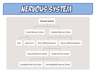
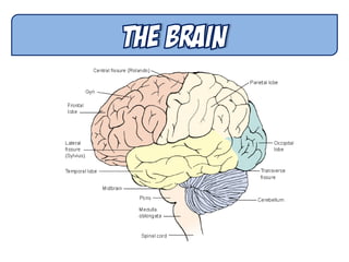
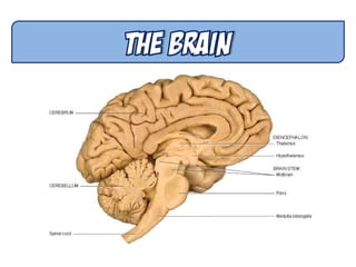
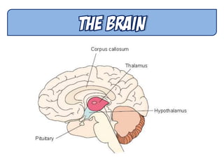
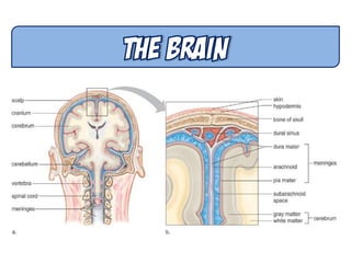
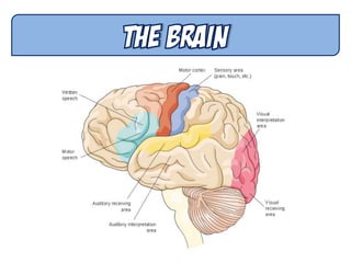
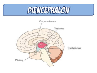
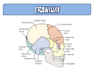
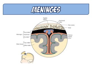
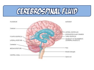
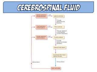
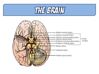
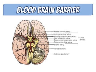
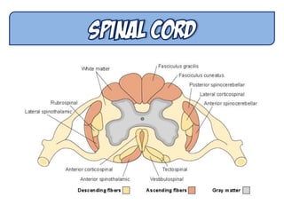
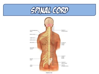
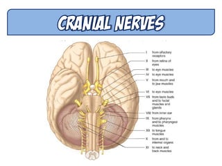
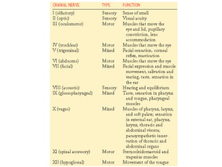
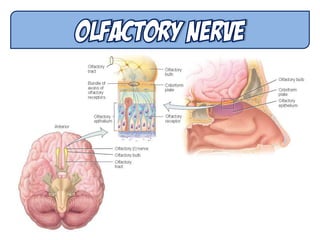
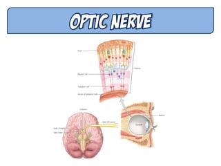
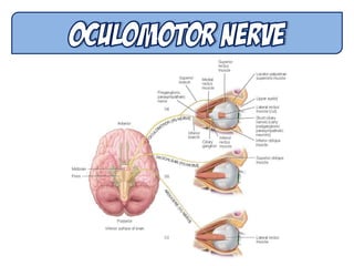
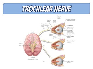
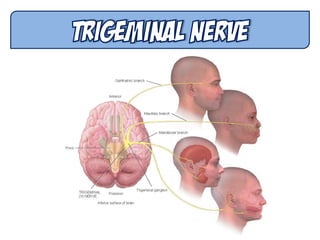
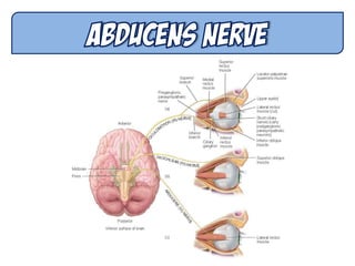
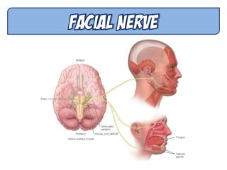
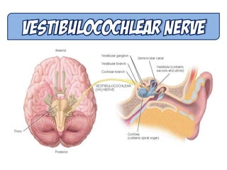
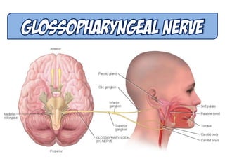
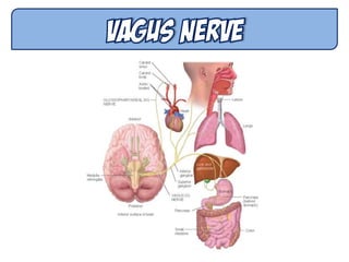
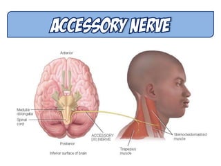
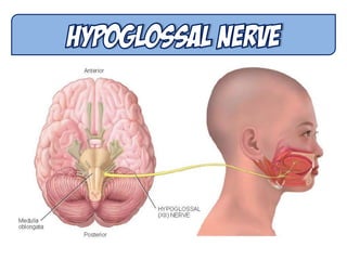
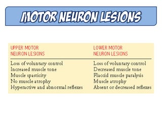
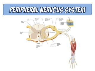
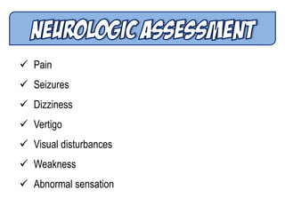
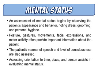
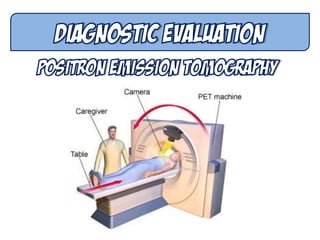
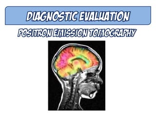
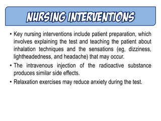
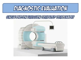
JOFRED M. MARTINEZ, RN
On completion of this chapter, the learner will be able to:
1. Describe the structures and functions of the central and
peripheral nervous systems.
2. Differentiate between pathologic changes that affect motor
control and those that affect sensory pathways.
3. Compare the functioning of the sympathetic and
parasympathetic nervous systems.
4. Describe the significance of physical assessment to the
diagnosis of neurologic dysfunction.
Continued…
5. Describe changes in neurologic function associated with
aging and their impact on neurologic assessment findings.
6. Describe diagnostic tests used for assessment of
suspected neurologic disorders and the related nursing
implications.
1. MOTOR NEURONS
Take nerve impulses from the CNS to muscles, organs, or
glands
2. SENSORY NEURONS
Take nerve impulses from sensory receptors to the CNS
3. INTERNEURONS
Convey nerve impulses between various parts of the CNS
There are two types of receptors: direct and indirect.
• DIRECT RECEPTORS are also known as inotropic because
they are linked to ion channels and allow passage of ions
when opened. They can be excitatory or inhibitory and are
rapid-acting (measured in milliseconds).
• INDIRECT RECEPTORS affect metabolic processes in the
cell, which can take from seconds to hours to occur
 Over average lifetime, brain shrinks 10%
 Most cell death occurs in temporal lobes
 By age 90, frontal cortex has lost half its neurons
 Number of dendritic branches decreases
 Decreased levels of neurotransmitters
 Fading memory
 Slowed responses and reflexes
 Increased risk of falling
 Changes in sleep patterns that result in fewer sleeping
hours
 Pain
 Seizures
 Dizziness
 Vertigo
 Visual disturbances
 Weakness
 Abnormal sensation
• An assessment of mental status begins by observing the
patient’s appearance and behavior, noting dress, grooming,
and personal hygiene.
• Posture, gestures, movements, facial expressions, and
motor activity often provide important information about the
patient.
• The patient’s manner of speech and level of consciousness
are also assessed.
• Assessing orientation to time, place, and person assists in
evaluating mental status.
• A person with an average IQ can repeat seven digits
without faltering and can recite five digits backward.
• The examiner might ask the patient to count backward from
100 or to subtract 7 from 100, then 7 from that, and so forth
(called serial 7s).
• The capacity to interpret well-known proverbs tests
abstract reasoning, which is a higher intellectual function.
• Questions designed to assess the integrative capacity
include the ability to recognize similarities.
• Can the patient make judgments about situations.
• Are the patient’s thoughts spontaneous, natural, clear,
relevant, and coherent?
• Does the patient have any fixed ideas, illusions, or
preoccupations?
• What are his or her insights into these thoughts?
• Preoccupation with death or morbid events, hallucinations,
and paranoid ideation are examples of unusual thoughts or
perceptions that require further evaluation.
• Is the patient’s affect (external manifestation of mood)
natural and even, or irritable and angry, anxious, apathetic
or flat, or euphoric?
• Does his or her mood fluctuate normally, or does the
patient unpredictably swing from joy to sadness during the
interview?
• Is affect appropriate to words and thought content?
• Are verbal communications consistent with nonverbal
cues?
• Agnosia is the inability to interpret or recognize objects
seen through the special senses.
• Placing a familiar object (eg, key, coin) in the patient’s hand
and having him or her identify it with both eyes closed is an
easy way to assess tactile interpretation.
• Assessment of cortical motor integration is carried out by
asking the patient to perform a skilled act.
• Successful performance requires the ability to understand
the activity desired and normal motor strength.
• Failure signals cerebral dysfunction.
• The person with normal neurologic function can understand
and communicate in spoken and written language.
• Does the patient answer questions appropriately?
• Can he or she read a sentence from a newspaper and
explain its meaning?
• Can the patient write his or her name or copy a simple
figure that the examiner has drawn?
• A deficiency in language function is called aphasia.
• Issues to consider include the limitations imposed on the
patient by any deficit and the patient’s role in society,
including family and community roles.
• The plan of care that the nurse develops needs to address
and support adaptation to the neurologic deficit and
continued function to the extent possible within the
patient’s support system.
• Interpretation and documentation of neurologic
abnormalities, particularly mental status abnormalities,
should be specific and nonjudgmental.
• Lengthy descriptions and the use of terms such as
―inappropriate‖ or ―demented‖ should be avoided.
• The examiner records and reports specific observations
regarding orientation, level of consciousness, emotional
state, or thought content, all of which permit comparison by
others over time.
• The patient is instructed to walk across the room while the
examiner observes posture and gait.
• The muscles are inspected, and palpated if necessary, for
their size and symmetry. Any evidence of atrophy or
involuntary movements (tremors, tics) is noted.
• MUSCLE TONE (the tension present in a muscle at rest)
is evaluated by palpating various muscle groups at rest
and during passive movement.
• Resistance to these movements is assessed and
documented. Abnormalities in tone include spasticity,
rigidity, and flaccidity.
• Assessing the patient’s ability to flex or extend the
extremities against resistance tests muscle strength.
• The evaluation of muscle strength compares the sides of
the body to each other.
• Distal and proximal strength in both upper and lower
extremities is recorded using the five-point scale.
• Cerebellar influence on the motor system is reflected in
balance control and coordination.
• Coordination in the hands and upper extremities is tested by
having the patient perform rapid, alternating movements and
point-to-point testing.
• First, the patient is instructed to pat his or her thigh as fast
as possible with each hand separately. Then the patient is
instructed to alternately pronate and supinate the hand as
rapidly as possible. Lastly, the patient is asked to touch each
of the fingers with the thumb in a consecutive motion.
• Speed, symmetry, and degree of difficulty are noted.
• Coordination in the lower extremities is tested by having the
patient run the heel down the anterior surface of the tibia of
the other leg.
• Ataxia is defined as incoordination of voluntary muscle
action, particularly of the muscle groups used in activities
such as walking or reaching for objects.
• The presence of ataxia or tremors during these movements
suggests cerebellar disease.
• It is advisable to perform a simple screening of the upper
and lower extremities by having the patient perform either
rapid, alternating movements or point-to-point testing.
• The Romberg test is a screening test for balance. The
patient stands with feet together and arms at the side, first
with eyes open and then with both eyes closed for 20 to 30
seconds.
• Slight swaying is normal, but a loss of balance is abnormal
and is considered a positive Romberg test.
• Additional cerebellar tests for balance in the ambulatory
patient include hopping in place, alternating knee bends,
and heel-to-toe walking (both forward and backward).
• Deep tendon reflex responses are often graded on a scale
of 0 to 4+.
• A 4+ indicates a hyperactive reflex, often indicating
pathology; 3+ indicates a response that is more brisk than
average but may be normal or indicative of disease; 2+
indicates an average or normal response; 1+ indicates a
hypoactive or diminished response; and 0 indicates no
response.
• When reflexes are very hyperactive, a phenomenon called
clonus may be elicited.
• The major superficial reflexes include corneal, gag or
swallowing, upper/lower abdominal, cremasteric (men only),
plantar, and perianal.
• These reflexes are graded differently than the motor reflexes
and are noted to be present (+) or absent (-). Of these, only
three are tested commonly.
• Assessment of the sensory system involves tests for tactile
sensation, superficial pain, vibration, and position sense
(proprioception).
• Tactile sensation is assessed by lightly touching a cotton
wisp to corresponding areas on each side of the body.
• Determining the patient’s sensitivity to a sharp object can
assess superficial pain perception.
• Vibration and proprioception are transmitted together in the
posterior part of the cord.
• Vibration may be evaluated through the use of a low-
frequency (128- or 256-Hz) tuning fork.
• Position sense or proprioception may be determined by
asking the patient to close both eyes and indicate, as the
great toe is alternately moved up and down, in which
direction movement has taken place.
• Integration of sensation in the brain may be performed by
testing two-point discrimination—when the patient is
touched with two sharp objects simultaneously, are they
perceived as two or as one
• Another test of higher cortical sensory ability is
stereognosis. The patient is instructed to close both eyes
and identify a variety of objects that are placed in one hand
by the examiner.
• Preparation includes teaching the patient about the need to
lie quietly throughout the procedure.
• A review of relaxation techniques may be helpful for
claustrophobic patients.
• Sedation can be used if agitation, restlessness, or confusion
will interfere with a successful study.
• If a contrast agent is used, the patient must be assessed
before the CT scan for an iodine/shellfish allergy, as the
contrast agent is iodine-based.
• An intravenous line for injection of the contrast agent and a
period of fasting (usually 4 hours) are required prior to the
study.
• Patients who receive an intravenous or inhalation contrast
agent are monitored during and after the procedure for
allergic reactions and other side effects, including flushing,
nausea, and vomiting.
• Key nursing interventions include patient preparation, which
involves explaining the test and teaching the patient about
inhalation techniques and the sensations (eg, dizziness,
lightheadedness, and headache) that may occur.
• The intravenous injection of the radioactive substance
produces similar side effects.
• Relaxation exercises may reduce anxiety during the test.
• Teaching about what to expect before the test can allay
anxiety and ensure patient cooperation during the test.
• Premenopausal women are advised to practice effective
contraception before and for several days after testing, and
the woman who is breastfeeding is instructed to stop nursing
for the period of time recommended by the nuclear medicine
department.
• The nurse may need to accompany and monitor the patient
during transport to the nuclear medicine department for the
scan.
• Patients are monitored during and after the procedure for
allergic reactions to the radiopharmaceutical agent
• Patient preparation should include teaching relaxation
techniques and informing the patient that he or she will be
able to talk to the staff by means of a microphone located
inside the scanner.
• Before the patient enters the room where the MRI is to be
performed, all metal objects and credit cards are removed.
No metal objects may be brought into the room where the
MRI is located.
• A patient history is obtained to determine the presence of
any metal objects (eg, aneurysm clips, orthopedic hardware,
pacemakers, artificial heart valves, intrauterine devices).
• These objects could malfunction, be dislodged, or heat up
as they absorb energy.
• Cochlear implants will be inactivated by MRI; therefore,
other imaging procedures are considered.
• The scanning process is painless, but the patient hears loud
thumping of the magnetic coils as the magnetic field is being
pulsed.
• Patients may experience claustrophobia; sedation may be
prescribed in these circumstances.
• The patient should be well hydrated, and clear liquids are
usually permitted up to the time of a regular arteriogram or
DSA.
• Before going to the x-ray department, the patient is
instructed to void.
• The locations of the appropriate peripheral pulses are
marked with a felt-tip pen.
• The patient is instructed to remain immobile during the
angiogram process and is told to expect a brief feeling of
warmth in the face, behind the eyes, or in the jaw, teeth,
tongue, and lips, and a metallic taste when the contrast
agent is injected.
• Nursing care after cerebral angiography includes
observation for signs and symptoms of altered cerebral
blood flow.
• Signs of minor arterial blockage include alterations in the
level of responsiveness and consciousness, weakness on
one side of the body, motor or sensory deficits, and speech
disturbances.
• The injection site is observed for hematoma formation, and
an ice bag may be applied intermittently to the puncture site
to relieve swelling and discomfort.
• Because a hematoma at the puncture site or embolization to
a distant artery affects the peripheral pulses, these pulses
are monitored frequently.
• The color and temperature of the involved extremity are
assessed to detect possible embolism.
• The patient is informed about what to expect during the
procedure and should be aware that changes in position
may be made during the procedure.
• A sedative may be prescribed to help the patient cope with
this lengthy test.
• After myelography, the patient lies in bed with the head of
the bed elevated 30 to 45 degrees for 3 hours or as
prescribed by the physician.
• The patient is encouraged to drink liberal amounts of fluid
for rehydration and replacement of CSF and to decrease the
incidence of post lumbar puncture headache.
• The blood pressure, pulse, respiratory rate, and temperature
are monitored, as well as the patient’s ability to void.
• Untoward signs include headache, fever, stiff neck,
photophobia (sensitivity to light), seizures, and signs of
chemical or bacterial meningitis.
• The patient is informed that this is a noninvasive test, that a
hand-held transducer will be placed over the neck and orbits
of the eyes, and that some type of water-soluble jelly is used
on the transducer.
• To increase the chances of recording seizure activity, it is
sometimes recommended that the patient be deprived of
sleep on the night before the EEG.
• Antiseizure agents, tranquilizers, stimulants, and
depressants should be withheld 24 to 48 hours before an
EEG.
• Coffee, tea, chocolate, and cola drinks are omitted in the
meal before the test because of their stimulating effect.
• The patient is informed that the standard EEG takes 45 to
60 minutes, 12 hours for a sleep EEG.
• The patient is assured that the procedure does not cause an
electric shock and that the EEG is a diagnostic test, not a
form of treatment.
• Sedation is not advisable as it may lower the seizure
threshold in patients with a seizure disorder and alter brain
wave activity in all patients.
• Routine EEGs use a water-soluble lubricant for electrode
contact, which at the conclusion of the study can be wiped
off and removed by shampooing.
• Sleep EEGs involve the use of collodion glue for electrode
contact, which requires acetone for removal.
• In evoked potential studies, electrodes are applied to the
scalp and an external stimulus is applied to peripheral
sensory receptors to elicit changes in the brain waves.
• Evoked changes are detected with the aid of computerized
devices that extract the signal, display it on an
oscilloscope, and store the data on magnetic tape or disk.
• In clinical practice, the visual, auditory, and somatosensory
systems are most often tested.
• There is no specific patient preparation other than to explain
the procedure and to reassure the patient and encourage
him or her to relax.
• The patient is advised to remain perfectly still throughout the
recording to prevent artifacts (signals not generated by the
brain) that interfere with the recording and interpretation of
the test.
• The procedure is explained and the patient is warned to
expect a sensation similar to that of an intramuscular
injection as the needle is inserted into the muscle.
• The muscles examined may ache for a short time after the
procedure.
• Nerve conduction studies are performed by stimulating a
peripheral nerve at several points along its course and
recording the muscle action potential or the sensory
action potential that results.
• Surface or needle electrodes are placed on the skin over
the nerve to stimulate the nerve fibers.
• This test is useful in the study of peripheral neuropathies.
• A lumbar manometric test (Queckenstedt’s test) may be
performed by compressing the jugular veins on each side
of the neck during the lumbar puncture.
• The increase in pressure caused by the compression is
noted; then the pressure is released and pressure
readings are made at 10-second intervals.
• A slow rise and fall in pressure indicates a partial block
due to a lesion compressing the spinal subarachnoid
pathways.
• If there is no pressure change, a complete block is
indicated.
• This test is not performed if an intracranial lesion is
suspected.
• The CSF should be clear and colorless. Pink, blood-
tinged, or grossly bloody CSF may indicate a cerebral
contusion, laceration, or subarachnoid hemorrhage.
• Usually, specimens are obtained for cell count, culture,
and glucose and protein testing.
• The specimens should be sent to the laboratory
immediately because changes will take place and alter the
result if the specimens are allowed to stand.
• It is a throbbing bifrontal or occipital headache, dull and
deep in character. It is particularly severe on sitting or
standing but lessens or disappears when the patient lies
down.
• Post–lumbar puncture headache may be avoided if a small
gauge needle is used and if the patient remains prone
after the procedure.
• When a large volume of fluid (more than 20 mL) is
removed, the patient is positioned prone for 2 hours, then
flat in a side-lying position for 2 to 3 hours, and then
supine or prone for 6 more hours.
• The postpuncture headache is usually managed by bed
rest, analgesic agents, and hydration.
• If the headache persists, the epidural blood patch
technique may be used.
• Herniation of the intracranial contents, spinal epidural
abscess, spinal epidural hematoma, and meningitis are
rare but serious complications of lumbar puncture.
• Other complications include temporary voiding problems,
slight elevation of temperature, backache or spasms, and
stiffness of the neck.

Neurologic Nursing 1

  • 1.
  • 2.
    On completion ofthis chapter, the learner will be able to: 1. Describe the structures and functions of the central and peripheral nervous systems. 2. Differentiate between pathologic changes that affect motor control and those that affect sensory pathways. 3. Compare the functioning of the sympathetic and parasympathetic nervous systems. 4. Describe the significance of physical assessment to the diagnosis of neurologic dysfunction.
  • 3.
    Continued… 5. Describe changesin neurologic function associated with aging and their impact on neurologic assessment findings. 6. Describe diagnostic tests used for assessment of suspected neurologic disorders and the related nursing implications.
  • 6.
    1. MOTOR NEURONS Takenerve impulses from the CNS to muscles, organs, or glands 2. SENSORY NEURONS Take nerve impulses from sensory receptors to the CNS 3. INTERNEURONS Convey nerve impulses between various parts of the CNS
  • 9.
    There are twotypes of receptors: direct and indirect. • DIRECT RECEPTORS are also known as inotropic because they are linked to ion channels and allow passage of ions when opened. They can be excitatory or inhibitory and are rapid-acting (measured in milliseconds). • INDIRECT RECEPTORS affect metabolic processes in the cell, which can take from seconds to hours to occur
  • 51.
     Over averagelifetime, brain shrinks 10%  Most cell death occurs in temporal lobes  By age 90, frontal cortex has lost half its neurons  Number of dendritic branches decreases  Decreased levels of neurotransmitters  Fading memory  Slowed responses and reflexes  Increased risk of falling  Changes in sleep patterns that result in fewer sleeping hours
  • 52.
     Pain  Seizures Dizziness  Vertigo  Visual disturbances  Weakness  Abnormal sensation
  • 53.
    • An assessmentof mental status begins by observing the patient’s appearance and behavior, noting dress, grooming, and personal hygiene. • Posture, gestures, movements, facial expressions, and motor activity often provide important information about the patient. • The patient’s manner of speech and level of consciousness are also assessed. • Assessing orientation to time, place, and person assists in evaluating mental status.
  • 54.
    • A personwith an average IQ can repeat seven digits without faltering and can recite five digits backward. • The examiner might ask the patient to count backward from 100 or to subtract 7 from 100, then 7 from that, and so forth (called serial 7s). • The capacity to interpret well-known proverbs tests abstract reasoning, which is a higher intellectual function. • Questions designed to assess the integrative capacity include the ability to recognize similarities. • Can the patient make judgments about situations.
  • 55.
    • Are thepatient’s thoughts spontaneous, natural, clear, relevant, and coherent? • Does the patient have any fixed ideas, illusions, or preoccupations? • What are his or her insights into these thoughts? • Preoccupation with death or morbid events, hallucinations, and paranoid ideation are examples of unusual thoughts or perceptions that require further evaluation.
  • 56.
    • Is thepatient’s affect (external manifestation of mood) natural and even, or irritable and angry, anxious, apathetic or flat, or euphoric? • Does his or her mood fluctuate normally, or does the patient unpredictably swing from joy to sadness during the interview? • Is affect appropriate to words and thought content? • Are verbal communications consistent with nonverbal cues?
  • 57.
    • Agnosia isthe inability to interpret or recognize objects seen through the special senses. • Placing a familiar object (eg, key, coin) in the patient’s hand and having him or her identify it with both eyes closed is an easy way to assess tactile interpretation.
  • 59.
    • Assessment ofcortical motor integration is carried out by asking the patient to perform a skilled act. • Successful performance requires the ability to understand the activity desired and normal motor strength. • Failure signals cerebral dysfunction.
  • 60.
    • The personwith normal neurologic function can understand and communicate in spoken and written language. • Does the patient answer questions appropriately? • Can he or she read a sentence from a newspaper and explain its meaning? • Can the patient write his or her name or copy a simple figure that the examiner has drawn? • A deficiency in language function is called aphasia.
  • 62.
    • Issues toconsider include the limitations imposed on the patient by any deficit and the patient’s role in society, including family and community roles. • The plan of care that the nurse develops needs to address and support adaptation to the neurologic deficit and continued function to the extent possible within the patient’s support system.
  • 63.
    • Interpretation anddocumentation of neurologic abnormalities, particularly mental status abnormalities, should be specific and nonjudgmental. • Lengthy descriptions and the use of terms such as ―inappropriate‖ or ―demented‖ should be avoided. • The examiner records and reports specific observations regarding orientation, level of consciousness, emotional state, or thought content, all of which permit comparison by others over time.
  • 66.
    • The patientis instructed to walk across the room while the examiner observes posture and gait. • The muscles are inspected, and palpated if necessary, for their size and symmetry. Any evidence of atrophy or involuntary movements (tremors, tics) is noted. • MUSCLE TONE (the tension present in a muscle at rest) is evaluated by palpating various muscle groups at rest and during passive movement. • Resistance to these movements is assessed and documented. Abnormalities in tone include spasticity, rigidity, and flaccidity.
  • 67.
    • Assessing thepatient’s ability to flex or extend the extremities against resistance tests muscle strength. • The evaluation of muscle strength compares the sides of the body to each other. • Distal and proximal strength in both upper and lower extremities is recorded using the five-point scale.
  • 69.
    • Cerebellar influenceon the motor system is reflected in balance control and coordination. • Coordination in the hands and upper extremities is tested by having the patient perform rapid, alternating movements and point-to-point testing. • First, the patient is instructed to pat his or her thigh as fast as possible with each hand separately. Then the patient is instructed to alternately pronate and supinate the hand as rapidly as possible. Lastly, the patient is asked to touch each of the fingers with the thumb in a consecutive motion. • Speed, symmetry, and degree of difficulty are noted.
  • 70.
    • Coordination inthe lower extremities is tested by having the patient run the heel down the anterior surface of the tibia of the other leg. • Ataxia is defined as incoordination of voluntary muscle action, particularly of the muscle groups used in activities such as walking or reaching for objects. • The presence of ataxia or tremors during these movements suggests cerebellar disease. • It is advisable to perform a simple screening of the upper and lower extremities by having the patient perform either rapid, alternating movements or point-to-point testing.
  • 71.
    • The Rombergtest is a screening test for balance. The patient stands with feet together and arms at the side, first with eyes open and then with both eyes closed for 20 to 30 seconds. • Slight swaying is normal, but a loss of balance is abnormal and is considered a positive Romberg test. • Additional cerebellar tests for balance in the ambulatory patient include hopping in place, alternating knee bends, and heel-to-toe walking (both forward and backward).
  • 72.
    • Deep tendonreflex responses are often graded on a scale of 0 to 4+. • A 4+ indicates a hyperactive reflex, often indicating pathology; 3+ indicates a response that is more brisk than average but may be normal or indicative of disease; 2+ indicates an average or normal response; 1+ indicates a hypoactive or diminished response; and 0 indicates no response.
  • 75.
    • When reflexesare very hyperactive, a phenomenon called clonus may be elicited. • The major superficial reflexes include corneal, gag or swallowing, upper/lower abdominal, cremasteric (men only), plantar, and perianal. • These reflexes are graded differently than the motor reflexes and are noted to be present (+) or absent (-). Of these, only three are tested commonly.
  • 76.
    • Assessment ofthe sensory system involves tests for tactile sensation, superficial pain, vibration, and position sense (proprioception). • Tactile sensation is assessed by lightly touching a cotton wisp to corresponding areas on each side of the body. • Determining the patient’s sensitivity to a sharp object can assess superficial pain perception. • Vibration and proprioception are transmitted together in the posterior part of the cord. • Vibration may be evaluated through the use of a low- frequency (128- or 256-Hz) tuning fork.
  • 77.
    • Position senseor proprioception may be determined by asking the patient to close both eyes and indicate, as the great toe is alternately moved up and down, in which direction movement has taken place. • Integration of sensation in the brain may be performed by testing two-point discrimination—when the patient is touched with two sharp objects simultaneously, are they perceived as two or as one • Another test of higher cortical sensory ability is stereognosis. The patient is instructed to close both eyes and identify a variety of objects that are placed in one hand by the examiner.
  • 80.
    • Preparation includesteaching the patient about the need to lie quietly throughout the procedure. • A review of relaxation techniques may be helpful for claustrophobic patients. • Sedation can be used if agitation, restlessness, or confusion will interfere with a successful study. • If a contrast agent is used, the patient must be assessed before the CT scan for an iodine/shellfish allergy, as the contrast agent is iodine-based.
  • 81.
    • An intravenousline for injection of the contrast agent and a period of fasting (usually 4 hours) are required prior to the study. • Patients who receive an intravenous or inhalation contrast agent are monitored during and after the procedure for allergic reactions and other side effects, including flushing, nausea, and vomiting.
  • 85.
    • Key nursinginterventions include patient preparation, which involves explaining the test and teaching the patient about inhalation techniques and the sensations (eg, dizziness, lightheadedness, and headache) that may occur. • The intravenous injection of the radioactive substance produces similar side effects. • Relaxation exercises may reduce anxiety during the test.
  • 88.
    • Teaching aboutwhat to expect before the test can allay anxiety and ensure patient cooperation during the test. • Premenopausal women are advised to practice effective contraception before and for several days after testing, and the woman who is breastfeeding is instructed to stop nursing for the period of time recommended by the nuclear medicine department. • The nurse may need to accompany and monitor the patient during transport to the nuclear medicine department for the scan. • Patients are monitored during and after the procedure for allergic reactions to the radiopharmaceutical agent
  • 91.
    • Patient preparationshould include teaching relaxation techniques and informing the patient that he or she will be able to talk to the staff by means of a microphone located inside the scanner. • Before the patient enters the room where the MRI is to be performed, all metal objects and credit cards are removed. No metal objects may be brought into the room where the MRI is located. • A patient history is obtained to determine the presence of any metal objects (eg, aneurysm clips, orthopedic hardware, pacemakers, artificial heart valves, intrauterine devices).
  • 92.
    • These objectscould malfunction, be dislodged, or heat up as they absorb energy. • Cochlear implants will be inactivated by MRI; therefore, other imaging procedures are considered. • The scanning process is painless, but the patient hears loud thumping of the magnetic coils as the magnetic field is being pulsed. • Patients may experience claustrophobia; sedation may be prescribed in these circumstances.
  • 94.
    • The patientshould be well hydrated, and clear liquids are usually permitted up to the time of a regular arteriogram or DSA. • Before going to the x-ray department, the patient is instructed to void. • The locations of the appropriate peripheral pulses are marked with a felt-tip pen. • The patient is instructed to remain immobile during the angiogram process and is told to expect a brief feeling of warmth in the face, behind the eyes, or in the jaw, teeth, tongue, and lips, and a metallic taste when the contrast agent is injected.
  • 95.
    • Nursing careafter cerebral angiography includes observation for signs and symptoms of altered cerebral blood flow. • Signs of minor arterial blockage include alterations in the level of responsiveness and consciousness, weakness on one side of the body, motor or sensory deficits, and speech disturbances. • The injection site is observed for hematoma formation, and an ice bag may be applied intermittently to the puncture site to relieve swelling and discomfort.
  • 96.
    • Because ahematoma at the puncture site or embolization to a distant artery affects the peripheral pulses, these pulses are monitored frequently. • The color and temperature of the involved extremity are assessed to detect possible embolism.
  • 98.
    • The patientis informed about what to expect during the procedure and should be aware that changes in position may be made during the procedure. • A sedative may be prescribed to help the patient cope with this lengthy test. • After myelography, the patient lies in bed with the head of the bed elevated 30 to 45 degrees for 3 hours or as prescribed by the physician. • The patient is encouraged to drink liberal amounts of fluid for rehydration and replacement of CSF and to decrease the incidence of post lumbar puncture headache.
  • 99.
    • The bloodpressure, pulse, respiratory rate, and temperature are monitored, as well as the patient’s ability to void. • Untoward signs include headache, fever, stiff neck, photophobia (sensitivity to light), seizures, and signs of chemical or bacterial meningitis.
  • 102.
    • The patientis informed that this is a noninvasive test, that a hand-held transducer will be placed over the neck and orbits of the eyes, and that some type of water-soluble jelly is used on the transducer.
  • 104.
    • To increasethe chances of recording seizure activity, it is sometimes recommended that the patient be deprived of sleep on the night before the EEG. • Antiseizure agents, tranquilizers, stimulants, and depressants should be withheld 24 to 48 hours before an EEG. • Coffee, tea, chocolate, and cola drinks are omitted in the meal before the test because of their stimulating effect. • The patient is informed that the standard EEG takes 45 to 60 minutes, 12 hours for a sleep EEG.
  • 105.
    • The patientis assured that the procedure does not cause an electric shock and that the EEG is a diagnostic test, not a form of treatment. • Sedation is not advisable as it may lower the seizure threshold in patients with a seizure disorder and alter brain wave activity in all patients. • Routine EEGs use a water-soluble lubricant for electrode contact, which at the conclusion of the study can be wiped off and removed by shampooing. • Sleep EEGs involve the use of collodion glue for electrode contact, which requires acetone for removal.
  • 106.
    • In evokedpotential studies, electrodes are applied to the scalp and an external stimulus is applied to peripheral sensory receptors to elicit changes in the brain waves. • Evoked changes are detected with the aid of computerized devices that extract the signal, display it on an oscilloscope, and store the data on magnetic tape or disk. • In clinical practice, the visual, auditory, and somatosensory systems are most often tested.
  • 107.
    • There isno specific patient preparation other than to explain the procedure and to reassure the patient and encourage him or her to relax. • The patient is advised to remain perfectly still throughout the recording to prevent artifacts (signals not generated by the brain) that interfere with the recording and interpretation of the test.
  • 109.
    • The procedureis explained and the patient is warned to expect a sensation similar to that of an intramuscular injection as the needle is inserted into the muscle. • The muscles examined may ache for a short time after the procedure.
  • 110.
    • Nerve conductionstudies are performed by stimulating a peripheral nerve at several points along its course and recording the muscle action potential or the sensory action potential that results. • Surface or needle electrodes are placed on the skin over the nerve to stimulate the nerve fibers. • This test is useful in the study of peripheral neuropathies.
  • 112.
    • A lumbarmanometric test (Queckenstedt’s test) may be performed by compressing the jugular veins on each side of the neck during the lumbar puncture. • The increase in pressure caused by the compression is noted; then the pressure is released and pressure readings are made at 10-second intervals. • A slow rise and fall in pressure indicates a partial block due to a lesion compressing the spinal subarachnoid pathways.
  • 113.
    • If thereis no pressure change, a complete block is indicated. • This test is not performed if an intracranial lesion is suspected.
  • 114.
    • The CSFshould be clear and colorless. Pink, blood- tinged, or grossly bloody CSF may indicate a cerebral contusion, laceration, or subarachnoid hemorrhage. • Usually, specimens are obtained for cell count, culture, and glucose and protein testing. • The specimens should be sent to the laboratory immediately because changes will take place and alter the result if the specimens are allowed to stand.
  • 115.
    • It isa throbbing bifrontal or occipital headache, dull and deep in character. It is particularly severe on sitting or standing but lessens or disappears when the patient lies down. • Post–lumbar puncture headache may be avoided if a small gauge needle is used and if the patient remains prone after the procedure.
  • 116.
    • When alarge volume of fluid (more than 20 mL) is removed, the patient is positioned prone for 2 hours, then flat in a side-lying position for 2 to 3 hours, and then supine or prone for 6 more hours. • The postpuncture headache is usually managed by bed rest, analgesic agents, and hydration. • If the headache persists, the epidural blood patch technique may be used.
  • 117.
    • Herniation ofthe intracranial contents, spinal epidural abscess, spinal epidural hematoma, and meningitis are rare but serious complications of lumbar puncture. • Other complications include temporary voiding problems, slight elevation of temperature, backache or spasms, and stiffness of the neck.
  • 118.
    • Herniation ofthe intracranial contents, spinal epidural abscess, spinal epidural hematoma, and meningitis are rare but serious complications of lumbar puncture. • Other complications include temporary voiding problems, slight elevation of temperature, backache or spasms, and stiffness of the neck.