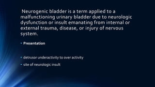
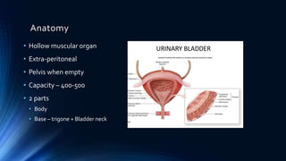
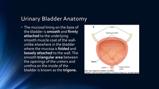
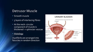
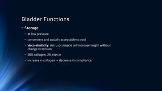
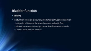
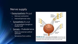
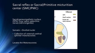
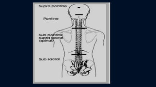
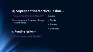
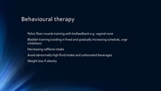
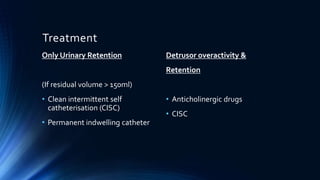
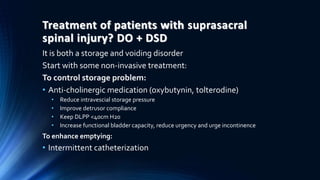
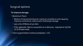
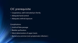
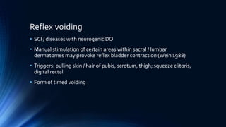
Neurogenic Bladder
DR. JUNISH BAGGA
Neurogenic bladder is a term applied to a
malfunctioning urinary bladder due to neurologic
dysfunction or insult emanating from internal or
external trauma, disease, or injury of nervous
system.
• Presentation
• detrusor underactivity to over activity
• site of neurologic insult
Anatomy
• Hollow muscular organ
• Extra-peritoneal
• Pelvis when empty
• Capacity – 400-500
• 2 parts
• Body
• Base – trigone + Bladder neck
Urinary Bladder Anatomy
• The mucosal lining on the base of
the bladder is smooth and firmly
attached to the underlying
smooth muscle coat of the wall-
unlike elsewhere in the bladder
where the mucosa is folded and
loosely attached to the wall.The
smooth triangular area between
the openings of the ureters and
urethra on the inside of the
bladder is known as the trigone.
Detrusor Muscle
• Smooth muscle
• 3 layers of interlacing fibres
• At the neck- circular
component of muscle is
thickened -> sphincter vesicae
• Histology
myofibrils are arranged into
fascicles in random direction
Bladder Functions
• Storage
• at low pressure
• convenient and socially acceptable to void
• visco-elasticity: detrusor muscle cell increase length without
change in tension
• 50% collagen, 2% elastin
• Increase in collagen -> decrease in compliance
Bladder function
• Voiding
• Micturition relies on a neurally mediated detrusor contraction
• initiated by inhibition of the striated sphincter and pelvic floor
• followed some seconds later by a contraction of the detrusor muscle.
• Causes a rise in detrusor pressure
Nerve supply
• Parasympathetic (S2,3,4)
1. Detrusor contraction
2. Internal Sphincter relax.
• Sympathetic (L1,2,3)
• To sympathetic Ganglia on
bladder wall
• Somatic – Pudendal nerve
• Motor to external urethral
sphincter
• Sensations from urethra
Control of Micturation
• 1.Cortical micturition centre
• 2.Pontine micturition centre
• 3.Spinal micturition centre
• 4. Peripheral nerves(S2,3,4)
Sympathetic
(T11 –L2)
Parasympathetic
( S2,3,4)
Cortical micturation centre(CMC)
Location: Paracentral lobule in the medial aspect of the
frontoparietal cotex
Function: Inhibitory to PMC
Dysfunction – loss of social control of bladder
The brain’s control of the PMC is part of the social
training that children experience at age 2 - 4 years
Pontine Micturition Centre (PMC)
Also called Barrington’s nucleus
Lateral region
• Function - continence, storage urine
• stimulation results in a powerful contraction of the urethral sphincter
Medial region
• Function - micturition center
• stimulation results in decrease in urethral pressure and silence of
pelvic floor EMG signal, followed by a rise in detrusor pressure.
Sacral reflex or Sacral/Primitive micturition
center (SMC/PMC)
Sacral parasympathetic nucleus
(SPN): S234- pelvic splanchnic
nerves (nervi erigentes)
Somatic – Onufoid nuclei
• Collection of external urethral
sphinter motoneurones
Levator Ani Motoneurones
Types according to the level of bladder
dysfunction
a) Suprapontine/cortical lesion –
“Uninhibited /Cortical bladder”
Severe urgency, frequency & urge
incontinence
b) Pontine lesion –
“ Reflex / Automatic bladder”
Causes
• Stroke
• Tumors
• Dementia
Spinal (subpontine/
suprasacral)
“ Spastic Bladder”
Disorders of storage and
emptying
Detrusor Sphincter
Dyssynergia
Detrusor Hyperreflexia
d) Sacral and subsacral lesions
I) Afferent fibres involved only –
• “Atonic /Areflexic bladder”
• Overflow incontinence
• Straining for micturition
II) Both afferent and efferent involved –
• “Autonomous bladder”
• Small capacity , acting of its own. No
DSD/DH
Hinman syndrome: Non –neurogenic neurogenic
bladder. Severe bladder sphincter dyssynergia.
Trabeculated bladder develops a high pressure
state with B/L VUR and large PVR akin to a
neurogenic bladder without any obvious
neurological abnormality. May lead to renal
failure.
Approach
• Detailed medical history
• h/o trauma
• h/o pelvic surgery
• h/o neurologic disease
• h/o urologic symptoms (incontinence, UTI)
Physical exam
• Perineal sensation (pudendal afferent limb)
• Anal sphinctor tone (distinguish suprasacral-increased/ sacral lesion-
reduced)
• Lower extremeity spasticity
• Bulbocavernosus reflex
• Test integrity of sacral micturition center S2-4, pudental afferent/efferent limb
• The S2 S4 reflex arc can be elicited by squeezing the glans in males or clitoris in females and
looking for contraction of the anal sphincter S2-S4
• Morbid obesity and mobility
• Lack of adequate hand function
• Palpable bladder
Investigations
• Urine analysis
• Ultrasound – PVRU,
• In out catheterisation
• UroDynamic Study (gold standard)
• Uroflowmetry
• Cystometry
• Urethral pressure profilometry
• Urethral EMG – bladder neck
Imaging
• Neuroimaging –
Cauda equina & conus lesions,
spinal,
supra pontine and pontine lesions
• CYSTOSCOPY
Goals of Bladder Management
• Protect upper tract (low pressure storage)
• Complete bladder emptying (prevent UTI and stone)
• Preserve continence
• Maintain quality living
Behavioural therapy
• Pelvic floor muscle training with biofeedback e.g. vaginal cone
• Bladder training (voiding in fixed and gradually increasing schedule, urge
inhibition)
• Decreasing caffeine intake
• Avoid abnormally high fluid intake and carbonated beverages
• Weight loss if obesity
Treatment
Only Urinary Retention
(If residual volume > 150ml)
• Clean intermittent self
catheterisation (CISC)
• Permanent indwelling catheter
Detrusor overactivity &
Retention
• Anticholinergic drugs
• CISC
Treatment of patients with suprasacral
spinal injury? DO + DSD
It is both a storage and voiding disorder
Start with some non-invasive treatment:
To control storage problem:
• Anti-cholinergic medication (oxybutynin, tolterodine)
• Reduce intravescial storage pressure
• Improve detrusor compliance
• Keep DLPP <40cm H20
• Increase functional bladder capacity, reduce urgency and urge incontinence
To enhance emptying:
• Intermittent catheterization
Surgical options
To improve storage:
• BotulinumToxin
• Reduce intravesical pressure, improve compliance and capacity,
improve continence, reduce anti-cholinergic dosage
• 300 units of Botox at 30 sites
• If for sphincter :Not as successful as in detrusor, Injected at 3,6,9 &
12 O O’clock clock
• Clam augmentation enterocystoplasty + CIC
Surgical options
To improve voiding:
• External sphincterotomy
• Urethral stent- Memokath, Alloy of NiTi , Deploy hot water 55°C, Removal cold
water 5°C
To abolished the autonoic desreflexia + coordinate muscle
contraction:
• Detrusor myectomy
• SARS with dorsal rhizotomy – not suitable for patient who is still
walking or incomplete SCI
External Sphincterotomy
Colling’s electrocautery knife
Anteromedian incision (12 o’clock)
Proximal part of verumontanum ->
Corpus spongiosum of the bulbous
urethra
Plane of periurethral venous
sinuses
Large bore catheter (24-48 hours)
Bladder irrigation
External sphincterotomy
Results
• 70% successful rate
• Resolution of hydronephrosis /
improvedVUR
• Reduced febrile UTIs
• Reduced autonomic dysreflexia
• Reduction in PVR
• Reduction in mean voiding
pressure
Complications
• Bleeding (clot retention)
• Severe infection
• Impotence
• Reoperation (50%)
• Laser sphincterotomy has
better results
• Not done often now –
irreversible
Transurethral Surgery
• Botulinum A toxin injection
• Balloon dilatation
• Endourethral stent
• Comparable outcomes
• Less transfusion
• Stricture formation
• Encrustation / migration
Sacral Anterior Roots Stimulation (SARS)
Sacral Nerve Neuromodulation
• Procedures to enhance detrusor
contractility, usually
accompanied by with Dorsal
Sacral Rhizotomy (abolish hyper-
reflexia )
• Suitable for patient wheelchair
bound and complete SCI
• Connection of anterior motor
roots to implant slots and implant
(“The Finetech-Brindley bladder
controller”) placed
Treatment for infrasacral lesion
Detrusor underactivity
• Intermittent catheterization
• Indwelling catheter
• Suprapubic catheter
• Valsava manuever: contraindicated in VUR or hydronephrosis
• Reflex voiding
• Cholinergic agonist: no randomized trials have demonstrated
efficay over placebo
Surgical management
Ileovesicostomy
• Low pressure conduit for
preferential drainage
(<10cmH2O)
• Native bladder as
continent reservoir
• Native ureterovesical
junction
• Easy stoma care
CIC- clean intermittent self- catheterization
• GOLD standard for Mx of NLUTD (EAU guidelines)
• Jack Lapides 1972
• Promoted & popularized CIC
• First applying concept to large groups of pts with voiding dysfunction
• Demonstrated safety & long term efficacy
• Most effective & practical means for attaining catheter free state in
SCI
• Effective method for pts with emptying failure, esp after failed
attempts ↑ Pves / ↓ outlet resistance
• Helps to prevent UTI & protect upper tract
CIC prerequisite
• Cooperative, well-motivated pt / family
• Adequate hand control
• Adequate urethral exposure
Complications
• Urethral false passage
• Bladder perforation
• Silent deterioration of upper tracts
• Bacteriuria common (not symptomatic infection )
Reflex voiding
• SCI / diseases with neurogenic DO
• Manual stimulation of certain areas within sacral / lumbar
dermatomes may provoke reflex bladder contraction (Wein 1988)
• Triggers: pulling skin / hair of pubis, scrotum, thigh; squeeze clitoris,
digital rectal
• Form of timed voiding
Neurogenic bladder

Neurogenic bladder

  • 1.
  • 2.
    Neurogenic bladder isa term applied to a malfunctioning urinary bladder due to neurologic dysfunction or insult emanating from internal or external trauma, disease, or injury of nervous system. • Presentation • detrusor underactivity to over activity • site of neurologic insult
  • 3.
    Anatomy • Hollow muscularorgan • Extra-peritoneal • Pelvis when empty • Capacity – 400-500 • 2 parts • Body • Base – trigone + Bladder neck
  • 4.
    Urinary Bladder Anatomy •The mucosal lining on the base of the bladder is smooth and firmly attached to the underlying smooth muscle coat of the wall- unlike elsewhere in the bladder where the mucosa is folded and loosely attached to the wall.The smooth triangular area between the openings of the ureters and urethra on the inside of the bladder is known as the trigone.
  • 5.
    Detrusor Muscle • Smoothmuscle • 3 layers of interlacing fibres • At the neck- circular component of muscle is thickened -> sphincter vesicae • Histology myofibrils are arranged into fascicles in random direction
  • 6.
    Bladder Functions • Storage •at low pressure • convenient and socially acceptable to void • visco-elasticity: detrusor muscle cell increase length without change in tension • 50% collagen, 2% elastin • Increase in collagen -> decrease in compliance
  • 7.
    Bladder function • Voiding •Micturition relies on a neurally mediated detrusor contraction • initiated by inhibition of the striated sphincter and pelvic floor • followed some seconds later by a contraction of the detrusor muscle. • Causes a rise in detrusor pressure
  • 8.
    Nerve supply • Parasympathetic(S2,3,4) 1. Detrusor contraction 2. Internal Sphincter relax. • Sympathetic (L1,2,3) • To sympathetic Ganglia on bladder wall • Somatic – Pudendal nerve • Motor to external urethral sphincter • Sensations from urethra
  • 10.
    Control of Micturation •1.Cortical micturition centre • 2.Pontine micturition centre • 3.Spinal micturition centre • 4. Peripheral nerves(S2,3,4) Sympathetic (T11 –L2) Parasympathetic ( S2,3,4)
  • 11.
    Cortical micturation centre(CMC) Location:Paracentral lobule in the medial aspect of the frontoparietal cotex Function: Inhibitory to PMC Dysfunction – loss of social control of bladder The brain’s control of the PMC is part of the social training that children experience at age 2 - 4 years
  • 12.
    Pontine Micturition Centre(PMC) Also called Barrington’s nucleus Lateral region • Function - continence, storage urine • stimulation results in a powerful contraction of the urethral sphincter Medial region • Function - micturition center • stimulation results in decrease in urethral pressure and silence of pelvic floor EMG signal, followed by a rise in detrusor pressure.
  • 13.
    Sacral reflex orSacral/Primitive micturition center (SMC/PMC) Sacral parasympathetic nucleus (SPN): S234- pelvic splanchnic nerves (nervi erigentes) Somatic – Onufoid nuclei • Collection of external urethral sphinter motoneurones Levator Ani Motoneurones
  • 14.
    Types according tothe level of bladder dysfunction
  • 16.
    a) Suprapontine/cortical lesion– “Uninhibited /Cortical bladder” Severe urgency, frequency & urge incontinence b) Pontine lesion – “ Reflex / Automatic bladder” Causes • Stroke • Tumors • Dementia
  • 17.
    Spinal (subpontine/ suprasacral) “ SpasticBladder” Disorders of storage and emptying Detrusor Sphincter Dyssynergia Detrusor Hyperreflexia
  • 18.
    d) Sacral andsubsacral lesions I) Afferent fibres involved only – • “Atonic /Areflexic bladder” • Overflow incontinence • Straining for micturition II) Both afferent and efferent involved – • “Autonomous bladder” • Small capacity , acting of its own. No DSD/DH
  • 19.
    Hinman syndrome: Non–neurogenic neurogenic bladder. Severe bladder sphincter dyssynergia. Trabeculated bladder develops a high pressure state with B/L VUR and large PVR akin to a neurogenic bladder without any obvious neurological abnormality. May lead to renal failure.
  • 20.
    Approach • Detailed medicalhistory • h/o trauma • h/o pelvic surgery • h/o neurologic disease • h/o urologic symptoms (incontinence, UTI)
  • 21.
    Physical exam • Perinealsensation (pudendal afferent limb) • Anal sphinctor tone (distinguish suprasacral-increased/ sacral lesion- reduced) • Lower extremeity spasticity • Bulbocavernosus reflex • Test integrity of sacral micturition center S2-4, pudental afferent/efferent limb • The S2 S4 reflex arc can be elicited by squeezing the glans in males or clitoris in females and looking for contraction of the anal sphincter S2-S4 • Morbid obesity and mobility • Lack of adequate hand function • Palpable bladder
  • 22.
    Investigations • Urine analysis •Ultrasound – PVRU, • In out catheterisation • UroDynamic Study (gold standard) • Uroflowmetry • Cystometry • Urethral pressure profilometry • Urethral EMG – bladder neck
  • 23.
    Imaging • Neuroimaging – Caudaequina & conus lesions, spinal, supra pontine and pontine lesions • CYSTOSCOPY
  • 24.
    Goals of BladderManagement • Protect upper tract (low pressure storage) • Complete bladder emptying (prevent UTI and stone) • Preserve continence • Maintain quality living
  • 25.
    Behavioural therapy • Pelvicfloor muscle training with biofeedback e.g. vaginal cone • Bladder training (voiding in fixed and gradually increasing schedule, urge inhibition) • Decreasing caffeine intake • Avoid abnormally high fluid intake and carbonated beverages • Weight loss if obesity
  • 26.
    Treatment Only Urinary Retention (Ifresidual volume > 150ml) • Clean intermittent self catheterisation (CISC) • Permanent indwelling catheter Detrusor overactivity & Retention • Anticholinergic drugs • CISC
  • 27.
    Treatment of patientswith suprasacral spinal injury? DO + DSD It is both a storage and voiding disorder Start with some non-invasive treatment: To control storage problem: • Anti-cholinergic medication (oxybutynin, tolterodine) • Reduce intravescial storage pressure • Improve detrusor compliance • Keep DLPP <40cm H20 • Increase functional bladder capacity, reduce urgency and urge incontinence To enhance emptying: • Intermittent catheterization
  • 28.
    Surgical options To improvestorage: • BotulinumToxin • Reduce intravesical pressure, improve compliance and capacity, improve continence, reduce anti-cholinergic dosage • 300 units of Botox at 30 sites • If for sphincter :Not as successful as in detrusor, Injected at 3,6,9 & 12 O O’clock clock • Clam augmentation enterocystoplasty + CIC
  • 29.
    Surgical options To improvevoiding: • External sphincterotomy • Urethral stent- Memokath, Alloy of NiTi , Deploy hot water 55°C, Removal cold water 5°C To abolished the autonoic desreflexia + coordinate muscle contraction: • Detrusor myectomy • SARS with dorsal rhizotomy – not suitable for patient who is still walking or incomplete SCI
  • 30.
    External Sphincterotomy Colling’s electrocauteryknife Anteromedian incision (12 o’clock) Proximal part of verumontanum -> Corpus spongiosum of the bulbous urethra Plane of periurethral venous sinuses Large bore catheter (24-48 hours) Bladder irrigation
  • 31.
    External sphincterotomy Results • 70%successful rate • Resolution of hydronephrosis / improvedVUR • Reduced febrile UTIs • Reduced autonomic dysreflexia • Reduction in PVR • Reduction in mean voiding pressure Complications • Bleeding (clot retention) • Severe infection • Impotence • Reoperation (50%) • Laser sphincterotomy has better results • Not done often now – irreversible
  • 32.
    Transurethral Surgery • BotulinumA toxin injection • Balloon dilatation • Endourethral stent • Comparable outcomes • Less transfusion • Stricture formation • Encrustation / migration
  • 33.
    Sacral Anterior RootsStimulation (SARS) Sacral Nerve Neuromodulation • Procedures to enhance detrusor contractility, usually accompanied by with Dorsal Sacral Rhizotomy (abolish hyper- reflexia ) • Suitable for patient wheelchair bound and complete SCI • Connection of anterior motor roots to implant slots and implant (“The Finetech-Brindley bladder controller”) placed
  • 34.
    Treatment for infrasacrallesion Detrusor underactivity • Intermittent catheterization • Indwelling catheter • Suprapubic catheter • Valsava manuever: contraindicated in VUR or hydronephrosis • Reflex voiding • Cholinergic agonist: no randomized trials have demonstrated efficay over placebo
  • 35.
    Surgical management Ileovesicostomy • Lowpressure conduit for preferential drainage (<10cmH2O) • Native bladder as continent reservoir • Native ureterovesical junction • Easy stoma care
  • 36.
    CIC- clean intermittentself- catheterization • GOLD standard for Mx of NLUTD (EAU guidelines) • Jack Lapides 1972 • Promoted & popularized CIC • First applying concept to large groups of pts with voiding dysfunction • Demonstrated safety & long term efficacy • Most effective & practical means for attaining catheter free state in SCI • Effective method for pts with emptying failure, esp after failed attempts ↑ Pves / ↓ outlet resistance • Helps to prevent UTI & protect upper tract
  • 37.
    CIC prerequisite • Cooperative,well-motivated pt / family • Adequate hand control • Adequate urethral exposure Complications • Urethral false passage • Bladder perforation • Silent deterioration of upper tracts • Bacteriuria common (not symptomatic infection )
  • 38.
    Reflex voiding • SCI/ diseases with neurogenic DO • Manual stimulation of certain areas within sacral / lumbar dermatomes may provoke reflex bladder contraction (Wein 1988) • Triggers: pulling skin / hair of pubis, scrotum, thigh; squeeze clitoris, digital rectal • Form of timed voiding