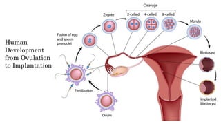
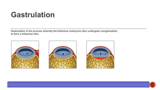
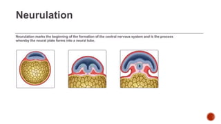
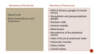
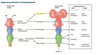
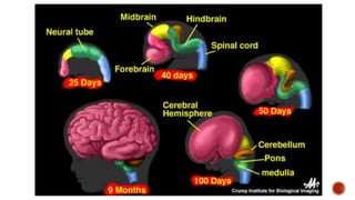
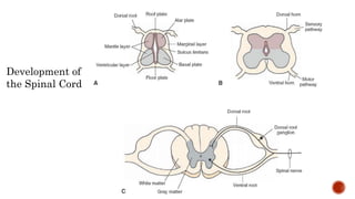
Human
Development
from Ovulation
to Implantation
Gastrulation
Neurulation
Derivatives of Neural tube
 Spinal cord
 Brain hemispheres and
brainstem
Derivatives of Neural crest
 DRG & Sensory ganglia of cranial
nerves
 Sympathetic and parasympathetic
ganglia
 Schwann cells
 Adrenal medulla
 Melanocytes
 Neurolemma of the peripheral
nerves
 Cells of the pia & arachnoid mater
 Intraocular muscles
 Ciliary bodies
 Carotid bodies
Development of
the Spinal Cord

Development of CNS

Editor's Notes

  • #2 The whole of the nervous system is derived from ectoderm except its blood vessels and some neuroglial elements.
  • #3 In midicycle due LH there is release of ovum. After fertilization , Zona Pellucida present around the ovum, completes its second meiotic division (metaphase phase). 23 chromosomes from female side and 23 from male .. Making it diploid cell. It then multiplies . Forming blastula and then morula. Grp of cells in center are called inner cell mass which eventually form the embryo and fetus and the outer cell mass forms the placenta. Zona Pellucida dissolves. Fluid fills in the cavity forming blastocyst. Inner cell mass is called embryoblast and outer called as trophoblast. Villi grows from the embryonic pole of trophoblast forming synctium trophoblast. One week of time has passed by now. Corpus leuteum continues the production of progesterone due to the signal comes by hcG now called corpus gravidum. Untill placenta takes over.
  • #4 2 week - Blastocyct form bilaminar disc . Upper layer is epiblast and lower is hypoblast, upper and lower layer are fused at point called procordal plate-future mouth of the baby. Some cells degenerate to form primitive streak with node. Cells in epiblast start moving downward under the surface of epiblast and forms the tube down to the primitive node called as notochord. Eventually converts to trilaminar disc. Above the notochord is ectoderm and below is endoderm and side is the mesoderm. Notochord present longitudinally , releases chemical substance and makes the ectoderm thickening known as Neural plate. Neural plate depresses downward and makes a fold with grooves called as neural grooves. Neural grooves fuse at anterior neuropore and posterior neuropore. On 25th day of gestation anterior neuropore fuses and on 27th posterior neuropore fuses forming neural tube. Neural crest cells will come out.
  • #5 Nucleus pulposus in intervertebral disc is the remnant of notochord in adult. Anencephaly when anterior neuropore is not fused. And if posterior neuropore does not close , gives rise to spinal bifida.
  • #7 In adult the fusion point of anterior neuropore is known as Lamina terminalis. Telencephalon are growing rapidly and goes far off and the inner part becomes Diencephalon.
  • #9 Near to the spinal canal are neuroblast which give rise anteriolaterally called as basal plate and posteriolaterally called as alar plates. Basal plate has motor neurons and alar plate has sensory neurons.