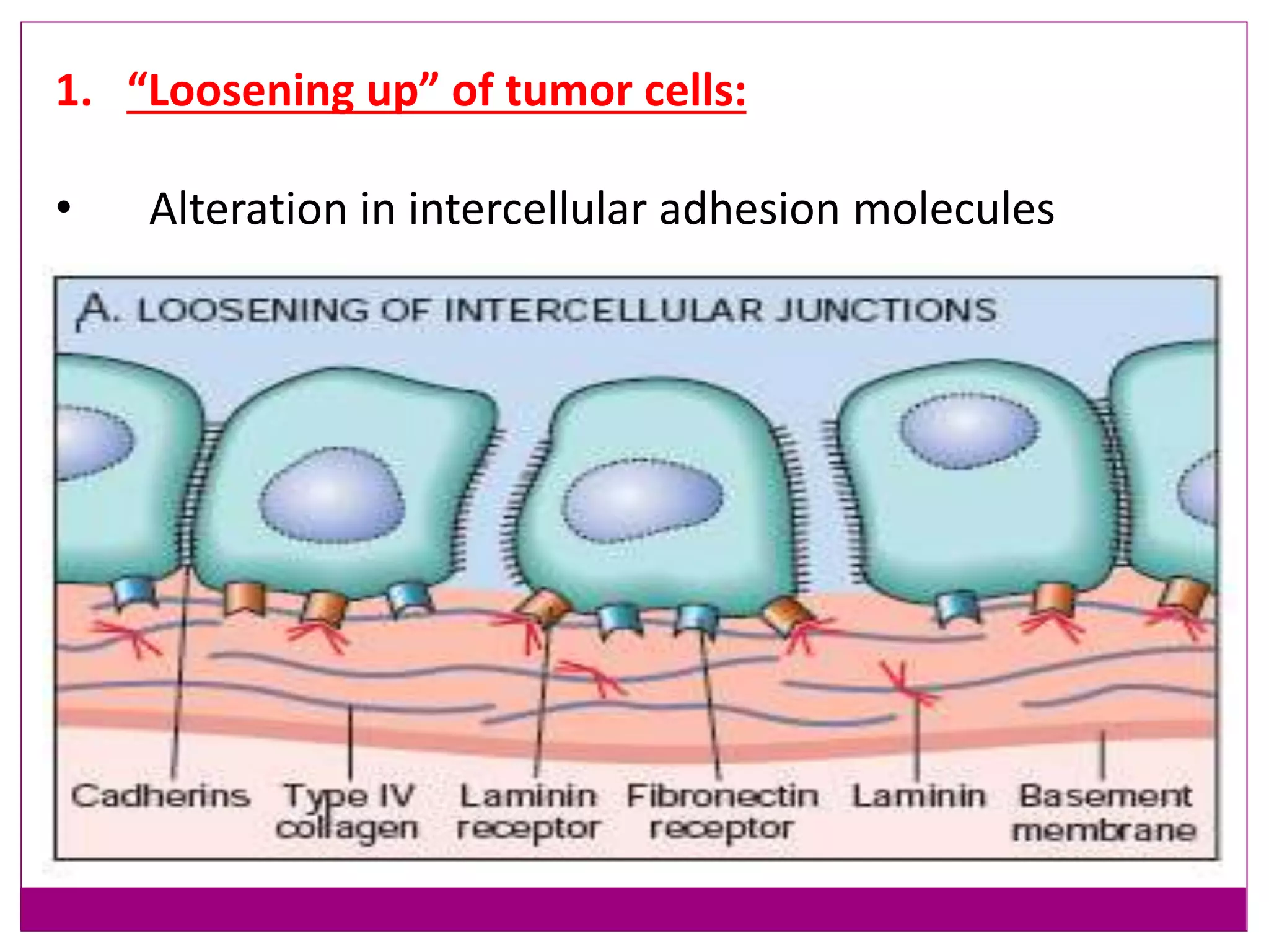
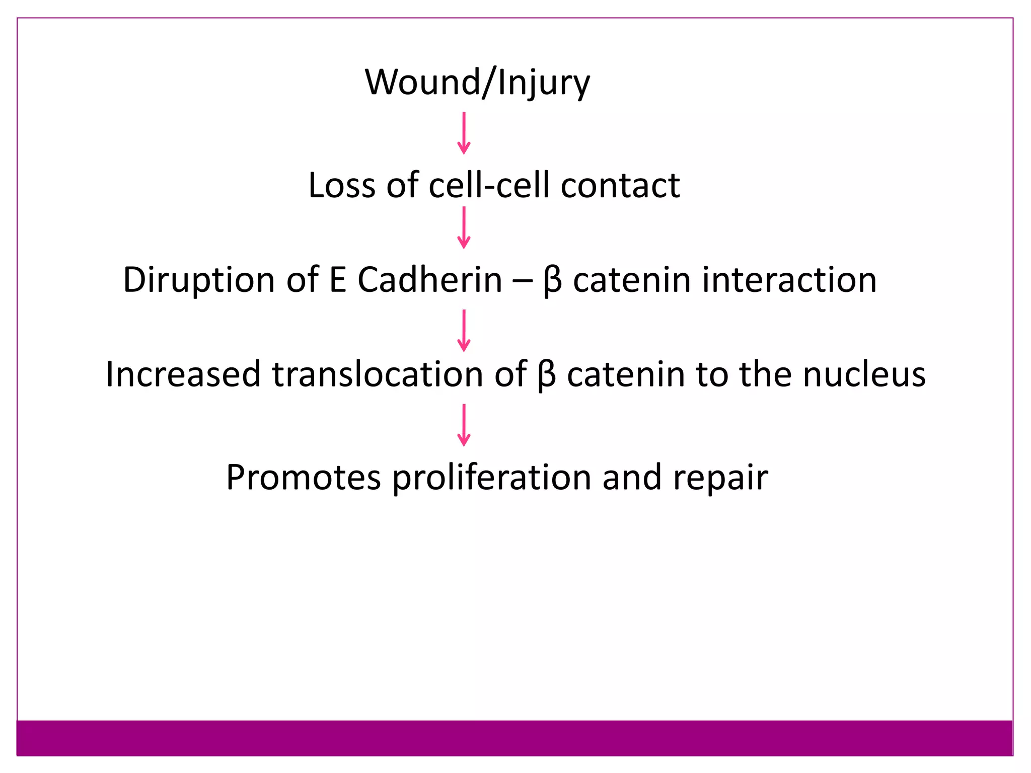
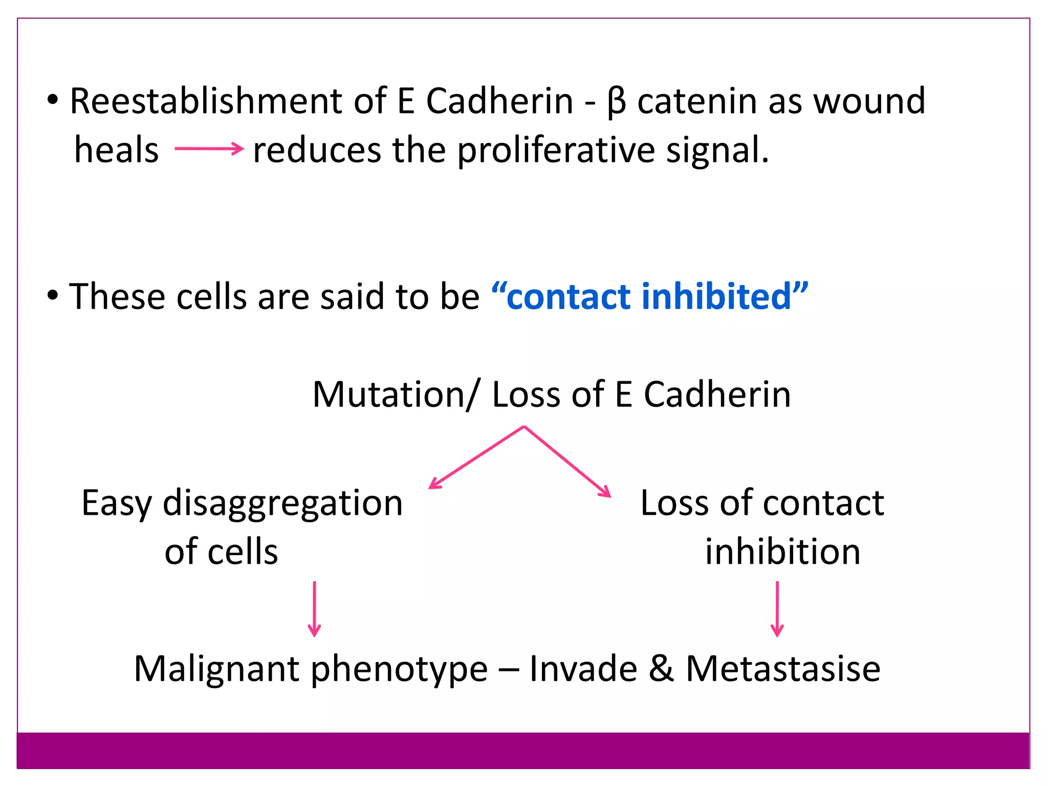
This document provides an overview of cancer metastasis and the epithelial-mesenchymal transition (EMT) process. It discusses the metastatic cascade, which involves tumor cell invasion, intravasation into blood vessels, transport through circulation, extravasation and homing to distant sites, and formation of secondary tumors. EMT is described as a key step in metastasis that allows epithelial cells to detach from primary tumors and migrate. The molecular and cellular changes involved in EMT include loss of epithelial markers like E-cadherin and gain of mesenchymal markers. Transcription factors such as Snail, Slug, Twist, and ZEB play important roles in inducing EMT. Understanding metastasis and EMT can help develop strategies to prevent cancer spread
An oncogene is a gene that has the potential to cause cancer. In tumor cells, they are mutated or expressed at high levels. Most normal cells undergo a programmed form of rapid cell death (apoptosis) when critical functions are altered.
Cancer is mainly caused by the conversion of proto-oncogenes into oncogenes. The process is known as oncogenesis.
This slide will help to get an idea about oncogenesis and also the proto-oncogenes which get converted.
The epigenetic regulation of DNA-templated processes has been intensely studied over the last 15
years. DNA methylation, histone modification, nucleosome remodeling, and RNA-mediated targeting regulate many biological processes that are fundamental to the genesis of cancer. Here, we
present the basic principles behind these epigenetic pathways and highlight the evidence suggesting that their misregulation can culminate in cancer. This information, along with the promising clinical and preclinical results seen with epigenetic drugs against chromatin regulators, signifies that it
is time to embrace the central role of epigenetics in cancer.
These hallmarks constitute an organizing principle for rationalizing the complexities of neoplastic disease. They include sustaining proliferative signaling, evading growth suppressors, resisting cell death, enabling replicative immortality, inducing angiogenesis, and activating invasion and metastasis.
ONCOGENE AND PROTOONCOGENE
P53 GENE AND ITS APPLICATION IN CANCER ETIOLOGY
TUMOUR SUPPRESSOR GENE AND BCA AND BAC GENE AND ITS APPLICATION ON THE APOPTOSIS AND DEATH RECEPTORS
This is a powerpoint explaining on the basics of neoplasia by Dr Libin Babu Cherian, MD, DNB (pathology), DM (oncopathology). It includes mechanisms of cancer evolutions, metastasis, radiation induced carcinogenes, virus induced carcinogenesis and paraneoplastic syndromes
Palestine last event orientationfvgnh .pptxRaedMohamed3
An EFL lesson about the current events in Palestine. It is intended to be for intermediate students who wish to increase their listening skills through a short lesson in power point.
Read| The latest issue of The Challenger is here! We are thrilled to announce that our school paper has qualified for the NATIONAL SCHOOLS PRESS CONFERENCE (NSPC) 2024. Thank you for your unwavering support and trust. Dive into the stories that made us stand out!
Acetabularia Information For Class 9 .docxvaibhavrinwa19
Acetabularia acetabulum is a single-celled green alga that in its vegetative state is morphologically differentiated into a basal rhizoid and an axially elongated stalk, which bears whorls of branching hairs. The single diploid nucleus resides in the rhizoid.
2024.06.01 Introducing a competency framework for languag learning materials ...Sandy Millin
http://sandymillin.wordpress.com/iateflwebinar2024
Published classroom materials form the basis of syllabuses, drive teacher professional development, and have a potentially huge influence on learners, teachers and education systems. All teachers also create their own materials, whether a few sentences on a blackboard, a highly-structured fully-realised online course, or anything in between. Despite this, the knowledge and skills needed to create effective language learning materials are rarely part of teacher training, and are mostly learnt by trial and error.
Knowledge and skills frameworks, generally called competency frameworks, for ELT teachers, trainers and managers have existed for a few years now. However, until I created one for my MA dissertation, there wasn’t one drawing together what we need to know and do to be able to effectively produce language learning materials.
This webinar will introduce you to my framework, highlighting the key competencies I identified from my research. It will also show how anybody involved in language teaching (any language, not just English!), teacher training, managing schools or developing language learning materials can benefit from using the framework.
Introduction to AI for Nonprofits with Tapp NetworkTechSoup
Dive into the world of AI! Experts Jon Hill and Tareq Monaur will guide you through AI's role in enhancing nonprofit websites and basic marketing strategies, making it easy to understand and apply.
The Roman Empire A Historical Colossus.pdfkaushalkr1407
The Roman Empire, a vast and enduring power, stands as one of history's most remarkable civilizations, leaving an indelible imprint on the world. It emerged from the Roman Republic, transitioning into an imperial powerhouse under the leadership of Augustus Caesar in 27 BCE. This transformation marked the beginning of an era defined by unprecedented territorial expansion, architectural marvels, and profound cultural influence.
The empire's roots lie in the city of Rome, founded, according to legend, by Romulus in 753 BCE. Over centuries, Rome evolved from a small settlement to a formidable republic, characterized by a complex political system with elected officials and checks on power. However, internal strife, class conflicts, and military ambitions paved the way for the end of the Republic. Julius Caesar’s dictatorship and subsequent assassination in 44 BCE created a power vacuum, leading to a civil war. Octavian, later Augustus, emerged victorious, heralding the Roman Empire’s birth.
Under Augustus, the empire experienced the Pax Romana, a 200-year period of relative peace and stability. Augustus reformed the military, established efficient administrative systems, and initiated grand construction projects. The empire's borders expanded, encompassing territories from Britain to Egypt and from Spain to the Euphrates. Roman legions, renowned for their discipline and engineering prowess, secured and maintained these vast territories, building roads, fortifications, and cities that facilitated control and integration.
The Roman Empire’s society was hierarchical, with a rigid class system. At the top were the patricians, wealthy elites who held significant political power. Below them were the plebeians, free citizens with limited political influence, and the vast numbers of slaves who formed the backbone of the economy. The family unit was central, governed by the paterfamilias, the male head who held absolute authority.
Culturally, the Romans were eclectic, absorbing and adapting elements from the civilizations they encountered, particularly the Greeks. Roman art, literature, and philosophy reflected this synthesis, creating a rich cultural tapestry. Latin, the Roman language, became the lingua franca of the Western world, influencing numerous modern languages.
Roman architecture and engineering achievements were monumental. They perfected the arch, vault, and dome, constructing enduring structures like the Colosseum, Pantheon, and aqueducts. These engineering marvels not only showcased Roman ingenuity but also served practical purposes, from public entertainment to water supply.
2. What is cancer metastasis?
• Cancer is defined as
•A population of cells that have lost their normal
controls of growth and differentiation
•Proliferating without check.
• Metastasis
•The process by which a tumor cell leaves the primary
tumor Travels to a distant site via the
circulatory system Establishes a secondary
tumor.
3. Why do we need to know the metastatic
cascade?
• It is estimated that metastasis is responsible for
about 90% of cancer deaths.
• About 1,500 people continue to die each day from
cancer due to the failure in managing the disease once it
disseminates through the body.
• Metastasis remains a final frontier in the search for a
cure for cancer.
4. • Every tumor’s:
- potential to metastasise is different
- metastatic sites are different
- same histological grade of tumor may show different
metastatic behaviour
• Study metastasis in detail to outline strategies to curb it.
5. Metastasic
Cascade
• Invasion and passage
through
basement membrane
• Migration through
extracellular matrix
• Migration into vessels
(intravasation)
6. • Adhesion to vascular
endothelium (usually in lung
or liver)
• Extravasation and migration
into tissue
• Establishment and growth
at new site
7. STEP I - Invasion
1. “Loosening up” of tumor cell-tumor cell interactions.
2. Degradation of ECM
3. Attachment to novel ECM components
4. Migration and invasion of tumor cells
8. 1. “Loosening up” of tumor cells:
• Alteration in intercellular adhesion molecules
9. Role of E Cadherin in invasion
-Cell surface protein – Intercellular adhesiveness
-β catenin protein
binds to the
cytoplasmic tail of
E Cadherin
10. Wound/Injury
Loss of cell-cell contact
Diruption of E Cadherin – β catenin interaction
Increased translocation of β catenin to the nucleus
Promotes proliferation and repair
11. • Reestablishment of E Cadherin - β catenin as wound
heals reduces the proliferative signal.
• These cells are said to be “contact inhibited”
Mutation/ Loss of E Cadherin
Loss of contact
inhibition
Easy disaggregation
of cells
Malignant phenotype – Invade & Metastasise
13. MMP’s
• Multigene family of zinc dependent extracellular matrix
(ECM) remodeling endopeptidases
• Produced as inactive precursors (Zymogens)
• Activated by cleavage of a propeptide
• Rapidly inhibited by specific tissue inhibitors of
metalloproteinase (TIMP’s) – Mesenchymal cells
15. Other functions of MMPs in cancer
• MMPs Affect Growth Signals – like TGF β, EGFR
• MMPs Regulate Apoptosis – cleavege of ligands (Fas)
that induce apoptosis.
• Tumor angiogenesis – MMP 9 has distinct role by
regulating the bioavailability of vascular derived
endothelial growth factors (VEGF).
16. Cathepsins
• Diverse group
• Serine proteases
• Cysteine proteases
• Aspartic proteases
• Synthesized as inactive precursors.
• Activated at acidic pH in the lysosomes.
• Contribution to invasion is well documented.
• Exact mechanism still not clear.
17. Cathepsin Cancer
A Malignant melanoma
B Breast, Lung, Gastric, Pancreatic, Bladder
D Thyroid, Renal, Ovarian, SCC
E Panreatic ductal carcinoma, Gastric
F Cervical carcinoma
G Breast
H Breast, Colorectal, Prostate
K Gastric, SCC, BCC
L Breast, Lung, Gastric
S Astrocytoma, Gastric, Hepatocellular
X Prostate, Gastric, Malignant melanoma
Z Melanomas, Gastric, Hepatocellular
18. Plasmin-Plasminogen activator
• Plasmin - ability to degrade several matrix components
like gelatin, fibronectin, laminin.
- activates MMP’s by propeptide cleavage.
- synthesized in its inactive form plasminogen
- conversion needs plasminogen activator
19. • Plasminogen activator – Two types
1. Urokinase (uPA)
2. Tissue (tPA)
-Plasminogen activator inhibitor (PAI) 1&2
- Involved in regulation of plasmin and pro MMP’s
20. Plasminogen
Plasmin
Pro MMP Active MMP
• Dissolution of basement membrane
& interstitial matrix
• Cell signaling and migration
• Angiogenesis
uPA/tPA
21. • Matrix is modified to promote invasion.
• Cleavage of basemement membrane proteins
- Collagen IV and laminin by MMP 2 or 9
- Generates novel sites
- Bind to receptors on tumor cell and stimulate
migration.
3. Attachment to novel ECM components:
23. • It is a multistep process.
• Cells must attach to the matrix at leading edge
• Detach from the trailing edge
• Contract the cytoskeleton to rachet forward.
• This is accomplished by epithelial to mesenchymal
transition (EMT).
24. • An orchestrated series of events
• Cell-cell and cell-extracellular matrix (ECM)
interactions are altered.
• Releasing epithelial cells from the surrounding tissue.
• The cytoskeleton is reorganized to allow movement in 3
dimensions in the ECM.
• New transcriptional program is induced to maintain the
mesenchymal phenotype.
Epithelial Mesenchymal Transition
25. • At completion of EMT:
- degradation of underlying basement membrane
- formation of a mesenchymal cell
- can migrate away from the epithelial layer in
which it originated.
26.
27. STEP II - Vascular dissemination of
tumor cells
• In circulation
- Tend to form clumps
- homotypic adhesion among tumor cells
- heterotypic adhesion with blood cells esp. platelets
• Platelet tumor aggregate may enhance tumor cell
survival and implantability.
28. • In circulation tumor cells are vulnerable to destruction
- mechanical shear stress
- apoptosis stimulated by loss of adhesion to basement
membrane (Anoikis)
- immune defense mechanisms
29. Mechanisms by which tumor cells escape immune
recognition
• Low immunogenecity:
- No peptide – No MHC ligand
- No adhesion molecules
- No co stimulatory molecules
30. • Tumor treated as self antigen:
- Tumor antigens taken up and
presented by APC’s are tolerated
by T- cells.
31. • Antigenic modulation:
- Antibody against tumor
cell surface antigens induce
endocytosis and degardation
of antigen.
- Immune selection of antigen
loss variants.
32. • Tumor induced immune suppression:
- Factors (TGF-β) secreted by tumor
cells directly inhibit T-cells.
- Expression of lymphocyte
death receptor ligands
(FasL, TRAIL) by tumor cells.
33. • Tumor induced privilege site:
- Factors secreted by tumor cells create
a physical barrier to the immune system.
- Recruitment and activation of
regulatory T-cells (Tregs – CD4+ CD25+)
34. STEP III - Homing of tumor cells
• Arrest and extravasation of tumor emboli at distant site
- adhesion to the endothelium
- egress through basement membrane
• Site at which circulating tumor cells leave the capillaries
to form secondary deposits
- anatomic location of primary tumor
- vascular drainage of primary tumor
- tropism of particular tumor to specific tissues
35. Preferential metastatic sites
Primary Tumor Common distant sites
Breast adenocarcinoma Bone, Brain, Lung, Adrenal
Prostate adenocarcinoma Bone
Lung small cell carcinoma Bone, Brain, Liver
Skin cutaneous melnoma Brain, Liver, GIT
Thyroid adenocarcinoma Bone
Renal cell carcinoma Bone, Liver, Thyroid
Bladder carcinoma Brain
36. Reason for organ selectivity
• Mechanistic theory: determined by the pattern of
blood flow.
•“Seed and soil” theory: the provision of a fertile
environment in which
compatible tumor cells could
grow.
37. Determining factors for organ tropism
• Compatible adhesion sites on endothelial luminal
surface of target organ.
• Selective chemokine secretion by target tissue for
metastasis.
- Breast carcinoma cells express receptors for chemokine
CXC4 and CCR7.
• Appropriate environment.
- Although well vascularised skeletal muscle and spleen
are rare sites for metastasis.
38. STEP IV - Establishment at distant site
• Accomplished by mesenchymal to epithelial transition
• Colonisation of tumor cells Micrometastasis
• Proliferation and angiogenesis Macroscopic
metastasis
39. Molecular Genetics of Metastasis
development
1. Clonal evolution model:
• Mutations accumulate in genetically unstable cancer
cells, leading to heterogenous population.
• Tumor cell subclones develop gene expression
permissive for EMT and hence to metastasis.
40. 2. Metastasis signature:
• Gene expression studies in breast carcinoma with
increased risk of metastasis found a phenotype which
signified EMT. Genotype ~ Metastasis signature
• These cells with “metastatic signature” have
predilection for metastasis during early stages of
carcinogenesis.
45. Why Epithelial Mesenchymal Transition ?
• EMT is the primary step in the process of metastasis
• Stopping this transition can curb metastasis
-tumors can be resected surgically
- amenable treatment
46. • Has been extensively investigated in past decade
- facilitate development of early detection strategies
- improve therapeutic targeting of malignant tumors
48. Characteristics of Epithelial cells:
• Tightly packed cells usually arranged in layers.
• Regularly spaced cell junctions and adhesions between
neighboring cells.
• Tight adhesion between cells resulting in inhibition of
movement away from the monolayer.
• Epithelial cell is polarised i.e. different ends of
cell do different things.
49. • Characterised by specialised membrane
domains:
1- Basal domain - interacts with basement
membrane (BM).
2- Apical domain - depends on the functional
needs of cell.
3- Lateral domain - form adherence and tight
junctions.
50. • A characteristic example is the enterocyte :
- Apical domain: Brush border essential for
resorptive activity.
51.
52. • Apical compartment protein complexes:
•Partitioning defective (PAR) complex
(PAR6, PAR3, atypical protein kinase C (aPKC))
• Crumbs (CRB) complex
• Protein associated with Lin-7 1 (PALS1)
• PALS1 associated tight junction protein (PATJ)
53.
54. -Basal domain: Laminin 5 mediates adhesion to BM
through interaction with integrins &
provides signalling cues from ECM.
61. Characteristics of Mesenchymal cells:
• They lack a regimented structure.
• Very few intracellular adhesions.
• Weak adhesions allow for ease of mobility.
• Front rear cytoplasmic polarity.
• No specific membrane domains.
62. • Cells are potentially mobile
- cytoskeleton composed of vimentin
- myofibroblasts of smooth muscle actin.
• Their secretory activity is targeted towards
production of extracellular matrix components.
63. EPITHELIAL CELL MESENCHYMAL CELL
Arranged in a layers Lack a regimented structure
Many cell junctions and adhesions Very few cell junctions and adhesions
Stationary Ability to migrate
Apical basal polarity Front rear polarity
64. • First observed and defined by Elizabeth Hay in late
1960’s using a model of chicken primitive streak in
embryogenesis.
- Epithelial Mesenchymal Transformation
• Process is reversible with unstable intermediate
EMT Metastable MET
- Hence the term “transition”
Epithelial to Mesenchymal Transition
66. • Types of EMT
• Three types of EMT:
- Type 1
-Type 2
- Type 3
67. Types of EMT
• Type 1 – EMT during implantation,
embryogenesis and organ development
68. • Type 2 – EMT associated with tissue
regeneration and organ fibrosis
69. • Type 3 – EMT associated with cancer
progression and metastasis
70.
71. Phenotypic modifications associated
with EMT
• Invitro morphology and function
- Stellate or spindle shape
- Resistance to anoikis
- Increased migration
- Invasion into collagen matrix
72. • Down – regulation of epithelilal proteins:
- E Cadherin
- Cytokeratin
- Occludin
- Claudin
Epithelial to Mesenchymal transition
73. • Up – regulation of Mesenchymal proteins:
- N Cadherin
- Vimentin
- Fibronectin
- α Smooth muscle actin
- MMP’s (2, 3, 9)
- Integrin αvβ6
Epithelial to Mesenchymal transition
76. Molecular events comprising EMT
• Deconstruction of cell junctions
• Dissolution of tight junctions
- decreased claudin & occludin expression
- diffusion of zona occludens 1 (ZO1 aka TJP1)
• Destabilization of adherence junctions
- Cleavage of E Cadherin
- Increased nuclear translocation of β catenin
77. • Decreased expression of E Cadherin
- represssion of polarity complex proteins
- loss of apical basal polarity
• Apical basal polarity - organised by polarity complexes
- integrated with cell junctions
• Loss of apical basal polarity
78. • Cytoskeletal changes and motility
• Reorganisation of cortical actin cytoskeleton to enable
cell elongation and directional motility
•Formation of actin rich membrane projections
- Lamellipodia: sheet like
- Filopodia: spike like
- Invadopodia: proteolytic
80. • To achieve directional polarity:
- PAR, PATJ, Crumbs & Scribble complexes relocalize
to leading edge of cell
- RAC1 & CDC42 activate actin polymerization and
membrane protrusion formation
- RHO A localizes at the rear end & promotes
disassembly of adhesion complexes & cell retraction
• Induces cell shape changes and front rear polarity
essential for migration
84. SNAI family of transcription factors
• Three vertebrate SNAI factors
- SNAI 1 aka SNAIL
- SNAI 2 aka SLUG
- SNAI 3 aka SMUC
• Repress epithelial genes by binding to E- box DNA
sequences through carboxy terminal zinc finger
domains.
• Central role in EMT
85. Twist transcripton factor
• Part of basic helix-loop helix (bHLH) transcription
factors.
• Implicated in cell lineage determination and
differentiation.
• Role in Breast carcinoma
86. ZEB transcription factors
• Two vertebrate ZEB factors
- ZEB1 aka TCF8
- ZEB2
• Bind regulatory gene sequences at E boxes
• Repress or activate transcription
• Role Lung carcinoma
87. Signalling pathways in EMT
RECEPTOR LIGAND SIGNALLING
MOLECULES
INTERMEDIATE
SIGNALLING
END POINT
EFFECT
TGF- β
receptor
TGF- β Rho A
Smad family
ZEB Migration
Receptor
Tyrosine
Kinae
FGF
HGF
EGF
Sarc
Ras, MAPK
SNAI2 -Cytoskeleton
activation
-Migration
-Focal adhesion -
rearrangement
Integrins Collagens
Fibronectin
FAK, Paxillin,
Rac
SNAI2 Increased
migration
Frizzled Wnt APC, axin, GSK3β,
β-catenin
SNAI1 E Cadherin down
regulation
Reduced cell
adhesion
88.
89. • Complex interactions between the signalling pathways
during EMT
- gravitate towards downregulation of E Cadherin
- disassembly of junctional complexes.
• Loss of E cadherin expression is associated with
nuclear expression of β catenin via the Wnt
signaling pathway.
• This is associated with the acquisition of stem cell
properties by the cancer cell.
90. Role of E cadherin in EMT
Downregulation of E Cadherin
Disassembly of junctional complexes
Liberates β catenin from
E Cadherin- Catenin
complex
Activation of Wnt
pathway through
LEF/TCF4.
Signals Integrins
93. SNAI1 & SNAI2 are the central
regulators
Interaction of FGF, EGF or HGF with their
respective RTK’s
Activates members of GTPase family
(Ras, Rho,Rac)
Activation of of Wnt pathway
95. SNAI1 and SNAI2
Modify pattern of genes for remodelling
cytoskeleton
Upregulation
of Vimentin
expression
Modification
of Integrin
expressionActivation
of contractile
apparatus
96. Epithelial plasticity is bi-directional
• The phenotypic plasticity afforded by an EMT is
revealed by the occurrence of a reverse process –
Mesenchymal epithelial transition.
97. • Disseminated cancer cells undergo MET at the site
of metastasis.
• Explained as EMT derived migratory cancer cells
typically establish secondary colonies at distant sites
that histopathologically resemble the primary tumor
from which they arose.
98.
99. Practical aspects of EMT
• The recent past has seen extensive research on the
different elements of EMT.
100.
101.
102.
103.
104. • The results of these research have lead to the use of
Epithelial Mesenchymal Transition as a powerful tool in:
-Surgical pathology for highlighting tumor invasiveness
- Prognostication of various tumors
- Prediction of therapy resistance
- Potential target for antineoplastic therapies
105. EMT In Surgical Pathology
• Immunohistochemical Markers
• Loss of epithelial markers
- E Cadherin : Colon, Breast, Lung, Ovary,
Esophagus, Prostate, Cervix
- Claudin : Ovary
- Occludin : Esophagus, Breast
Loss of E Cadherin in Breast CA
106. • Loss of E-cadherin expression is associated with
• The switch from noninvasive adenoma to invasive
carcinoma in a transgenic mouse model of
pancreatic β-cell carcinogenesis.
107. • Expression of mesenchymal markers
- N Cadherin : Ovary, Prostate
- Vimentin : Breast, Esophagus, Cervix
- β catenin (Nuclear) : Breast, Cervix
Vimentin in Breast CA
109. • Immunofluoresence markers
-E Cadherin
- Vimentin
- HGF
- TGF β
- NOTCH 1
- Wnt-3a
- BMP 7
Loss of E Cadherin in Breast CA
110. • The single-cell infiltration pattern
• Observed in some lobular carcinomas
• Linked to protein truncation mutations in the CDH1
gene encoding for E-cadherin.
• Tumor cell budding
• Morphologic hallmark of invasive tumor
phenotype and tumor aggressiveness in colorectal
cancer tissue specimens
• Loss of membraneous β-catenin
Histomorphological phenotype associated with EMT
111. EMT and patient prognosis
• The metastatic spread of malignant tumors accounts
for the majority of cancer- specific deaths
• Therefore possible correlations between EMT markers
and patient prognosis have been intensely studied in
multiple tumor entities.
112. • In colon cancer
• Upregulation of genes involved in EMT/matrix
remodeling defines a molecularly distinct subtype
with very unfavorable prognosis.
• Downregulation of E-cadherin in patient samples,
seems to be associated with high TNM stages and
distant metastasis.
113. •In prostate cancer
• Levels of Twist and Vimentin assessed by
immunohistochemistry in radical prostatectomy
specimens
• Are independent predictors for biochemical
recurrence
• Correlate with resurgence in serum prostate-
specific antigen (PSA) levels following surgery.
114. • Additionally, loss of membraneous E-cadherin
staining is associated with
• Increased Gleason score.
• Advanced clinical stage.
• Poor prognosis in prostate cancer.
• In basal-like, triple-negative breast cancers
• Upregulation of Vimentin associated a poor prognosis.
115. EMT in therapy resistance
• EMT-like cellular phenotype in both surgical
specimens and cell lines is associated with
increased resistance to most conventional
approaches
• Chemotherapy
• Radiotherapy
• Hormone withdrawal
116. • In non-small cell lung cancer cells
• Mesenchymal-like cells (that express Vimentin or
Fibronectin) are less sensitive to EGFR kinase
inhibitors.
• Also in urinary bladder, head and neck, pancreas, and
colorectal carcinoma
• An EMT-like phenotype of the tumor cells is
associated with resistance to anti EGFR therapy.
117. • The resistance to antineoplastic therapies:
• Might be due to stem-cell like properties of tumor
cells that have undergone EMT
• Allowing for self-renewing of a proportion of cells
within the tumor based on the activation of
central signaling pathways. ( Wnt, Notch etc)
118. EMT as potential target for
antineoplastic therapies
• Since the population of stem cell-like tumor cells will
always bear considerable resistance to conventional
therapies efforts have been made to develop
antineoplastic therapies that directly target EMT.
119. Current therapeutic approaches aiming at EMT
Tumors Target Drug Mechanism Effect
Breast LYN Kinase Dasatinib
Metformin
Transcriptional
repression
- Invasion
+E Cadherin
Urothelial uPA Sorafenib Inhibition of
MAPK signalling
-uPA
+E Cadherin
Hepatocellular ILK Kinase activated
ILK
( S343A)
Reduction of
AKT signalling
Increased
senstivity to anti
EGFR therapy
Pancreatic Hedgehog genes Cyclopamine
Resveratol
Transcriptional
repression
-Metastasis
+E Cadherin
Lung HAT/HDAC Vorinostat
Romidepsin
+HAT
-HDAC
Repression of
EMT genes
120. In the era of molecular pathology a good understanding
of EMT and metastasis is essential to better patient
management.