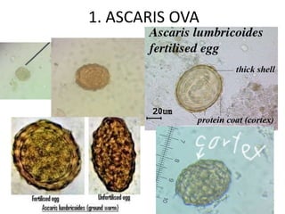
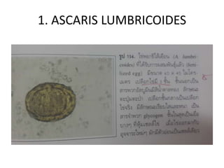
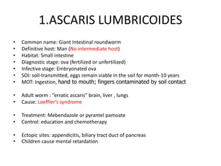
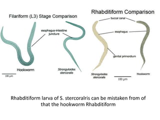
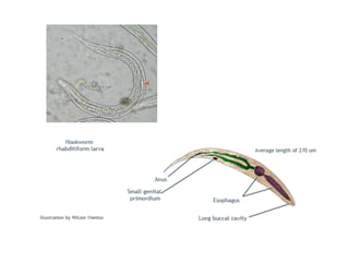
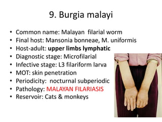
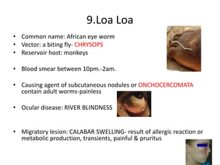
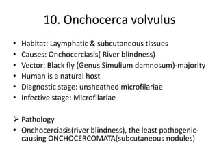
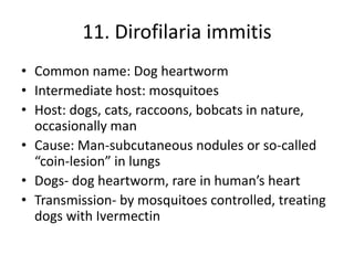
4. 1.ASCARIS LUMBRICOIDES
• Common name: Giant Intestinal roundworm
• Definitive host: Man (No intermediate host)
• Habitat: Small intestine
• Diagnostic stage: ova (fertilized or unfertilized)
• Infective stage: Embryonated ova
• SOI: soil-transmitted, eggs remain viable in the soil for month-10 years
• MOT: Ingestion, hand to mouth; fingers contaminated by soil contact
• Adult worm : “erratic ascaris” brain, liver , lungs
• Cause: Loeffler’s syndrome
• Treatment: Mebendazole or pyrantel pamoate
• Control: education and chemotherapy
• Ectopic sites: appendicitis, biliary tract duct of pancreas
• Children cause mental retardation
5. Ascaris worm
Toxocara cati, T. canis
• Cause: visceral larva migrans or Toxocariasis
• Eggs are threat to human
• Children more prone to infect because of a contact
• Migration produces hemorrhage, necrosis, granulomas
• Eosinophillia, liver damage, pulmonary inflammation,
ocular problem will be observed.
• Take note: they do not develop further than larva stage
10. Hookworm in human
• Ancylostoma duodenale and Necator
americanus
• Infection occurs when filariform (LARVAE)
penetrates the skin of man
• Causing “ground itch” or “dewy itch”
maculopapular lesion
• Iron deficiency anemia, hypoalbuminemia
11. Hookworm in Human
• Common name: Old world hookworm( Ancylostoma
duodenale), New world hookworm (Necator americanus)
• Definitive host: Human (no intermediate host)
• Body curvature: “C” shaped and “S” shaped
• Dental pattern: 2 pairs of teeth, semilunar-cutting plate
• Habitat: Small intestine
• Diagnostic stage: Ova
• Infective stage: L3 (filariform)
• Mode of transmission: skin penetration, transmammary and
purely percutaneous
• Disease: Hookworm infection, Ancylostomiasis
12. Animal Hookworm
• Ancylostoma caninum and Ancylostoma
braziliense
• Causing Cutaneous Larva Migrans (CLM)
produces linear, pruritic, papulovesicular lesions
• Condition referred to serpiginous dermatitis or
creeping eruption
13. 2. Hookworm
• Infective stage: filariform larva(L3)
• Skin: site of entry of filariform
• Lung: Larva migration- bronchitis, pneumonitis
• Small intestine: habitat of adult worm-
steatorrhea, diarrhea w/ blood and mucus
• Eosinophil 30-60%
• Treatment: mebendazole (DOC) and pyrantel
pamoate
14. Characteristic of hookworm dentition
• A: Necator americanus, semi-luna cutting plate
• B: Ancylostoma braziliense: 2 pair of teeth
• C: Ancylostoma caninum: 3 pairs of teeth
• D: Ancylostoma duodenale: 2 pair of teeth
A B C D
16. Hookworm
• Laboratory diagnosis: based on the identification
of eggs
1. DFS (only in heavy infection)
2. Kato technique or Kato-katz method: detection
rate, quantitative diagnosis
3. Concentration method like ZnSO4 contrifugal
floatation& formalin-ether concentration
method, increase positive finding many folds
4. Harada-Mori culture allow hatching larva from
eggs on strips of filter paper with one end
immersed in water.
19. 3. Enterobius vermicularis
• Common name: Pinworm, seatworm, society worm
• Final host: man (without intermediate host and reservoir
host)
• Habitat: large intestine (caecum and colon)
• Diagnostic stage: ova
• Infective stage: Embryonated ova
• Source of infection: contact borne
• Mode of transmission: ingestion, inhalation
Take note: Larva develop and the eggs become infected
within 4-6 hours
Newly hatched larva migrate back to anus- Retroinfection
20. 3. Enterobius vermicularis or Pinworm
• Causing enterobiasis, oxyuriasis or seatworm infection
• Lab: scotch tape method early in the morning before taking
a bath
• Familial disease
• Treatment:
1. pyrantel pamoate 10mg/kg w/ a second dose 2-4 wks later
2. Albendazole(400mg) mebendazole (500mg) single dose
26. 5. Trichuris trichiura
• Common name: whip worm
• Final host: human
• Habitat: Large intestine-attached
• Diagnostic stage: Ova
• Infective stage: Embryonated ova
• Source of infection: Soil-transmitted helminthes
• Mode of transmission: Ingestion
• Portal of entry: mouth
27. 5. Trichuris trichiura
• Egg Morphology; hatched in Large intestine
Trichuris trichiura eggs are 'football' or 'barrel'
shaped with clear, mucoid-appearing polar
plugs at each end.
28. 5. Trichuris trichiura
Whipworm
• Infective stage: fully embryonated egg found in
brackish
• Stool exam: reveals bile-stained eggs w/ polar
plugs
• Pathology: Rectal prolapsed in children,
appendicitis, Iron deficiency anemia, diarrhea and
eosinophilia (seen in severe infection)
• Treatment : mebendazole(DOC)
29. To declare free from
Ascaris lumbricoides and Trichuris trichiura
• Three specimen- negative
• Stool examination negative for eggs-
interpreted as
1. All male parasites
2. Female worms are immature(migration
stage)
3. Unfertilized eggs (all females and males-
immature)
31. • Common name: Pudoc worm
• Final host: man/other vertebrate; fish-eating birds
• Intermediate host(IH): glass fish, fresh brackish-water fish
• Diagnostic stage: Ova in stool;peanut-shaped(unembryonated
egg)
• Infective stage: encysted larva (larva in IH)
• Habitat: small intestine
• Source of infection: food borne
• Mode of transmission: eat raw brackish water fish/
contaminated fish
• Portal of entry: mouth
6. Capillaria philippinensis or
Pudoc worm
32. 6. Capillaria philippinensis
Autoinfection and hyperinfection
Ova(diagnostic stage):
Produced by typical female
Moderately thick striated egg sheath with
flattened bipolar plug
33. 6. Capillaria philppinensis or
Pudoc worm
Pathology
Decrease secretion of xylose, low e’lyte(K+)
Cause micro ulceration, depression of intestinal villi (cause
malabsorption of fluid, protein and electrolytes)
Borborygmi (Gurgling stomach)
Malabsorption syndrome(Fat&sugar)
Laboratory test
DFS (direct fecal stool exam)
Concentration technique(FECT)
Examination of duodenal aspirate
34. 6. Capillaria philippinensis or
Pudoc worm
• Treatment:
1. Albendazole(DOC) 400 mg once daily for 10 days
2. Mebendazole 200 mg 2 tab once daily for 20 days
Relapse may occur if the treatment regimen is not
followed. If dis. Not treated soon after severe
manifestation – die
Parasites can be found in small intestine by:
“duodenal aspiration”
35. 7. Strongyloides stercularis
• Common name: Threadworm, smallest nematode of
man
• Final host: man
• Diagnostic stage: Rhabditiform Larva
• Infective stage: Filarliform Larva(L3)
• Mode of transmission: Skin penetration of infective
larva; AUTOINFECTION
• Causative agent of : Cochin China Diarrhea,
malabsorption syndrome
Take note: Hyperinfection are limited to Lungs and GIT
36. Rhabditiform larva of S. stercoralris can be mistaken from of
that the hookworm Rhabditiform
41. 9.Loa Loa
• Common name: African eye worm
• Vector: a biting fly- CHRYSOPS
• Reservoir host: monkeys
• Blood smear between 10pm.-2am.
• Causing agent of subcutaneous nodules or ONCHOCERCOMATA
contain adult worms-painless
• Ocular disease: RIVER BLINDNESS
• Migratory lesion: CALABAR SWELLING- result of allergic reaction or
metabolic production, transients, painful & pruritus
42. 10. Onchocerca volvulus
• Habitat: Laymphatic & subcutaneous tissues
• Causes: Onchocerciasis( River blindness)
• Vector: Black fly (Genus Simulium damnosum)-majority
• Human is a natural host
• Diagnostic stage: unsheathed microfilariae
• Infective stage: Microfilariae
Pathology
• Onchocerciasis(river blindness), the least pathogenic-
causing ONCHOCERCOMATA(subcutaneous nodules)
43. 11. Dirofilaria immitis
• Common name: Dog heartworm
• Intermediate host: mosquitoes
• Host: dogs, cats, raccoons, bobcats in nature,
occasionally man
• Cause: Man-subcutaneous nodules or so-called
“coin-lesion” in lungs
• Dogs- dog heartworm, rare in human’s heart
• Transmission- by mosquitoes controlled, treating
dogs with Ivermectin
44. 12. Dracunculus medinensis
• “Little dragon of Medina”- ancient worm infection; “fiery serpant”
noted by moses with the Isralites at the Red Sea
• Reservoir host: dogs, fur-bearing animals drink contaminated water
containing infective Cyclops
• Human infection- result of ingestion of water from so-called
“stepwell” where people stand or bathe in the water , at which gravid
female worm discharges lava from lesion on the arms, legs, feet,
ankles to infect cyclops in the water.
• Take note: a filarial worm but a tissue invading nematodes. They are
not appeared in the blood
• Immediate host: fresh water microcutaneous(copped) of genus
Cyclops
45. 12. Dracunculus medinensis
• Laboratory diagnosis: observation of typical ulcer
& flooding the ulcer with water to recover&
discharge the larval form
• Occasionally, x-ray examination reveals worms in
various part of the body
• Treatment: Surgical removal, wrapping the worm
on a twig
• Tiridazole(DOC), alternative drug:metronidazole,
thiabendazole
46. Take Note
• Filariform larva:-infective stage of hookworm ,
ascaris and other nematodes, long, thread-like
often “designed” for penetration
• Filarial worm: any of a group of parasitic
worms of family Filariaidae (Phylum
Nematodes) requires 2 hosts, an
arthropod(Intermediate host) and a vertebrae
(primary host) to complete the life cycle