Download to read offline
![Loutfy H Madkour, BAOJ Nanotech 2017, 3: 1
3: 015
BAOJ Nanotechnology
Research
BAOJ Nanotech, an open access journal Volume 3; Issue 1; 015
*Corresponding author: Loutfy H Madkour, Chemistry Department,
Faculty of Science and Arts, Baljarashi, Al Baha University, Baljarashi
65635, Saudi Arabia, Tel; +966 533899075; Fax: +966 77247272; E-
mail: loutfy_madkour@yahoo.com / lha.madkour@gmail.com / Lmad-
kour@bu.edu.sa
Sub Date: April 13, 2017, Acc Date: May 3, 2017, Pub Date: May 3,
2017.
Citation:LoutfyHMadkour(2017)AdvancedAuNMsasNanomedicine’s
central Goals Capable of Active Targeting in Both Imaging and Therapy
in Biomolecules. BAOJ Nanotech 3: 015.
Copyright: © 2017 Loutfy H Madkour. This is an open-access article
distributed under the terms of the Creative Commons Attribution Li-
cense, which permits unrestricted use, distribution, and reproduction
in any medium, provided the original author and source are credited.
Advanced AuNMs as Nanomedicine’scentral Goals Capable of Active Targeting in Both
Imaging and Therapy in Biomolecules
Loutfy H Madkour*
Chemistry Department, Faculty of Science and Arts, Baljarashi, Al Baha University, Baljarashi 65635, Saudi Arabia
Abstract
The stability and dispersity of AuNMs in solution play a key
role for the many applications. Most inorganic nonmaterial’s are
not well dispersed in physiological buffers and require function-
alization by thiols or surfactants to offer the stabilization forces.
Furthermore, sufficient blood circulation time is critical for both
imaging and in vivo drug delivery. Localized surface Plasmon
resonance (LSPR) is one of the most significant features of
AuNMs. The AuNMs as reporters have been broadly applied
into lateral flow immunechromatographical assay (LFICA)and
enzyme-linked immunosorbent assay (ELISA),which is a well-
established technology for analysis of the target analytes in food
safety, clinical diagnosis, environmental monitoring, and medical
science and soon. Au based nonmaterial’s (AuNMs) are known to
possess many attractive features such as unique electrical, optical
and catalytic properties as well as excellent biocompatibility. In this
review, we summarize the current advancement on application of
AuNMs in analytical sciences based on their local surface Plasmon
resonance, fluorescence and electrochemistry properties. AuNMs
based imaging and therapy in bimolecular is explained. As one of
the most reliable imaging modes, computed tomography (CT),
X-ray and SERS imaging has been widely used owing to its high
spatial and density resolution. We end the review by a discussion
of the conjugation between gold nanoparticles with other kinds
of nanoparticles such as other metals and carbon nanostructures.
Finally, future development in this research area is also prospected.
Introduction
In 2017 Madkour, L.H. [1] presents a vision for life sciences:
through interfaces between nanoelectronic and biological
systems in order to the prevention and treatment of disease in
the human body. In this article, we review the applications of gold
nanoparticles in medicine. Generally, the recognition elements,
which are applied into colorimetric sensors based on AuNMs, are
categorized as follows: (1) antibodies or proteins modified AuNMs
with immuno reaction, (2) chemically-modified AuNMs with non-
covalent bond recognition, and (3) aptamers modified AuNMs
with conformational change. Furthermore, some novel techniques
have been tried to improve the selectivity of colorimetric sensing,
such as click-chemistry-based assay.
Physics, chemistry and biology of Au based nonmaterial’s (AuNMs)
have emerged as a broad and new sub discipline in the community
of colloids and surfaces. The specific size and shape dependent
physiochemical properties and remarkable bio/chemical inertness
of AuNMs have
made themselves the ideal candidates for both fundamental
and technical study including crystal growth, electron-transfer
mechanism, localized electro-magnetic theory, catalysis, DNA
assay, bioimaging and therapy, and so on [2]. Among those rich
properties, the optical characteristics originated from the giant
electromagnetic field near the surface of AuNMs are particularly
intriguing and thus broadly applied in analytical science, e.g.
colorimetric assay, surface enhanced Raman spectroscopy (SERS)
and surface Plasmon resonance (SPR)spectroscopy, as well as
bioimaging. Another interesting optical property of AuNMs is
that fluorescence appears with their size shrinkage to below 2 nm,
which allows development of luminescence-based analysis. Also,
electrochemical (EC) sensors could be constructed based on the
redox feature of Au NMs (Figure 1).Analytical science has been
playing a primary role in our daily life, for instance, food safety
control [3], biomedical diagnosis [4], medico legal appraisement
[5], anti-terrorism alert [6], and environmental pollution](https://image.slidesharecdn.com/nanotechnology15-180530122541/85/Nanotechnology15-1-320.jpg)
![Loutfy H Madkour, BAOJ Nanotech 2017, 3: 1
3: 015
BAOJ Nanotechnology
Research
BAOJ Nanotech, an open access journal Volume 3; Issue 1; 015
*Corresponding author: Loutfy H Madkour, Chemistry Department,
Faculty of Science and Arts, Baljarashi, Al Baha University, Baljarashi
65635, Saudi Arabia, Tel; +966 533899075; Fax: +966 77247272; E-
mail: loutfy_madkour@yahoo.com / lha.madkour@gmail.com / Lmad-
kour@bu.edu.sa
Sub Date: April 13, 2017, Acc Date: May 3, 2017, Pub Date: May 3,
2017.
Citation:LoutfyHMadkour(2017)AdvancedAuNMsasNanomedicine’s
central Goals Capable of Active Targeting in Both Imaging and Therapy
in Biomolecules. BAOJ Nanotech 3: 015.
Copyright: © 2017 Loutfy H Madkour. This is an open-access article
distributed under the terms of the Creative Commons Attribution Li-
cense, which permits unrestricted use, distribution, and reproduction
in any medium, provided the original author and source are credited.
Advanced AuNMs as Nanomedicine’scentral Goals Capable of Active Targeting in Both
Imaging and Therapy in Biomolecules
Loutfy H Madkour*
Chemistry Department, Faculty of Science and Arts, Baljarashi, Al Baha University, Baljarashi 65635, Saudi Arabia
Abstract
The stability and dispersity of AuNMs in solution play a key
role for the many applications. Most inorganic nonmaterial’s are
not well dispersed in physiological buffers and require function-
alization by thiols or surfactants to offer the stabilization forces.
Furthermore, sufficient blood circulation time is critical for both
imaging and in vivo drug delivery. Localized surface Plasmon
resonance (LSPR) is one of the most significant features of
AuNMs. The AuNMs as reporters have been broadly applied
into lateral flow immunechromatographical assay (LFICA)and
enzyme-linked immunosorbent assay (ELISA),which is a well-
established technology for analysis of the target analytes in food
safety, clinical diagnosis, environmental monitoring, and medical
science and soon. Au based nonmaterial’s (AuNMs) are known to
possess many attractive features such as unique electrical, optical
and catalytic properties as well as excellent biocompatibility. In this
review, we summarize the current advancement on application of
AuNMs in analytical sciences based on their local surface Plasmon
resonance, fluorescence and electrochemistry properties. AuNMs
based imaging and therapy in bimolecular is explained. As one of
the most reliable imaging modes, computed tomography (CT),
X-ray and SERS imaging has been widely used owing to its high
spatial and density resolution. We end the review by a discussion
of the conjugation between gold nanoparticles with other kinds
of nanoparticles such as other metals and carbon nanostructures.
Finally, future development in this research area is also prospected.
Introduction
In 2017 Madkour, L.H. [1] presents a vision for life sciences:
through interfaces between nanoelectronic and biological
systems in order to the prevention and treatment of disease in
the human body. In this article, we review the applications of gold
nanoparticles in medicine. Generally, the recognition elements,
which are applied into colorimetric sensors based on AuNMs, are
categorized as follows: (1) antibodies or proteins modified AuNMs
with immuno reaction, (2) chemically-modified AuNMs with non-
covalent bond recognition, and (3) aptamers modified AuNMs
with conformational change. Furthermore, some novel techniques
have been tried to improve the selectivity of colorimetric sensing,
such as click-chemistry-based assay.
Physics, chemistry and biology of Au based nonmaterial’s (AuNMs)
have emerged as a broad and new sub discipline in the community
of colloids and surfaces. The specific size and shape dependent
physiochemical properties and remarkable bio/chemical inertness
of AuNMs have
made themselves the ideal candidates for both fundamental
and technical study including crystal growth, electron-transfer
mechanism, localized electro-magnetic theory, catalysis, DNA
assay, bioimaging and therapy, and so on [2]. Among those rich
properties, the optical characteristics originated from the giant
electromagnetic field near the surface of AuNMs are particularly
intriguing and thus broadly applied in analytical science, e.g.
colorimetric assay, surface enhanced Raman spectroscopy (SERS)
and surface Plasmon resonance (SPR)spectroscopy, as well as
bioimaging. Another interesting optical property of AuNMs is
that fluorescence appears with their size shrinkage to below 2 nm,
which allows development of luminescence-based analysis. Also,
electrochemical (EC) sensors could be constructed based on the
redox feature of Au NMs (Figure 1).Analytical science has been
playing a primary role in our daily life, for instance, food safety
control [3], biomedical diagnosis [4], medico legal appraisement
[5], anti-terrorism alert [6], and environmental pollution](https://image.slidesharecdn.com/nanotechnology15-180530122541/75/Nanotechnology15-1-2048.jpg)
![Citation: Loutfy H Madkour (2017) Advanced AuNMs as Nanomedicine’s central Goals Capable of Active Targeting in Both Imaging
and Therapy in Biomolecules. BAOJ Nanotech 3: 015.
Page 2 of 18
BAOJ Nanotech, an open access journal Volume 3; Issue 1; 015
monitoring [7]. Efficient detection technologies depend greatly
on the analytical method in terms of analysis time, signal-to-noise
(S/N) ratio, selectivity, and limit of detection (LOD) [8]. Therefore,
a great of efforts should be paid to develop new analytical methods
with the advantages of rapid determination, high analysis flux,
satisfactory sensitivity and good robustness. Thanks to the
above-mentioned optical and electrochemical features, many
functionalized AuNMs have been successfully synthesized [9–14]
for improving performances of analytical techniques.
Fig. 1. Application of AuNMs in analytical science.
Small Organic Molecules and Metal Ions
For the sensitive detection of small organic molecules, the
AuNMs play an important role in improving sensitivity of the
EC sensors. Especially, AuNMs composited with conducting
polymers or carbon nonmaterial’s have been used to increase
electro-chemical efficiency. It deserves noting that the decorated
carbon nanomaterials with metal NPs are of special interest due
to the integrated property of two components with better catalytic
activity and enhanced electrical conductivity [15,16]. Zhu et al.
fabricated EC sensors by using spherical AuNPs combined with
conducting polymer (poly-[2, 5-di-(2- thienyl)-1H-pyrrole-1-
(p-benzoicacid)]) [17]. To further improve electron transfer,
synergistic effect of multiple nanocomposite layers, namely,
chitosan-AuNPs, graphene-AuNPs and multi-walled carbon
nanotubes (MWCNTs)-cobalt phthalocyanine, was exploited
by Sun et al. [18]. Roushaniet al. [19] fabricated a sensitive and
selective EC apta sensor for detection of cocaine based on the
conformational change of the aptamer-functionalized AuNPs onto
CNTs-based nanocomposite.
Detection of organic small molecules and metal ions is very
important for controlling food quality, environmental protection
and antiterrorism. In order to realize detection of trace amount
of analytes at extremely low concentrations, various efforts for
increasing the sensitivity of SPR biosensors have been paid. Most
of these methods are based on changing the refractive index at
the sensor surfaces by means of a variety of (bio) chemical or NP
agents, which are captured by the sensor surfaces subsequent to
the binding of the target analyte to the primary biorecognition
elements. These methods include the use of secondary and tertiary
antibodies, antibodies labeled with enzymes, and dielectric or
metallic NPs [20]. In particular, AuNMs of diameters ranging from
5 to 40 nm have been widely used to enhance the response of SPR
biosensors [21,22].
Metal Ions
Some EC sensor methods based on AuNMs have been developed
for detection of heavy metal ions. For examples, Shen et al.[23]
proposed an EC DNAzyme sensor based on AuNPs for sensitive
and selective detection of Pb2+
. A label-free EC sensor was
developed for the highly sensitive and selective detection of Hg2+
by Tang et al [24]. In their study, the catalytic HAuCl4
/NH2
OH
reaction was utilized for formation of AuNPs as signal reporter
after capturing Hg2+
on the modified electrode surface through the
specific thymine-Hg2+
-thymine (T-Hg2+
-T) coordination. Com-
pared with the traditional metal NPs-based method, this sensor
avoided the labeling of the DNA probe with NP tags, and only
one unlabeled T-rich DNA sequence was needed, which greatly
reduced the cost and simplified the sensing procedure. Thus, a
LOD as low as 0.06 nM could be obtained for Hg2+
. Furthermore,
in 2016, Wanget al. presented a sensitive, selective and reusable EC
biosensor for determination of Hg2+
based on thymine modified
AuNPs/reduced graphene oxide nanocomposites [25] (Fig. 2).
Notably, the developed EC sensors afforded excellent selectivity
for Hg2+
against other heavy metal ions including Zn2+
, Cd2+
, Pb2+
,
Cu2+
, Ni2+
and Co2+
.Wang et al. prepared an EC sensor based on
graphene and AuNPs for detection of trace Cu2+
with the anodic
stripping voltammeter analysis [26]. Yang et al. constructed a highly
sensitive electro-chemical DNA biosensor made of polyaniline
(PANI) and AuNPs nanocomposite (AuNPs@PANI) for detection
of trace concentration of Ag+
[27]. In the presence of Ag+, with
interaction of cytosine-Ag+-cytosine (C-Ag+
-C), cytosine-rich
DNA sequence immobilized onto the surface of AuNPs@PANI
had a self-hybridization and then formed a duplex-like structure.
The prepared EC sensors could detect Ag+
at a wide linear range of
0.01–100 nM with a LOD of 10 pM.
Synthesis of AuNMs
Traditionally, the tri-sodium citrate-based thermal reduction has-
been broadly recognized as a universal approach for preparing
spherical Au nanoparticles (AuNPs) of varied sizes ranging
from 15to 147 nm possessing strong SPR signals [28]. Although](https://image.slidesharecdn.com/nanotechnology15-180530122541/85/Nanotechnology15-2-320.jpg)
![Citation: Loutfy H Madkour (2017) Advanced AuNMs as Nanomedicine’s central Goals Capable of Active Targeting in Both Imaging
and Therapy in Biomolecules. BAOJ Nanotech 3: 015.
Page 3 of 18
BAOJ Nanotech, an open access journal Volume 3; Issue 1; 015
this preparation method simply involves controlling the ratios
of citrate to Au ions [29], the reaction often needs to be carried
out on a relatively large scale at the temperature of boiling water
and lacks the reproducible preparation of uniform AuNPs with
rather large sizes. After the breakthroughs reported by Schmid
[30, 31] and Brust et al. [32], a variety of methods have been
developed to synthesize AuNMs, and many excellent reviews
[33] are available now. In general, the reported synthesis methods
of spherical AuNMs can be categorized as followed: (1) citrate
reduction; (2) Brust-Schiffrin phase transfer synthesis; (3) other
sulfur-containing ligands method; (4) soft templates including
micro emulsion, reversed Micelles, surfactants, membranes and
polyelectrolyte’s; (5) seed-mediated growth; and (6) physical
methods [34]. In addition, non-spherical nanoparticles(NPs)
such as Au nanorods (AuNRs) [35], Au nanocages [36], Aucubes
[37], Au triangular prisms [38], as well as other unconventional
structures like hollow tubes and even branched nanocrystals
[39] have attracted significant research attention in the past two
decades, since the properties of AuNMs are known to be strongly
dependent on the size and shape of the particle [40]. Recently, a
lot of excellent reviews have summarized how to achieve shape-
and size-defined AuNMs with different techniques including solve
thermalsynthesis,seed-mediatedgrowth,homogeneousnucleation
and electrochemical method [41,42]. In this review, we mainly
introduce the latest and important advancement of synthesis of the
AuNMs, especially on how to control the morphology of AuNMs.
A great of efforts have been paid to achieve high-quality and shape-
controlled AuNMs for the purpose of highly-sensitive analysis.
Unfortunately, most of the synthetic methods are empirical, and
understanding their growth mechanism remains a challenging
task. Interestingly, introduction of Ag+
in growth solutions
significantly improves the yield of AuNRs, and Yang et al. prepared
AuNRs with controlled aspect ratio by using photochemistry in
the presence of Ag+
[43]. This process itself was highly promising
for producing uniform AuNRs, and more importantly it was useful
in deciphering the growth mechanism of anisotropic AuNPs due
to its simplicity and the relatively slow growth rate of the AuNRs.
Later, a versatile seed-mediated growth method was reported for
selective synthesis of single crystalline rhombic dodecahedral,
octahedral, and cubic AuNMs [44]. These results might provide
basis for gaining mechanistic insight into the growth of shape-
and structure-controlled noble metal nonmaterial. An even more
complex sample is nanostar, which is characteristic of uneven
arms with different lengths. To fully implement the advantageous
property of Au nanostars, a precise control over their symmetry
and uniformity is highly desired. In 2015, Niu et al. obtained Au
nanostars with excellent symmetry control by using a solution-
phase method in high yield and good monodispersity [45]. In
their study,icosahedral seeds were used to dictate the growth of
the evenly distributed arms in an Ih
symmetric manner. Benefiting
from their high symmetry, the Au nanostars exhibited much better
single-particle SERS performance compared with asymmetric
Au nanostars, in terms of both intensity and reproducibility.
Apart from large-sized Au particles, Au nanoclusters (AuNCs)
with the sizes of less than2 nm formed in dendrimers displays
visible luminescence of high quantum yield (QY) [46]. Ying et
al. reported a simple, one-pot and “green” synthetic route, based
on the reduction and stabilization capability of bovine serum
albumin (BSA) proteins. The AuNCs prepared at the physiological
temperature (37o
C) showed red emission with a QY of ∼6% [47].
In order to further control the luminescence property, synthesis of
atomically precise AuNCs has ignited numerous research efforts
[48,49]. In 2014, Yu et al. [50] synthesized Au22
(glutathione)18
clusters of the emission peak at ∼665 nm with a QY of ∼8%. Upon
comparison of their luminescence properties with those of Au(I)-
thiolate complexes, the enhanced luminescence of Au22
(SG)18
was ascribed to aggregation-induced emission. Nevertheless,
the origin of emission in AuNCs still keeps unclear, posing a
major hurdle for development of highly luminescent clusters.
In 2015, Pyo et al. prepared Au22
(glutathione)18
clusters with a
luminescence QY greater than 60% by rigidifying their Au shell
with tetraoctylammonium cations [51]. This study presents an
effective strategy to enhance the luminescence efficiencies of
AuNCs by optimizing the shell structure.
In order to precisely tune the size and shape of AuNMs, DNA
is recently adopted as a powerful programmable tool to realize
Fig. 2. Scheme of thymidine functionalized biosensor for Hg2+
detection. Printed with permission from [25]. Copyright 2016 Elsevier.](https://image.slidesharecdn.com/nanotechnology15-180530122541/85/Nanotechnology15-3-320.jpg)
![Citation: Loutfy H Madkour (2017) Advanced AuNMs as Nanomedicine’s central Goals Capable of Active Targeting in Both Imaging
and Therapy in Biomolecules. BAOJ Nanotech 3: 015.
Page 4 of 18
BAOJ Nanotech, an open access journal Volume 3; Issue 1; 015
above goals. Lu’s group systematically summarized morphology
controlling, spatial positioning and dynamic assembly of AuNMs
with DNA as a powerful tool [52]. In some details, their research
results demonstrated that DNA could be used to control the
morphologies of AuNMs during seed-mediated growth [53]. They
also developed a novel method based on DNA-encoded tuning
to achieve AuNMs from AuNR seeds with controlled geometric
and plasmonic properties [54]. Furthermore, they suggested
that the DNA molecules played important roles via influencing
diffusion of the Au precursors to the seed, and therefore the
AuNMs growth might be modulated through difference in DNA
desorption, density and mobility on the seed surfaces [55]. These
insights into the mechanism of DNA-guided AuNMs synthesis not
only provide deep understanding of the interactions between the
DNA and nonmaterial but also allow better control of the shapes
and surface properties of many nonmaterial. Ye et al. designed a
millifluidi creactor to synthesize a wide variety of AuNMs at high
concentrations [56]. The synthesized method based on millifluidic
reactors inspired other researchers to use the analogous reactors
for possible mechanism and application study. Recently, Krishna
et al. demonstrated adoption of a simple millifluidic chip for in sit
analysis of morphology- and dimension-controlled growth of Au
nano- and micro-structures with a time resolution of 5 ms [57]
(Figure 3). See from Fig. 3, gold structures with ∼3 µ m in diameter
that had corrugated metal-like shapes were formed after a time
interval of 5 h (Figure 3 d). When the time of flow was increased to9
h, these structures further transformed into micro-hemispherical
ones (Figure 3 c). This study offers a feasible way to obtain
dimension-and morphology-controlled AuNMs in a high yield.
Fig. 3. Scheme showing different stages of spatially (and time) resolved
growth process of AuNMs within millifluidic chip. Printed with permission
from [57]. Copyright 2013 American Chemical Society.
Noteworthy, some novel synthesis strategies, such as biosynthesis
[58,59], 2D immiscible oil/water interface [60], levitated leidenfrost
drop [61], sunlight-induced synthesis [62] and so on, have been
reported, and such green and economic synthesis methods for
preparation of uniform and controlled AuNMs provide the
opportunity for development of low-cost and sustainable analytical
techniques. However, the above technologies need to be further
improved and standardized.
Surface Modification of AuNMs
Owing to the large surface to volume ratio, modification and
fictionalization of AuNMs surfaces with biomolecules, DNA or
chemicals becomes imperative for their application in analytical
science.
It is well known that free AuNMs have high surface energy and tend
to aggregate and fuse. As a result, the intriguing properties observed
for the AuNPs would disappear, and the difficulty arises for long
term storage, processing, and applications. There-fore, great efforts
have been devoted to develop novel strategies to stabilize AuNMs
[63], and the most common approach is to coat AuNMs with either
organic or inorganic shells. These shells not only endow AuNPs
with high stability but also offer them additional functionalities.
As an example, in addition to good stability and biocompatibility,
the mesoporous silica shells that are currently broadly used have
high surface area and tunable pore size and volume, which can
accommodate analytes and drug molecules [64].
Biomolecules (DNA, protein and cell)
AuNMs have been widely used in SERS based immunoassays
of biomolecules such as DNA, protein and cell. However,
challenges still remain with amplification of SERS signals due
to the extremely small cross-section of Raman scattering [65].
Yan et al. introduced a new strategy based on nano rolling-circle
amplification (nanoRCA) and nano hyper branched rolling circle
amplification(nanoHRCA) to increase “hot spot” groups for
protein microarrays [66]. Li et al. constructed a SERS immune
sensor for detection of biomarkers [67]. In their study, the capture
antibody was immobilized on the Au triangle nanoarray chip,
while the detection antibody was conjugated with the SERS probe
(Au@Ramanreporter@SiO2
sandwich NP). The antigen (analyte)
was sandwiched between the capture antibody and the detection
antibody. Under light excitation, a lot of “hot spots” were created
betweentheAutrianglesinnanoarray,andthusthedevelopedSERS
immunosensor could be used for sensitive biomarker detection.
Luo et al [68]. Reported a facile immunoassay for porcine circovirus
type 2 (PCV2) based on SERS using multi-branched AuNPs(mb-
AuNPs) as substrates. The mb-AuNPs in the immunosensor act
as Raman reporters, which were prepared via Tris base-induced
reduction and subsequent reaction with p-mercaptobenzoic acid.
The modified mb-AuNPs were covalently conjugated to the mono-](https://image.slidesharecdn.com/nanotechnology15-180530122541/85/Nanotechnology15-4-320.jpg)
![Citation: Loutfy H Madkour (2017) Advanced AuNMs as Nanomedicine’s central Goals Capable of Active Targeting in Both Imaging
and Therapy in Biomolecules. BAOJ Nanotech 3: 015.
Page 5 of 18
BAOJ Nanotech, an open access journal Volume 3; Issue 1; 015
clonal antibody (McAb) against the PCV2 cap protein to form SER
Simmuno nanoprobes. These were captured in a microtiterplate via
an immunoreactions in the presence of target antigens. Compared
to conventional detecting methods such as those based on PCR,
themethod was demonstrated to be rapid, facile and very sensitive.
Tang et al. developed a simple and reproducible SERS chip for
highly sensitive and selective screening of active ricin in complex
matrices [69]. They fabricated single strand oligodeoxynucleotides
modified AuNPs in order to amplify recognition and reaction by
virtue of their cooperative binding property. To improve sensitivity
of this method, a Ag nano shell was deposited on post-reacted
poly(21dA)-AuNPs, which lowered LOD to 8.9 ng mL−1
.
The utilization of SERS-based techniques in targeting specific
DNA and RNA sequences is generally realized by combination
with AuNMs and Raman-active molecules [70,71]. Wang et al.
raised a concept to use enzyme controlled plasmonic coupling as
SERS nanosensors for DNA demethylation [72]. The nanosensors
were constructed by decorating AuNPs with Raman reporters and
hemi ethylated DNA probes. The enzymatic degradation of DNA
substrate probes was utilized to induce aggregation of AuNPs, so
the reproducible and sensitive SERS signals were achieved from
biological recognition events. Development of SERS-bar-coded
particles, which contain multiple Raman-active molecules and
are insensitive to photo bleaching, is particularly intriguing for
high-throughput and multiplex DNA screening [73]. Recently,
Chenet al. prepared a SERS-bar-coded nanosensor to detect
Bacillusthuringiens is (Bt) gene transformed rice expressing
insecticidal proteins [74]. The bar-coded sensor was designed
by encapsulation of AuNPs with silica and conjugation of
oligonucleotide strands for targeting DNA strands. The transition
between the cry1A (b) andcry1A(c) fusion gene sequence was
used to construct a specific SERS-based detection method with
a LOD of 0.1 pg/mL. In order to build the determination models
to screen transgene, a series mixture of Bt rice and normal rice
were prepared for SERS assay, and the LOD was 0.1% (w/w)
was explored for transgenic Bt rice relative to normal rice. The
sensitivity and accuracy of the SERS-based assay were comparable
with real-time PCR. Recently, Ye et al. proposed an asymmetric
signal amplification method for simultaneously detecting multiple
biomarkers with significantly different levels [75]. The bio barcode
consisted of a large number of signals DNA (Cy3-DNA for Cy3-
bio barcode or Rox-DNA for Rox-bio barcode) and capture DNA
immobilized onto the Au NPs. The AuNPs simultaneously act both
as a Raman-signal-enhancing substrate and a Raman signal carrier.
Using these bifunctional probes, a linear amplification mode was
obtained with high-concentration markers, whereas quadratic
amplification mode was responsible to low-concentration markers.
Detection of circulating tumor cells (CTCs) in the blood of cancer
patients is significant for early cancer diagnosis, cancer prognosis,
evaluation of the treatment effect of chemotherapy drugs, and
choice of cancer treatment options [76]. In 2011, Wang et al.
introduced SERS technology for direct detection of target CTCs
in human peripheral blood. The LOD of this method ranged from
5 to 50CTCs in 1 mL of blood [77]. Recently, Wu et al. fabricated
SERSNPs for direct detection of CTCs in the blood with excellent
specificity and high sensitivity [78]. The AuNPs were encoded with
a Raman reporter molecule, 4-mercaptobenzoic acid (4-MBA),
and then functionalized with reductive bovine serum albumin
(rBSA)to stabilize the 4-MBA-encoded AuNPs (AuNP-MBA) and
decrease the nonspecific interaction with blood cells. Then AuNP-
MBA-rBSA-FA composite NPs were constructed with a targeted
ligand folic acid (FA) (Fig. 4(A) and (B)). The FA on the surface of
AuNP-MBA-rBSA-FA NPs was recognized by CTCs, which over
expressed late receptor alpha (FRα). The protection layer of rBSA
was much thinner than that of the reported PEG, resulting in a
stronger SERS signal (Fig.4(C)).
For detection of the biological macromolecules such as bacteria
pathogen and enzyme, SPR with the advantage of label free
provides a powerful platform [79,80]. There are many successful
examples of SPR sensing as a medical diagnostic tool, which have
been reported for biomarkers, pathogen detection and hormone
analysis with high sensitivity. For example, Mc Phillips et al.
employed aligned Au nano tube arrays to strengthen performance
of refractive index sensors in biomolecular binding reactions
[81]. Generally, sandwich [82] and competitive or inhibition
assay [83] are two major detection approaches in SPR biosensor.
In2012, Fernandez et al. demonstrated that AuNMs combined with
sandwich assays could improve detection sensitivity and selectivity
of protein detection [84]. Jung et al. [85] introduced a strategy
for enhancement of SPR signals by adopting AuNPs and a SiO2
layer on a Au surface. The modified surfaces showed significant
changes in SPR signal when biomolecules were attached to the
surface as compared with an unmodified Au surface. The LOD of
AuNPs immobilized on a SPR chip was 0.1 ng mL−1
for the prostate
specific antigen (PSA), a cancer marker. The enhanced shift of the
absorption curve resulted from coupling of the surface and particle
Plasmon by the SiO2
layer and the AuNPs on the gold surface. Now,
there were a lot of reports of SPR sensors based on spherical AuNMs
for detection of biomolecules [86,87]. The anti-E.coli O157:H7
polyclonal antibodies (pAb) were labeled with AuNPs, which were
used as secondary antibodies. The E.coli O157:H7 was detected
using direct assay and enhancing sandwich assay based on the two
channels SPR biosensor. By introducing AuNPs-PAb compound,
the LOD was determined to be 10 cfu/mL. The sensitivity was
100 times higher than that of direct detection [88]. Compared
with spherical AuNMs, AuNRs labels are more favorable for the](https://image.slidesharecdn.com/nanotechnology15-180530122541/85/Nanotechnology15-5-320.jpg)
![Citation: Loutfy H Madkour (2017) Advanced AuNMs as Nanomedicine’s central Goals Capable of Active Targeting in Both Imaging
and Therapy in Biomolecules. BAOJ Nanotech 3: 015.
Page 6 of 18
BAOJ Nanotech, an open access journal Volume 3; Issue 1; 015
Au-amplified SPR biosensors because of the tunable longitudinal
plasmonic peak enables an effective plasmonic coupling between
sensing film and NPs. Law et al. revealed the potential of applying
this “perfectly matched” nanotag in a well-established SPR sensing
system and immunoassay. Through detection of tumor necrosis
factor alpha antigen, 40-fold sensitivity enhancement using
wavelength-matched AuNRs was observed [89]. Recently, Xianget
al [90]. developed a SPR DNA biosensor array based on target-
primed rolling circle amplification (RCA) for isothermal and rapid
detection of two pathogenic mycobacterium, mycobacterium
tuberculosis complex (MTBC) and mycobacterium avium complex
(MAC) (Fig. 5). AuNPs were directly assembled on to the surface
of the sensor chip via hexanedithiol (HDT) for enhancement of
sensitivity as a label-free detection system. Experimental results
showed that the signal enhancement by the target-primed RCA
together with AuNPs-embedded surface caused at least 10-fold
increased sensitivity as compared with conventional RCA on bare
SPR chip method.
Besides, the chemical vapor deposition (CVD) method has been
adopsted by Ruoff’s group for large-scale preparation of high-
quality graphene sheets [91]. The strategy based on AuNMs
combined with graphene sheets could improve the conductivity
of SPR [92]. In 2013, Zhang et al. employed Au-graphene oxide
composite in the wavelength modulation SPR biosensor for rabbit
IgG detection [93]. In their study, the staphylococcal protein
A (SPA) modified Au-graphene oxide composite was directly
immobilized onto SPR chips without any additional chemical
treatment. The biosensor with the SPA modified Au-graphene
oxide composite as the enhanced sensing platform exhibited a
satisfactory response to rabbit IgG. The LOD obtained with the
composite was 16 times lower than that obtained with the SPA
modified chip. This study provided a simple and effective approach
for fabrication of sensitive SPR immunosensors and extended the
application of the Au-graphene oxide composite in immunoassays.
Molecularlyimprintedpolymers(MIPs)havebecomeacompetitive
tool in the field of molecular recognition, owing to their low-cost,
physical robustness, thermal stability and easy preparation over
biological receptors and other functionalized materials [3]. High
surface-to-volume ratio makes AuNPs a competitive candidate
as matrix material for novel nanosized MIPs. Xie et al. showed
a surface molecular self-assembly strategy for MIP in electro
polymerized aminothiophenol (ATP) membranes at the AuNP-
modified glass carbon electrode for electrochemical detection
of pesticidechlorpyrifos [94]. Yu et al. constructed an EC sensor
for determination of dopamine based on core-shell composite of
AuNPs and SiO2
MIPs through sol- gel technique [95]. Similarly,
Xue et al. prepared a highly sensitive and selective biomimetic
EC sensor foramperometric detection of trace dopamine (DA)
Fig. 4. (A) Schematic Illustration for the Design of SERS Nanoparticles. (B) Characterization of the AuNPs (control) and SERS nanoparticles. TEM images
of AuNPs (a) andAuNP-MBA5-rBSA2-FA3 (b). (c) Size distributions of AuNPs and AuNP-MBA5-rBSA2-FA3 in Milli-Q water at room temperature. (d) UV-
vis spectra of AuNPs and AuNP-MBA5-rBSA2-FA3. (C) Detection sensitivity of the AuNP-MBA5-rBSA2-FA3 nanoparticles for MCF-7 cells in the rabbit
blood. Printed with permission from [78]. Copyright 2015 American Chemical Society.](https://image.slidesharecdn.com/nanotechnology15-180530122541/85/Nanotechnology15-6-320.jpg)
![Citation: Loutfy H Madkour (2017) Advanced AuNMs as Nanomedicine’s central Goals Capable of Active Targeting in Both Imaging
and Therapy in Biomolecules. BAOJ Nanotech 3: 015.
Page 7 of 18
BAOJ Nanotech, an open access journal Volume 3; Issue 1; 015
in human serums by AuNPs doped MIPs (Fig 6). The LOD of the
proposed EC sensor for DA was 7.8 nmol/L [96]. Sun et al. [97]
presented an EC sensor for detection of 3-chloro-1, 2-propandiol
(3-MCPD) that was usually used as surfactant. They fabricated an
AuNPs-modified glassy carbon electrode coated with an MIP film
via electro polymerization. The LOD reached 3.8 × 10⁻¹⁸
mol/L.
impressively, the sensor showed high sensitivity, good selectivity,
excellent reproducibility and stability during the quantitative
determination of 3-MCPD.
Ultrasensitive detection of biomolecules (especially for disease
markers) is very important for early disease diagnosis. To realize
ultrasensitive detection of biomolecules, development of novel and
robust detection methods for signal amplification of EC sensors is
essential. In past work, a lot of photo- or electro-active indicators,
such as ferrocene [98], methylene blue [99] and enzymes [100],
were immobilized onto AuNPs to realize the detection signals
amplification. Zheng et al. prepared an ultrasensitive EC sensor
based on network-like thiocyanuric acid/AuNPs for detection of
thrombin [101]. Zhao et al. fabricated an ultrasensitive and highly
specific EC aptasensor for thrombin based on amplification of
aptamer-AuNPs-horseradish peroxidase conjugates [102]. He et
al. reported a label-free and sensitive signal-on electrochemical
assay for MTase quantification and activity analysis using AuNP
samplification [103]. In 2016, an ultrasensitive DNA biosensor
was developed through in situ labeling of electro active melamine-
Cu2+complex (Mel-Cu2+
) on the end of hairpin-like probe using
AuNPs as the signal amplification platform by our group [104]. In
this work, the highly conductive AuNPs were applied as the loading
platform of the Mel-Cu2+
tags. The proposed biosensor showed
high selectivity, wide linear range and low LOD for target DNA.
Except for the aforementioned amplification strategies, rolling
circleamplification(RCA)isoneofthemostpopularandimportant
amplification strategies. Therefore, the dual amplification strategies
combined RCA with DNA-AuNPs probe could tremendously
Fig. 5. (A) Principle of target-primed RCA-cleavage reaction-based AuNP-embedded SPR assay. (B) Mode of the padlock probe. Ta and Tb are asymmetric
target complementary regions in padlock probe. Each padlock probe contains a unique code sequence for multiplex hybridization. The dotted line
represents the bases in the linker sequence.Printed with permission from [90]. Copyright 2015 Elsevier.](https://image.slidesharecdn.com/nanotechnology15-180530122541/85/Nanotechnology15-7-320.jpg)
![Citation: Loutfy H Madkour (2017) Advanced AuNMs as Nanomedicine’s central Goals Capable of Active Targeting in Both Imaging
and Therapy in Biomolecules. BAOJ Nanotech 3: 015.
Page 8 of 18
BAOJ Nanotech, an open access journal Volume 3; Issue 1; 015
improve the sensitivity of bacterial detection. Zhu et al. reported
an electrochemical sensing strategy for ultrasensitive and rapid
detection of Salmonella by combining the RCA with DNA-
AuNPs probe [105]. As shown in Fig 7, the target DNA could be
specifically captured by probe 1 on the sensing interface. Then
the circularization mixture was added to form a typical sandwich
structure. In the presence of dNTPs and phi29 DNA polymerase,
the RCA was initiated to produce micrometer-long single-strand
DNA. Finally, the detection probe (DNA-AuNPs) recognized
RCA product to produce enzymatic electrochemical signal. The
developed method was successfully applied to detect Salmonella
with lower LOD of 6CFU mL−1
in real milk sample.
Both of graphene based materials and AuNPs are the frequently
used nonmaterial in the field of EC biosensors for detection of
biomolecules due to their excellent electrical signal amplification
and the versatile fictionalization chemistry. Accordingly, hybrid
nonmaterial sensors based on AuNPs distributing on the surface
of graphene oxide (GO) or reduce GO (rGO) also attract much
scientific interest [106,107]. The most frequently used technique
refers to reaction of AuCl3
with GO under reductive condition for in
situ anchoring AuNPs to GO or rGO. This method, however, often
lacks fine control over the size, uniformity and density of AuNPs
on the GO sheets in the reaction process [108]. AuNPs can be
decorated to GO surface based on NH-Au binding [109] or SH-Au
binding [110]. DNA [111] and proteins [112] have been also used
as the molecular linkers between AuNPs and GO. These methods
have greatly increased the uniformity and density of AuNPs. Liu
et al. presented stable label-free EC sensor for detection of cardiac
troponin-I (cTnI) in the early diagnosis of myocardial infarction
based on AuNPs and GO nanocomposites [113]. The EC sensor
demonstrated good selectivity and high sensitivity against human-
cTnI, and was capable of detecting cTnI at concentrations as low as
0.05 ng mL−1
, which was 100 times lower than that by conventional
methods. Karaboga et al. described a simple and disposable
immunosensor based on indium-tinoxide (ITO) sheets modified
with AuNPs to sensitively analyze heat shock protein70 (HSP70),
a potential biomarker that could be evaluated in diagnosis of some
carcinomas [114]. Wang et al. constructed an AuNPs/polyaniline/
chitosan-graphene sheets based electrochemical DNA sensor
with functional hairpin probe for detection of BCR/ABL fusion
gene in chronic myel-ogenous leukemia [115]. Recently, the
interest in layered transition metal chalcogenides, especially MoS2
,
has been growing quickly because they share many impressive
physicochemical properties of graphene. Su et al. have prepared an
EC sensor based on MoS2
layer for sensitive detection of proteins
[116]. In subsequent work, they further fabricated a MoS2
-based EC
aptasensor for simultaneous detection of thrombin and adenosine
triphosphate (ATP) based on AuNPs-MoS2
nano composites [117].
Development of novel ultrasensitive EC sensors provides a great
opportunity for quick and specific sensing of cancer molecular
markers (CMMs), which early detection is of great importance
for the success of cancer therapy [118]. Johari-Ahar et al. [119]
modified an Au electrode with mercapto propionic acid (MPA),
and then consecutively conjugated with silica coated AuNPs
(AuNPs@SiO2
), CdSe QDs and anti-CA-125 monoclonal antibody
(mAb). Successive conjugation of AuNP@SiO2
, CdSe QD and anti-
Fig. 6. Scheme of preparation of AuNPs@MIES. Printed with permission from [96]. Copyright 2013 Elsevier.](https://image.slidesharecdn.com/nanotechnology15-180530122541/85/Nanotechnology15-8-320.jpg)
![Citation: Loutfy H Madkour (2017) Advanced AuNMs as Nanomedicine’s central Goals Capable of Active Targeting in Both Imaging
and Therapy in Biomolecules. BAOJ Nanotech 3: 015.
Page 9 of 18
BAOJ Nanotech, an open access journal Volume 3; Issue 1; 015
CA-125 mAb onto the Au electrode resulted in sensitive detection
of CA-125 with a LOD of 0.0016 U mL−1
. Serafin et al. presented
an immuno reagent label-free strategy to construct a dual EC
immunosensor using car-bon nanotube screen printed electrodes
modified with AuNPs and PEDOT NPs for the multiplexed
determination of human growth hormone and prolactin [120].
The ECL of luminol promoted by AuNMs was reported by Cuiet al
[121]. The ECL intensity of luminol was found to be enhanced by
∼2 to 3 orders with catalysis of AuNPs. Li et al. proposed a simple
and sensitive sandwich-type ECL immunosensor for detection of
cancer antigen 125 on a nonporous Au modified glassy carbon
electrode [122]. Gao et al. prepared an ultrasensitive luminol ECL
immunosensor by using carboxyl group functionalized MWCNTs
as platform and glucose oxidase supported on AuNPs decorated
MWCNTs as labels [123]. The results indicated that this developed
sensor exhibited sensitive and stable response for detection of α
-1-fetoprotein (AFP), ranging from 0.0001 to 80 ng mL−1
with a
LOD down to 0.03 pg mL−1
. Gui et al. constructed a simple and
sensitive ECL biosensor for determination of β-lactamase with
Ru(phen)2
(cpaphen)2+
linked-ampicillin (Ru-Amp) [124]. In their
work, Ru-Amp complex act not only as a specific recognition
element for β-lactamase but also as the ECL luminescent reagent.
The biosensor displayed excellent sensitivity of a concentration
variation from 50 pg mL−1
to 100 ng mL−1
with a high sensitivity
of 37 pg mL−1
. Beside the organic dye, noble metal NPs could
greatly affect ECL behavior of semiconductor QDs [125]. The ECL
enhancement by LSPR of AuNPs was investigated and termed
as surface enhanced ECL (SEECL) [126]. The LSPR of AuNPs
was found to increase both the excitation rate and the emission
factor of luminophores, and thus the ECL intensity was enhanced
greatly. Based on this SEECL effect, a method was developed for
Fig. 7. Scheme of the designed strategy for detection of Salmonella.
Printed with permission from [105]. Copyright 2014 Elsevier.
ultrasensitive detection of Hg2+
in drinking water [127]. Moreover,
a SEECL sensor was prepared for detection of carcinoembryonic
antigen(CEA). In this sensor, Ru(bpy)3
2+
-doped SiO2
NPs (Ru@
SiO2
) act as ECL luminophores, while AuNPs were used as LSPR
source to enhance the ECL signal. Two different types of aptamers
specific to CEA were modified on the surface of Ru@SiO2
and
AuNPs, respectively. A LOD of 1.52 × 10⁻⁶
ng/mL of CEA in human
serum was achieved [128].
Electrochemical kinase analysis is of special interest in
monitoring biological activity in process of life. Measurements of
phosphorylation reactions based on oxidation current of electro
active species such as tyrosine [129], ferrocene [130], etc., which are
conjugated on the substrate during the phosphorylation processes,
have been reported [131]. To simplify the detection procedure,
AuNPs labeled phosphorylation process for the kinase assay
was designed by measuring the redox currents of AuNPs [132].
However, the sensitivity of the developed method was relatively
low. In order to improve the analytical performance, a DNA-based
strategy was also described by the chronocoulometric response of
[Ru(NH3
)6
]3+
absorbed on the DNA-AuNPs that linked with the
phosphory lated peptide by Zr4+
[133]. Despite the improvement of
these methods, it still remains great challenge to develop sensitive,
accurate and rapid methods for the profiling of kinase activity
and inhibition. Most recently, Wang et al. built a highly sensitive
EC sensor for monitoring kinase activity based on DNA induced
AuNPs polymeric network block signal amplification [134]. In
this strategy, the current signal of EC sensor was significantly
amplified to afford a highly sensitive electrochemical analysis
of kinase activity, due to its excellent electro activity and high
accommodation of the DNA AuNPs polymeric network block
for [Ru(NH3
)6
]3+
. Notably, the proposed EC sensor presented a
low LOD of 0.03 U mL−1
forprotein kinase A (PKA) activity and
excellent stability even in celllysates and serum samples. Jing et
al. developed a sensitive and selective electrochemical method
for detection of DNA methylation as well as determination of
DNA methyl transferase (MTase) activity and screening of MTase
inhibitor [135]. In this work, methyleneblue (MB) was employed as
electrochemical indicator and DNA-modified AuNPs were used as
signalamplificationunitbecausetheDNAstrandsinthiscomposite
had strong adsorption ability toward MB (Fig. 8). The differential
pulse voltammetry signal demonstrated a linear relationship with
logarithm of DNA methylation concentration ranging from 0.075
to 30 U/mL, achieving a LOD of 0.02 U/mL.
Overall, the LSPR feature and excellent biocompatibility of AuNMs
providegreatchanceforimprovingtheanalyticalperformanceofEC
sensors. Currently, only spherical AuNPs have been extensively ap-
plied into fabrication of EC sensor. Certainly, other shaped AuNMs
possesses specific physiochemical property, and may offer addition-](https://image.slidesharecdn.com/nanotechnology15-180530122541/85/Nanotechnology15-9-320.jpg)
![Citation: Loutfy H Madkour (2017) Advanced AuNMs as Nanomedicine’s central Goals Capable of Active Targeting in Both Imaging
and Therapy in Biomolecules. BAOJ Nanotech 3: 015.
Page 10 of 18
BAOJ Nanotech, an open access journal Volume 3; Issue 1; 015
al application option in signal amplification of ECL or EC sensors.
AuNMs Based Imaging
Recently, AuNMs have been highlighted in biological imaging as a
contrast agent due to their unique optical property [136]. Biological
imaging with simultaneous diagnosis and therapy will provide the
multimodality needed for accurate targeted therapy [137]. AuNMs
have been considered as one of the best contrast agents for disease
diagnosis, and functionalization of AuNMs becomes essential for
application of AuNMs in computed tomography (CT), X-ray and
SERS imaging [138].
As one of the most reliable imaging modes, CT has been widely
used owing to its high spatial and density resolution. For CT
imaging, contrast agents are generally required to increase the
density of the area to be imaged to improve the diagnostic accuracy.
The commercially available CT imaging agents used in clinics are
usually iodinated small molecules (e.g. omnipaque) that have
severe drawbacks such as renal toxicity at a high concentration,
short imaging time and non-specificity [139]. For improved CT
imaging, some inorganic NPs such as AuNPs [140,141] have been
used as contrast agents because of their higher X-ray attenuation
coefficients than those of iodine-based small molecular agents.
AuNPs radio sensitization represents a novel technique in
enhancement of ionising radiation dose and its effect on biological
systems [142]. From the first demonstration of AuNPs as a
radiation contrast agent, AuNPs radio enhancement/sensitization
has become an increasing area of investigation as an approach to
increase the effectiveness of ionizing radiation in biological systems
[143]. For example, dendrimers of well-defined structure have
been used as templates or stabilizers to form dendrimer-entrapped
AuNPs [144,145] for CT imaging applications, especially for blood
pool imaging and tumor imaging. Peng et al. reported a facile
approach to synthesize the dendrimer-stabilized AuNPs through
use of amine-terminated fifth-generation poly(amidoamine)
(PAMAM) dendrimers modified by diatrizoic acid as stabilizers for
enhanced CT imaging [146]. Li et al. constructed Au-coated iron
oxide (Fe3
O4
@Au) nanoroses to integrate five distinct functions
including aptamer-based targeting, magnetic resonance imaging
(MRI), optical imaging, photo thermal therapy and chemotherapy
into one single probe [147]. Zhang et al. introduced design and
synthesis of branched polyethylene mine (PEI)-stabilized AuNPs
modified with polyethylene glycol (PEG) for blood pool, lymph
node and tumor CT imaging [148].
Despite recent advances on the different shapes of AuNPs in
Fig. 8. Scheme of the developed method for detection of DNA methylation and assay of DNA methyltransferase activity. Printed with permission from
[135]. Copyright2014 Elsevier.](https://image.slidesharecdn.com/nanotechnology15-180530122541/85/Nanotechnology15-10-320.jpg)
![Citation: Loutfy H Madkour (2017) Advanced AuNMs as Nanomedicine’s central Goals Capable of Active Targeting in Both Imaging
and Therapy in Biomolecules. BAOJ Nanotech 3: 015.
Page 11 of 18
BAOJ Nanotech, an open access journal Volume 3; Issue 1; 015
improving CT attenuation differences or radiotherapy damage
of tumors, few studies have systematically investigated the size
effect of spherical AuNPs on CT imaging and radio sensitization
[149,150]. In most cases, AuNPs concentration effect was examined
for evaluating enhancement performances [151]. Some published
results indicated that great difference existed even with the same
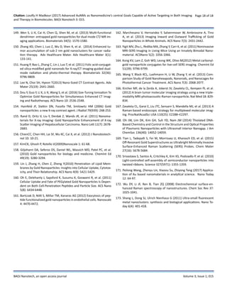
con-centration of AuNPs with different size [152]. In 2016, Dou et
al. explored and confirmed the distinctive size-dependent effects
based on a series of spherical AuNPs for enhanced CT imaging and
radiotherapy (Figure 9) [153].
The result indicated that AuNPs had great size-dependent
enhancement on CT imaging and radiotherapy (RT) in the size
range of 3–50 nm. Interestingly, AuNPs with a size of ∼13 nm could
Fig. 9. Monte Carlo simulation to evaluate size-dependent enhancement. (a) Scheme showing a phantom filled with a AuNP aqueous suspension that
may trigger completelydifferent secondary radiation depending on primary radiation energy irradiated from the X-ray point source for CT detection
or radiotherapy. A simulated “modeling vesicle”containing two particles randomly distributed, representative of the system inhomogeneity based
on particle sizes. (b) X-ray attenuation mainly coming from a photoelectriceffect under the kilovoltage energy radiation for CT imaging, with little
influence by other interactions including Rayleigh scattering, Compton scattering and electronpair effect. (c) Photoelectric effect generation. Inner-
shell electrons receive energy from the incident X-rays, which are subsequently ejected from the atom as a photoelectron,](https://image.slidesharecdn.com/nanotechnology15-180530122541/85/Nanotechnology15-11-320.jpg)
![Citation: Loutfy H Madkour (2017) Advanced AuNMs as Nanomedicine’s central Goals Capable of Active Targeting in Both Imaging
and Therapy in Biomolecules. BAOJ Nanotech 3: 015.
Page 12 of 18
BAOJ Nanotech, an open access journal Volume 3; Issue 1; 015
simultaneously possess superior CT contrastability and significant
radioactive disruption.
AuNPs have been studied as potential contrast agents for X-ray
imaging, because they are nontoxic and have a higher atomic
number and X-ray absorption coefficient compared with typical
iodine-based contrast agents [154]. Rand et al. proposed that the
enhanced sensitivity of the X-ray scatter imaging technique over
that of typical absorption based X-ray imaging could reduce the
amount of AuNPs required for visible contrast [155]. Therefore,
they developed an imaging technique for the early diagnosis of
hepato cellular carcinoma that utilized surface-modified AuNPs in
combination with X-ray imaging. Tissues labeled with these elec-
tron-dense particles showed enhanced X-ray scattering over nor-
mal tissues, distinguishing cells containing AuNPs from cells with-
out Au in X-ray scatter images. This approach could enable in vivo
detection of tumors as small as a few millimeters in size. In 2012,
Chien et al. used AuNPs as high-resolution X-ray imaging con-
trast agents for analysis of tumor-related micro-vasculature [156].
AuNMs hold great interest in imaging field thanks to their char-
acteristics such as monodispersity, stability, minimal toxicity and
excellent contrasting for transmission electron microscopy (TEM)
analysis, which enable tracking with unprecedented resolution on
dynamic of uptake, intracellular sorting and potential secretion
[157,158]. Recent results gathered on the interaction of AuNPs with
different synthetic lipid membranes [159] or cell lines demonstrate
that not only properties such as size, charge and chemical
functionality, but also the arrangement of organic ligands on NP
surface, dictate the internalization route [160]. Moreover, surface
functionalization may influence the fate of AuNP upon cell uptake,
for instance, HUVEC cells treated with AuNPs coated by different
peptides display distinct exocytos is profiles [161]. Recently,
Marchesano et al. observed inward and outward trafficking of
AuNPs at whole animal level using the small invertebrate Hydra
vulgaris [162].
AuNPs are also excellent candidates as contrast agents for SERS
imaging. Yigit et al. synthesized conjugates of AuNMs and
3,3-diethylthiatricarbocyanine iodide (AuNM-DTTC) that were
used as a bimodal contrast agent for in vivo MRI and Raman
spectroscopy [163]. The probe consisted of MRI-active super
paramagnetic iron oxide NPs, stably complexed with AuNM-
DTTC. The Au component served as a substrate for a Raman active
dye molecule to generate a SERS effect. The synthesized probe
produced T2 weighted contrast and was simultaneously used as a
SERS active material both in silico and in vivo. Kong et al. prepared
metal carbonyl based bio tags by combining osmium carbonyl
clusters and AuNPs, as an example of an organo metallic-AuNPs
(OM-AuNPs) conjugate used to cell SERS imaging [164]. It showed
clearly the advantage of transition-metal carbonyl compounds, for
which the CO stretching vibration signal was well separated from
other molecular vibrational modes of the cells in live-cell imaging.
Disadvantage of Using AuNMs
A big disadvantage of using AuNMs as the optical contrast
agents is their high photo thermal conversion efficiency under
resonance excitation, which may perturb or even damage
the biological species being imaged [165]. Thus, to minimize
unwanted heat-ing in bioimaging applications, laser irradiation
time is typically set to be in the range of 0.1–10 S, which either
lowers the SERS signal contrast or is simply not suited for image-
guided tumor resection in intraoperative settings, considering
the longer timescales (1–2 h) associated with such procedures
[166,167]. While resonant nanostructures are certainly desirable
for the ranostic applications, having the ability to tune the photo
thermal effect of SERS probes while preserving their high SERS
activity is critical to avoid unwanted heating during imaging. Oh
et al. designed and synthesized plasmonic AuNPs with ultra small
(typically, ∼1 nm)interior gap using thiolated DNA and obtained
the relationships between these noble metal nanogap structures
and plasmonic signals from these structures [168]. In 2015, Tian et
al. demonstratebioenabled synthesis of a novel class of ultra-bright
SERS probes with built-in and accessible electromagnetic hotspots
formed by densely packed satellite NPs grown on a plasmonic core
[169]. Through the rational choice of the shape of the core, the LSPR
wavelength of Au superstructures was tuned to be either off- or
on-resonant with the NIR excitation without sacrificing their high
SERS activity. Consequently, the photo thermal efficiency of these
ultra-bright SERS tags could be tuned to realize either contrast
agents with minimal heating and perturbation, or multifunctional
the ranostic agents that imaged and photo thermally killed the
targeted cells.
Conclusions
1. The outstanding characteristics of AuNMs make them promising
candidates as the signal reporters, enhancement materials
or others involved with bioassay, food safety, environmental
monitoring and medical science.
2. a series of novel and sophisticated synthesis methods for AuNMs
have been developed, but how to precisely control them on
dispersed size and morphology and to achieve high quality of
AuNMs with vivid color (LSPR) is still a crucial but challengeable
issue for their application in analytical science. The AuNMs
with precisely-controlled vivid color possess abundant optical
information and can encode biological or chemical recognition
units to develop robust analytical methods in food safety, clinic
diagnosis and so on. In addition, we are far from controllable
assembly of AuNMs into the desirable structures of the collective
properties [170].](https://image.slidesharecdn.com/nanotechnology15-180530122541/85/Nanotechnology15-12-320.jpg)
![Citation: Loutfy H Madkour (2017) Advanced AuNMs as Nanomedicine’s central Goals Capable of Active Targeting in Both Imaging
and Therapy in Biomolecules. BAOJ Nanotech 3: 015.
Page 13 of 18
BAOJ Nanotech, an open access journal Volume 3; Issue 1; 015
3. AuNMs have shown excellent performance in enhancement
of SERS signal to improve detection sensitivity. Therefore,
AuNMs in combination with other functional metal or organic
nonmaterial’s, such as Si, Al, MOFs and so on, to develop
multimodal composite nonmaterial become a new trend in high
sensitive SERS detection of various target analytes [171,172], as
well as in high resolution bioimaging in the future.
4- AuNMs offer a rapid, efficient, cost-effective and robust sensing
platform for detection of different chemicals and bio-markers
due to their unique chemical, physical and optical properties. It
is noted that the sensitive, stable and multiplex assay of target
analytes is highly desirable. To meet these requirements, new
multifunctional AuNMs must be developed and used as LFICA
label, colorimetric sensing readout and signal amplification of
EC sensor, etc.
5. AuNCs as excellent fluorescent probe materials have shown
great potential in analytical science [173]. However, extensive
efforts must be made for developing novel synthesis methods
to achieve high quality AuNCs with remarkable QY and
extraordinary stability. In addition, the coordination nature
of the AuNCs-protection group complexes implies that the
stability of metal NCs is affected by the presence of other strong
competing ligands in aqueous solution. The intracellular stability
of AuNCs hence becomes a serious issue. With development of
technologies of synthesis and surface modification, AuNCs will
be widely employed as alternatives to conventional fluorophores
in analytical science including biosensors, bioimaging and soon.
6. We notice that though many new analytical methods based on
AuNMs have been developed in the laboratories, such materials
are not used as much in an industrial setting. There must be
more scope for these platforms to improve, for example, cost of
preparing the AuNMs based sensors, reproducibility of different
batches for sensor production, and stability of long-term storage
of the AuNMs based sensors, and so on.
7. The mechanism of DNA-guided AuNMs synthesis not only
provide deep understanding of the interactions between the
DNA and nanomaterials but also allow better control of the
shapes and surface properties of many nanomaterials.
8.AuNMswithexcellentfeatureshavepromptedgreatdevelopment
of analytical sciences and have stood at a critical juncture, with a
vast amount of researches that form a solid foundation for future
work. Great deals of efforts need to be paid to push on to take
AuNMs-based analytical technologies from lab to market.
References
1. Madkour LH (2017) Vision for life sciences: interfaces between nano-
electronic and biological systems. Glob Drugs Therap 2(4): 1-4. doi:
10. 15761/GDT. 1000126
2. Sardar R, Funston AM, Mulvaney P, Murray RW (2009) Gold nanopar-
ticles: past, present, and future. Langmuir 25(24): 13840-13851.
3. Wang P, Sun X, Su X, Wang T (2016) Advancements of molecularly im-
printed polymers in the food safety field. Analysts 141: 3540-3553.
4. Zhang, L Wang E (2014) Nano Today 9: 132-157.
5. Ballou S, Goodpaster J, MacCrehan W, Reeder D (2003) Forensic anal-
ysis. Anal. Bioanal Chem 376(8): 1149-1150.
6. Burnworth M, Rowan SJ, Weder C (2007) Fluorescent sensors for the
detection of chemical warfare agents. Chem Eur J 13(28): 7828-7836.
7. Zhang Z, Liu J, Feng T, Yao Y, Gao L, et al. (2013) Time-Resolved Fluo-
roimmunoassay as an Advantageous Analytical Method for Assess-
ing the Total Concentration and Environmental Risk of Fluoroquino-
lones in Surface Waters Environ. Sci Technol 47(1): 454-462.
8. Anker JN, Hall WP, Lyandres O, Shah NC, Zhao J, et al. (2008) Biosens-
ing with plasmonic nanosensors. Nat Mater 7: 442-453.
9. Debouttie`re PJ, Roux S, Vocanson F, BilloteyC, Beuf O, et al. (2006)
Design of Gold Nanoparticles for Magnetic Resonance Imaging Tille-
ment AdvFunct Mater 16(18): 2330-2339.
10. Qian X, Li J, Nie S (2009) Stimuli-Responsive SERS Nanoparticles:
Conformational Control of Plasmonic Coupling and Surface Raman
Enhancement. J Am ChemSoc 131(22): 7540-7541.
11. Thomas KG, Kamat PV (2000) Making Gold Nanoparticles Glow: En-
hanced Emission from a Surface-Bound Fluoroprobe. J Am Chem Soc
122(11): 2655-2656.
12. Ipe BI, Yoosa f K, Thomas KG (2006) Functionalized Gold Nanopar-
ticles as Phosphorescent Nanomaterials and Sensors. J Am Chem
Soc 128(6): 1907-1913.
13. Kisailus D, Najarian M, Weaver JC, Morse DE (2005) Functionalized
Gold Nanoparticles Mimic Catalytic Activity of a Polysiloxane-Syn-
thesizing Enzyme Adv Mater 17(10): 1234-1239.
14. Qian X, Peng XH, Ansari DO, Yin Goen Q, Chen GZ, et al. (2008) In
vivo tumor targeting and spectroscopic detection with surface-en-
hanced Raman nanoparticle tags. Nat Biotechnol 26(1): 83-90.
15. Haghighi B, Bozorgzadeh S (2011) Enhanced Electrochemilumines-
cence From Luminol at Multi-Walled Carbon Nanotubes Decorated
With Palladium Nanoparticles: A Novel Route for the Fabrication of
an Oxygen Sensor and a Glucose Biosensor. Anal ChimActa 697(1-2):
90-97.
16. Tang J, Tang DP, Su BL, Huang JX, Qiu B, et al. (2011) Enzyme-free elec-
trochemical immunoassay with catalytic reduction of p-nitrophenol
and recycling of p-aminophenol using gold nanoparticles-coated
carbon nanotubes as nanocatalysts. BiosensBioelectron26(7): 3219-
3226.
17. Zhu YP, Chandra P, Song KM, Ban C, Shim YB (2012) Label-free detec-
tion of kanamycin based on the aptamer-functionalized conducting
polymer/gold nanocomposite. BiosensBioelectron 36(1): 29-34.
18. SunX, Li F, Shen G, Huang J, Wang X (2014) Aptasensor based on the
synergistic contributions of chitosan–gold nanoparticles, graphene–
gold nanoparticles and multi-walled carbon nanotubes-cobalt
phthalocyaninenanocomposites for kanamycin detection. Analyst
139: 299-308.](https://image.slidesharecdn.com/nanotechnology15-180530122541/85/Nanotechnology15-13-320.jpg)

The document discusses advancements in the application of gold nanoparticles (AuNPs) in nanomedicine and analytical sciences, highlighting their unique properties such as electrical, optical, and catalytic capabilities. It covers the importance of stability, targeting for imaging and therapy, and various synthesis methods to improve these nanoparticles' performance in biosensing for food safety, medical diagnostics, and environmental monitoring. Additionally, future research directions include enhancing the sensitivity of electrochemical sensors and improving the control of nanoparticle morphology and luminescence properties.


![ONFH[AVN HIP] -TRIPLE REGIME -A NOVAL SURGICAL CONCEPT .pptx](https://cdn.slidesharecdn.com/ss_thumbnails/onfhavnhip2026koaconcalicutdrgokuldevdrmashraf-260210064517-213ec005-thumbnail.jpg?width=640&height=640&fit=bounds)


