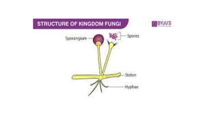
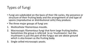
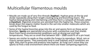
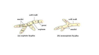
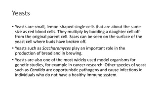
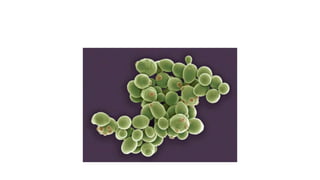
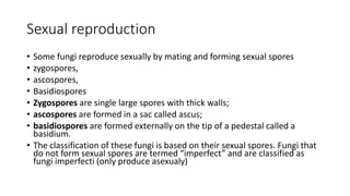
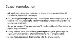
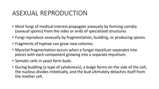
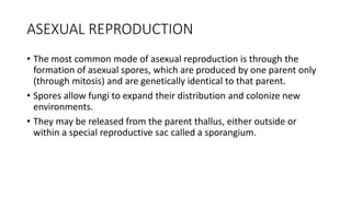
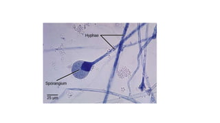
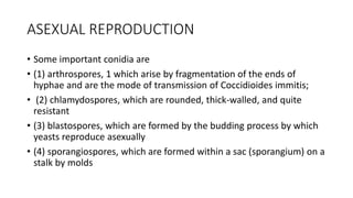
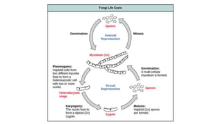
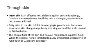
SlideShare a Scribd company logo
MYCOLOGY
• Fungi are eukaryotic organisms; i.e., their cells contain membrane-
bound organelles and clearly defined nuclei. Historically, fungi were
included in the plant kingdom; however, because fungi
lack chlorophyll and are distinguished by unique structural and
physiological features (i.e., components of the cell wall and cell
membrane), they have been separated from plants.
• Because fungi (yeasts and molds) are eukaryotic organisms, whereas
bacteria are prokaryotic, they differ in several fundamental respects
Structure
• fungal cell structures are important medically:
• (1) The fungal cell wall consists primarily of chitin (not peptidoglycan as in
bacteria); thus fungi are insensitive to certain antibiotics, such as penicillins
and cephalosporins, that inhibit peptidoglycan synthesis.
• (2) Chitin is a polysaccharide composed of long chains of N-
acetylglucosamine. The fungal cell wall contains other polysaccharides as
well, the most important of which is β-glucan, a long polymer of d-glucose.
The medical importance of β-glucan is that it is the site of action of the
antifungal drug caspofungin.
• (3) The fungal cell membrane contains ergosterol, in contrast to the human
cell membrane, which contains cholesterol.
Types of fungi
• Fungi are subdivided on the basis of their life cycles, the presence or
structure of their fruiting body and the arrangement of and type of
spores (reproductive or distributional cells) they produce.
• The three major groups of fungi are:
1. Multicellular filamentous moulds.
2. Macroscopic filamentous fungi that form large fruiting bodies.
Sometimes the group is referred to as ‘mushrooms’, but the
mushroom is just the part of the fungus we see above ground
which is also known as the fruiting body.
3. Single celled microscopic yeasts.
Multicellular filamentous moulds
• Moulds are made up of very fine threads (hyphae). Hyphae grow at the tip and
divide repeatedly along their length creating long and branching chains. The
hyphae keep growing and intertwining until they form a network of threads called
a mycelium. Digestive enzymes are secreted from the hyphal tip. These enzymes
break down the organic matter found in the soil into smaller molecules which are
used by the fungus as food.
• Some of the hyphal branches grow into the air and spores form on these aerial
branches. Spores are specialised structures with a protective coat that shields
them from harsh environmental conditions such as drying out and high
temperatures. They are so small that between 500 – 1000 could fit on a pin head.
• Spores are similar to seeds as they enable the fungus to reproduce. Wind, rain or
insects spread spores. They eventually land in new habitats and if conditions are
right, they start to grow and produce new hyphae. As fungi can’t move they use
spores to find a new environment where there are fewer competing organisms.
• Molds grow as long filaments (hyphae) and form a mat (mycelium).
Some hyphae form transverse walls (septate hyphae), whereas others
do not (nonseptate hyphae).
• Nonseptate hyphae are multinucleated (coenocytic). The growth of
hyphae occurs by extension of the tip of the hypha, not by cell
division all along the filament.
Macroscopic filamentous fungi
• Macroscopic filamentous fungi also grow by producing a mycelium
below ground. They differ from moulds because they produce visible
fruiting bodies (commonly known as mushrooms or toadstools) that
hold the spores. The fruiting body is made up of tightly packed
hyphae which divide to produce the different parts of the fungal
structure, for example the cap and the stem. Gills underneath the cap
are covered with spores and a 10 cm diameter cap can produce up to
100 million spores per hour.
Yeasts
• Yeasts are small, lemon-shaped single cells that are about the same
size as red blood cells. They multiply by budding a daughter cell off
from the original parent cell. Scars can be seen on the surface of the
yeast cell where buds have broken off.
• Yeasts such as Saccharomyces play an important role in the
production of bread and in brewing.
• Yeasts are also one of the most widely used model organisms for
genetic studies, for example in cancer research. Other species of yeast
such as Candida are opportunistic pathogens and cause infections in
individuals who do not have a healthy immune system.
Yeast cell structure
• Yeast cells possess ultrastructural features typical of other eukaryotic
cells, with the presence of membrane-bound organelles
• Cell wall compose of glycosylated glycoproteins (mannoproteins), two
types of β-glucans, and chitin.
• The surface plasma membrane of yeast is a lipid bilayer, The lipid
components comprise mainly phospholipids and sterol.
• The periplasmic space is a region external to the plasma membrane
that contains proteins unable to permeate the cell wall, such
as invertase and phosphatase.
Yeast cell structure
• The yeast cytoplasm is an aqueous, slightly acidic colloidal fluid that
contains proteins, glycogen, other soluble macromolecules and larger
macromolecular entities such as ribosomes, proteasomes, and lipid
particles. Many essential functions for cellular integrity are localized
in the cytoplasm.
• The cytoskeleton of yeast cells comprises microtubules
and microfilaments.
• In the center of the cell or slightly excentrically, the nuclear structure
is located, which is surrounded by a double membrane that separates
the nucleoplasm from the cytoplasm.
Yeast cell structure
• The outer nuclear membrane is contiguous with the membrane of
the endoplasmic reticulum.
• The nuclear chromosomes are in the nucleoplasm and packed into a
chromatin structure
• Yeast cell contains different organelles surrounded by individual
membranes, mostly the endoplasmic reticulum, the Golgi apparatus,
and transport vesicles that are necessary for the manufacturing and
trafficking of proteins, vacuoles, mitochondria, and microbodies.
• Several medically important fungi are thermally dimorphic (i.e., they form different
structures at different temperatures).
• They exist as molds in the environment at ambient temperature and as yeasts (or other
structures) in human tissues at body temperature.
• A true or primary fungal pathogen is a species that can invade and grow in a healthy,
noncompromised animal host. This behavior is contrary to the metabolism and
adaptation of fungi, most of which are inhibited by the relatively high temperature and
low oxygen tensions of a warm-blooded animal’s body.
• But a small number of fungi have the morphological and physiological adaptations
required to survive and grow in this habitat. By far, their most striking adaptation is a
switch from hyphal cells typical of the mycelial or mold phase to yeast cells typical of the
parasitic phase.
• This biphasic characteristic of the life cycle is termed thermal dimorphism because it is
initiated by changing temperature. In general, these organisms grow as molds at 30°C
and as yeasts at 37°C.
Reproduction
• Fungi reproduce sexually and/or asexually. Perfect fungi reproduce
both sexually and asexually, while imperfect fungi reproduce only
asexually (by mitosis).
• In both sexual and asexual reproduction, fungi produce spores that
disperse from the parent organism by either floating on the wind or
hitching a ride on an animal.
• Fungal spores are smaller and lighter than plant seeds. The giant
puffball mushroom bursts open and releases trillions of spores. The
huge number of spores released increases the likelihood of landing in
an environment that will support growth.
The release of fungal spores: The (a) giant puff ball mushroom releases (b) a cloud of
spores when it reaches maturity.
Sexual reproduction
• Some fungi reproduce sexually by mating and forming sexual spores
• zygospores,
• ascospores,
• Basidiospores
• Zygospores are single large spores with thick walls;
• ascospores are formed in a sac called ascus;
• basidiospores are formed externally on the tip of a pedestal called a
basidium.
• The classification of these fungi is based on their sexual spores. Fungi that
do not form sexual spores are termed “imperfect” and are classified as
fungi imperfecti (only produce asexualy)
Sexual reproduction
• Sexual reproduction introduces genetic variation into a population of
fungi. In fungi, sexual reproduction often occurs in response to
adverse environmental conditions.
• Two mating types are produced. When both mating types are present
in the same mycelium, it is called homothallic, or self-fertile.
• Heterothallic mycelia require two different, but compatible, mycelia
to reproduce sexually.
Sexual reproduction
• Although there are many variations in fungal sexual reproduction, all
include the following three stages.
• First, during plasmogamy (literally, “marriage or union of cytoplasm”), two
haploid cells fuse, leading to a dikaryotic stage where two haploid nuclei
coexist in a single cell.
• During karyogamy (“nuclear marriage”), the haploid nuclei fuse to form a
diploid zygote nucleus.
• Finally, meiosis takes place in the gametangia (singular, gametangium)
organs, in which gametes of different mating types are generated.
• At this stage, spores are disseminated into the environment.
ASEXUAL REPRODUCTION
• Most fungi of medical interest propagate asexually by forming conidia
(asexual spores) from the sides or ends of specialized structures
• Fungi reproduce asexually by fragmentation, budding, or producing spores.
• Fragments of hyphae can grow new colonies.
• Mycelial fragmentation occurs when a fungal mycelium separates into
pieces with each component growing into a separate mycelium.
• Somatic cells in yeast form buds.
• During budding (a type of cytokinesis), a bulge forms on the side of the cell,
the nucleus divides mitotically, and the bud ultimately detaches itself from
the mother cell.
ASEXUAL REPRODUCTION
• The most common mode of asexual reproduction is through the
formation of asexual spores, which are produced by one parent only
(through mitosis) and are genetically identical to that parent.
• Spores allow fungi to expand their distribution and colonize new
environments.
• They may be released from the parent thallus, either outside or
within a special reproductive sac called a sporangium.
ASEXUAL REPRODUCTION
• Some important conidia are
• (1) arthrospores, 1 which arise by fragmentation of the ends of
hyphae and are the mode of transmission of Coccidioides immitis;
• (2) chlamydospores, which are rounded, thick-walled, and quite
resistant
• (3) blastospores, which are formed by the budding process by which
yeasts reproduce asexually
• (4) sporangiospores, which are formed within a sac (sporangium) on a
stalk by molds
Pathogenesis
• The response to infection with many fungi is the formation of granulomas.
• Granulomas are produced in the major systemic fungal diseases (e.g.,
coccidioidomycosis, histoplasmosis, and blastomycosis, as well as several
others).
• The cell-mediated immune response is involved in granuloma formation.
• Acute suppuration, characterized by the presence of neutrophils in the
exudate, also occurs in certain fungal diseases such as aspergillosis and
sporotrichosis.
• Fungi do not have endotoxin in their cell walls and do not produce
bacterial-type exotoxins.
Through skin
• Intact skin is an effective host defense against certain fungi (e.g.,
Candida, dermatophytes), but if the skin is damaged, organisms can
become established.
• Fatty acids in the skin inhibit dermatophyte growth, and hormone-
associated skin changes at puberty limit ringworm of the scalp caused
by Trichophyton.
• The normal flora of the skin and mucous membranes suppress fungi.
When the normal flora is inhibited (e.g., by antibiotics), overgrowth of
fungi such as C. albicans can occur
Through respiratory tract
• In the respiratory tract, the important host defenses are the mucous
membranes of the nasopharynx, which trap inhaled fungal spores,
and alveolar macrophages.
• Circulating IgG and IgM are produced in response to fungal infection,
but their role in protection from disease is uncertain.
• The cell-mediated immune response is protective; its suppression can
lead to reactivation
Clinical significance
• In addition to mycotic infections, there are two other kinds of fungal
disease:
• (1) mycotoxicoses, caused by ingested toxins,
• (2) allergies to fungal spores
• Ingestion of Amanita mushrooms causes liver necrosis due to the
presence of two fungal toxins, amanitin and phalloidin.
• Amanitin inhibits the RNA polymerase that synthesizes cellular mRNA.
• Ingestion of peanuts and grains contaminated with A. flavus causes
liver cancer due to the presence of aflatoxin.
• Inhalation of the spores of Aspergillus fumigatus can cause allergic
bronchopulmonary aspergillosis. This is an IgE mediated immediate
hypersensitivity response
Laboratory diagnosis
• There are four approaches to the laboratory diagnosis of fungal
diseases:
• (1) direct microscopic examination
• (2) culture of the organism
• (3) DNA probe tests
• (4) serologic tests
direct microscopic examination
• Direct microscopic examination of clinical specimens such as sputum, lung biopsy
material, and skin scrapings depends on finding characteristic asexual spores,
hyphae, or yeasts in the light microscope.
• The specimen is either treated with 10% potassium hydroxide (KOH) to dissolve
tissue material, leaving the alkali-resistant fungi intact, or stained with special
fungal stains.
• Some examples of diagnostically important findings made by direct examination
are
• (1) the spherules of C. immitis
• (2) the wide capsule of Cryptococcus neoformans seen in India ink preparations
of spinal fluid.
• Calcofluor white is a fluorescent dye that binds to fungal cell walls and is useful in
the identification of fungi in tissue specimens.
• Methenamine silver stain is also useful in the microscopic diagnosis of fungi in
tissue.
culture of the organism
• Fungi are frequently cultured on Sabouraud’s agar, which facilitates
the appearance of the slow-growing fungi by inhibiting the growth of
bacteria in the specimen.
• Inhibition of bacterial growth is due to the low pH of the medium and
to the penicillin, streptomycin, and cycloheximide that are frequently
added.
• The appearance of the mycelium and the nature of the asexual spores
are frequently sufficient to identify the organism
DNA probe tests
• Tests involving DNA probes can identify colonies growing in culture at
an earlier stage of growth than can tests based on visual detection of
the colonies.
• As a result, the diagnosis can be made more rapidly.
• At present, DNA probe tests are available for Coccidioides,
Histoplasma, Blastomyces, and Cryptococcus.
serologic tests
• Tests for the presence of antibodies in the patient’s serum or spinal
fluid are useful in diagnosing systemic mycoses but less so in
diagnosing other fungal infections.
• As is the case for bacterial and viral serologic testing, a significant rise
in the antibody titer must be observed to confirm a diagnosis.
• The complement fixation test is most frequently used in suspected
cases of coccidioidomycosis, histoplasmosis, and blastomycosis. In
cryptococcal meningitis, the presence of the polysaccharide capsular
antigens of C. neoformans in the spinal fluid can be detected by the
latex agglutination test.
delayed hypersensitivity skin test
• Infection with the systemic fungi, such as Histoplasma and
Coccidioides, can be detected by using skin tests.
• An antigen extracted from the organism injected intradermally elicits
a delayed hypersensitivity reaction, manifested as induration
(thickening) of the skin.
• Note that a positive skin test only indicates that infection has
occurred, but it is not known whether that infection occurred in the
past or at the present time. Therefore, a positive skin test does not
indicate that the disease the patient has now is caused by that
organism.

More Related Content

Similar to MYCOLOGY.pptx

Zygomycotina
ZygomycotinaZygomycotina
Zygomycotina
vaishalidandge3
 
Ppt of fungi
Ppt of fungiPpt of fungi
Ppt of fungi
vaishalidandge3
 
Morphology, Classification, Cultivation and Reproduction of Fungi
Morphology, Classification, Cultivation and Reproduction of FungiMorphology, Classification, Cultivation and Reproduction of Fungi
Morphology, Classification, Cultivation and Reproduction of Fungi
Krutika Pardeshi
 
Kingdom of fungi by Bnp
Kingdom of fungi by BnpKingdom of fungi by Bnp
Kingdom of fungi by Bnp
College of pharmaceutical sciences
 
Introduction to fungus of all kinds.pptx
Introduction to fungus of all kinds.pptxIntroduction to fungus of all kinds.pptx
Introduction to fungus of all kinds.pptx
chandanabinu
 
Typical structure of Fungi cell.pptx
Typical structure of Fungi cell.pptxTypical structure of Fungi cell.pptx
Typical structure of Fungi cell.pptx
GalaanaTuluOromo
 
Fungi.pdf
Fungi.pdfFungi.pdf
Fungi.pdf
Anup Bajracharya
 
Morphologyoffungi 170226153026
Morphologyoffungi 170226153026Morphologyoffungi 170226153026
Morphologyoffungi 170226153026
ali albadry
 
Morphology of fungi
Morphology of fungiMorphology of fungi
Topic 3 fungi. yeasts
Topic 3 fungi. yeastsTopic 3 fungi. yeasts
Topic 3 fungi. yeasts
Viktor Stabnikov
 
Introduction mycology
Introduction mycologyIntroduction mycology
Introduction mycology
Sk. Mizanur Rahman
 
Microbes, Man and Environment (fungal replication) .pptx
Microbes, Man and Environment (fungal replication) .pptxMicrobes, Man and Environment (fungal replication) .pptx
Microbes, Man and Environment (fungal replication) .pptx
MidhatSarfraz
 
Bacterial growth
Bacterial growthBacterial growth
Bacterial growth
Ram Rosete Rmt
 
MYCOLOGY.pdf
MYCOLOGY.pdfMYCOLOGY.pdf
MYCOLOGY.pdf
AKBoudh
 
STRUCTURE OF FUNGI.pptx
STRUCTURE OF FUNGI.pptxSTRUCTURE OF FUNGI.pptx
STRUCTURE OF FUNGI.pptx
NithyaNandapal
 
Different modes of reproduction of fungi and reproductive structure and nutri...
Different modes of reproduction of fungi and reproductive structure and nutri...Different modes of reproduction of fungi and reproductive structure and nutri...
Different modes of reproduction of fungi and reproductive structure and nutri...
hassabbinahmed
 
Fungi
FungiFungi
Bacteriology3.pdf
Bacteriology3.pdfBacteriology3.pdf
Bacteriology3.pdf
FkFo
 
Class xi ch 2
Class xi ch   2Class xi ch   2
Class xi ch 2
RashmiSinha61
 
Mycology Introduction
Mycology IntroductionMycology Introduction
Mycology Introduction
Aeraaf Khuraishi
 

Similar to MYCOLOGY.pptx (20)

Zygomycotina
ZygomycotinaZygomycotina
Zygomycotina
 
Ppt of fungi
Ppt of fungiPpt of fungi
Ppt of fungi
 
Morphology, Classification, Cultivation and Reproduction of Fungi
Morphology, Classification, Cultivation and Reproduction of FungiMorphology, Classification, Cultivation and Reproduction of Fungi
Morphology, Classification, Cultivation and Reproduction of Fungi
 
Kingdom of fungi by Bnp
Kingdom of fungi by BnpKingdom of fungi by Bnp
Kingdom of fungi by Bnp
 
Introduction to fungus of all kinds.pptx
Introduction to fungus of all kinds.pptxIntroduction to fungus of all kinds.pptx
Introduction to fungus of all kinds.pptx
 
Typical structure of Fungi cell.pptx
Typical structure of Fungi cell.pptxTypical structure of Fungi cell.pptx
Typical structure of Fungi cell.pptx
 
Fungi.pdf
Fungi.pdfFungi.pdf
Fungi.pdf
 
Morphologyoffungi 170226153026
Morphologyoffungi 170226153026Morphologyoffungi 170226153026
Morphologyoffungi 170226153026
 
Morphology of fungi
Morphology of fungiMorphology of fungi
Morphology of fungi
 
Topic 3 fungi. yeasts
Topic 3 fungi. yeastsTopic 3 fungi. yeasts
Topic 3 fungi. yeasts
 
Introduction mycology
Introduction mycologyIntroduction mycology
Introduction mycology
 
Microbes, Man and Environment (fungal replication) .pptx
Microbes, Man and Environment (fungal replication) .pptxMicrobes, Man and Environment (fungal replication) .pptx
Microbes, Man and Environment (fungal replication) .pptx
 
Bacterial growth
Bacterial growthBacterial growth
Bacterial growth
 
MYCOLOGY.pdf
MYCOLOGY.pdfMYCOLOGY.pdf
MYCOLOGY.pdf
 
STRUCTURE OF FUNGI.pptx
STRUCTURE OF FUNGI.pptxSTRUCTURE OF FUNGI.pptx
STRUCTURE OF FUNGI.pptx
 
Different modes of reproduction of fungi and reproductive structure and nutri...
Different modes of reproduction of fungi and reproductive structure and nutri...Different modes of reproduction of fungi and reproductive structure and nutri...
Different modes of reproduction of fungi and reproductive structure and nutri...
 
Fungi
FungiFungi
Fungi
 
Bacteriology3.pdf
Bacteriology3.pdfBacteriology3.pdf
Bacteriology3.pdf
 
Class xi ch 2
Class xi ch   2Class xi ch   2
Class xi ch 2
 
Mycology Introduction
Mycology IntroductionMycology Introduction
Mycology Introduction
 

More from AhmadShafiq43

HIV & AIDs (Micro Group 1).pptx
HIV & AIDs (Micro Group 1).pptxHIV & AIDs (Micro Group 1).pptx
HIV & AIDs (Micro Group 1).pptx
AhmadShafiq43
 
FIBERS .pptx
FIBERS .pptxFIBERS .pptx
FIBERS .pptx
AhmadShafiq43
 
magmas and gel Pharmaceutcs.pptx
magmas and gel Pharmaceutcs.pptxmagmas and gel Pharmaceutcs.pptx
magmas and gel Pharmaceutcs.pptx
AhmadShafiq43
 
PESTICIDES Pharmacognosy.pptx
PESTICIDES Pharmacognosy.pptxPESTICIDES Pharmacognosy.pptx
PESTICIDES Pharmacognosy.pptx
AhmadShafiq43
 
Papaveraceae family (poppy capsule + sanguinaria) .pptx
Papaveraceae family (poppy capsule + sanguinaria) .pptxPapaveraceae family (poppy capsule + sanguinaria) .pptx
Papaveraceae family (poppy capsule + sanguinaria) .pptx
AhmadShafiq43
 
Diagnostic tools for disease evaluation.pdf
Diagnostic tools for disease evaluation.pdfDiagnostic tools for disease evaluation.pdf
Diagnostic tools for disease evaluation.pdf
AhmadShafiq43
 
Ristedaro ke haqooq.pptx
Ristedaro ke haqooq.pptxRistedaro ke haqooq.pptx
Ristedaro ke haqooq.pptx
AhmadShafiq43
 
CNS-stimulants.pptx
CNS-stimulants.pptxCNS-stimulants.pptx
CNS-stimulants.pptx
AhmadShafiq43
 
Digestive system (1) (1)_042113.ppt
Digestive system (1) (1)_042113.pptDigestive system (1) (1)_042113.ppt
Digestive system (1) (1)_042113.ppt
AhmadShafiq43
 
aloe vera 417.pdf
aloe vera 417.pdfaloe vera 417.pdf
aloe vera 417.pdf
AhmadShafiq43
 

More from AhmadShafiq43 (10)

HIV & AIDs (Micro Group 1).pptx
HIV & AIDs (Micro Group 1).pptxHIV & AIDs (Micro Group 1).pptx
HIV & AIDs (Micro Group 1).pptx
 
FIBERS .pptx
FIBERS .pptxFIBERS .pptx
FIBERS .pptx
 
magmas and gel Pharmaceutcs.pptx
magmas and gel Pharmaceutcs.pptxmagmas and gel Pharmaceutcs.pptx
magmas and gel Pharmaceutcs.pptx
 
PESTICIDES Pharmacognosy.pptx
PESTICIDES Pharmacognosy.pptxPESTICIDES Pharmacognosy.pptx
PESTICIDES Pharmacognosy.pptx
 
Papaveraceae family (poppy capsule + sanguinaria) .pptx
Papaveraceae family (poppy capsule + sanguinaria) .pptxPapaveraceae family (poppy capsule + sanguinaria) .pptx
Papaveraceae family (poppy capsule + sanguinaria) .pptx
 
Diagnostic tools for disease evaluation.pdf
Diagnostic tools for disease evaluation.pdfDiagnostic tools for disease evaluation.pdf
Diagnostic tools for disease evaluation.pdf
 
Ristedaro ke haqooq.pptx
Ristedaro ke haqooq.pptxRistedaro ke haqooq.pptx
Ristedaro ke haqooq.pptx
 
CNS-stimulants.pptx
CNS-stimulants.pptxCNS-stimulants.pptx
CNS-stimulants.pptx
 
Digestive system (1) (1)_042113.ppt
Digestive system (1) (1)_042113.pptDigestive system (1) (1)_042113.ppt
Digestive system (1) (1)_042113.ppt
 
aloe vera 417.pdf
aloe vera 417.pdfaloe vera 417.pdf
aloe vera 417.pdf
 

Recently uploaded

waterlessdyeingtechnolgyusing carbon dioxide chemicalspdf
waterlessdyeingtechnolgyusing carbon dioxide chemicalspdfwaterlessdyeingtechnolgyusing carbon dioxide chemicalspdf
waterlessdyeingtechnolgyusing carbon dioxide chemicalspdf
LengamoLAppostilic
 
bordetella pertussis.................................ppt
bordetella pertussis.................................pptbordetella pertussis.................................ppt
bordetella pertussis.................................ppt
kejapriya1
 
Eukaryotic Transcription Presentation.pptx
Eukaryotic Transcription Presentation.pptxEukaryotic Transcription Presentation.pptx
Eukaryotic Transcription Presentation.pptx
RitabrataSarkar3
 
EWOCS-I: The catalog of X-ray sources in Westerlund 1 from the Extended Weste...
EWOCS-I: The catalog of X-ray sources in Westerlund 1 from the Extended Weste...EWOCS-I: The catalog of X-ray sources in Westerlund 1 from the Extended Weste...
EWOCS-I: The catalog of X-ray sources in Westerlund 1 from the Extended Weste...
Sérgio Sacani
 
Authoring a personal GPT for your research and practice: How we created the Q...
Authoring a personal GPT for your research and practice: How we created the Q...Authoring a personal GPT for your research and practice: How we created the Q...
Authoring a personal GPT for your research and practice: How we created the Q...
Leonel Morgado
 
Equivariant neural networks and representation theory
Equivariant neural networks and representation theoryEquivariant neural networks and representation theory
Equivariant neural networks and representation theory
Daniel Tubbenhauer
 
The binding of cosmological structures by massless topological defects
The binding of cosmological structures by massless topological defectsThe binding of cosmological structures by massless topological defects
The binding of cosmological structures by massless topological defects
Sérgio Sacani
 
Basics of crystallography, crystal systems, classes and different forms
Basics of crystallography, crystal systems, classes and different formsBasics of crystallography, crystal systems, classes and different forms
Basics of crystallography, crystal systems, classes and different forms
MaheshaNanjegowda
 
在线办理(salfor毕业证书)索尔福德大学毕业证毕业完成信一模一样
在线办理(salfor毕业证书)索尔福德大学毕业证毕业完成信一模一样在线办理(salfor毕业证书)索尔福德大学毕业证毕业完成信一模一样
在线办理(salfor毕业证书)索尔福德大学毕业证毕业完成信一模一样
vluwdy49
 
Unlocking the mysteries of reproduction: Exploring fecundity and gonadosomati...
Unlocking the mysteries of reproduction: Exploring fecundity and gonadosomati...Unlocking the mysteries of reproduction: Exploring fecundity and gonadosomati...
Unlocking the mysteries of reproduction: Exploring fecundity and gonadosomati...
AbdullaAlAsif1
 
Applied Science: Thermodynamics, Laws & Methodology.pdf
Applied Science: Thermodynamics, Laws & Methodology.pdfApplied Science: Thermodynamics, Laws & Methodology.pdf
Applied Science: Thermodynamics, Laws & Methodology.pdf
University of Hertfordshire
 
Describing and Interpreting an Immersive Learning Case with the Immersion Cub...
Describing and Interpreting an Immersive Learning Case with the Immersion Cub...Describing and Interpreting an Immersive Learning Case with the Immersion Cub...
Describing and Interpreting an Immersive Learning Case with the Immersion Cub...
Leonel Morgado
 
The debris of the ‘last major merger’ is dynamically young
The debris of the ‘last major merger’ is dynamically youngThe debris of the ‘last major merger’ is dynamically young
The debris of the ‘last major merger’ is dynamically young
Sérgio Sacani
 
molar-distalization in orthodontics-seminar.pptx
molar-distalization in orthodontics-seminar.pptxmolar-distalization in orthodontics-seminar.pptx
molar-distalization in orthodontics-seminar.pptx
Anagha Prasad
 
The use of Nauplii and metanauplii artemia in aquaculture (brine shrimp).pptx
The use of Nauplii and metanauplii artemia in aquaculture (brine shrimp).pptxThe use of Nauplii and metanauplii artemia in aquaculture (brine shrimp).pptx
The use of Nauplii and metanauplii artemia in aquaculture (brine shrimp).pptx
MAGOTI ERNEST
 
Micronuclei test.M.sc.zoology.fisheries.
Micronuclei test.M.sc.zoology.fisheries.Micronuclei test.M.sc.zoology.fisheries.
Micronuclei test.M.sc.zoology.fisheries.
Aditi Bajpai
 
Topic: SICKLE CELL DISEASE IN CHILDREN-3.pdf
Topic: SICKLE CELL DISEASE IN CHILDREN-3.pdfTopic: SICKLE CELL DISEASE IN CHILDREN-3.pdf
Topic: SICKLE CELL DISEASE IN CHILDREN-3.pdf
TinyAnderson
 
Oedema_types_causes_pathophysiology.pptx
Oedema_types_causes_pathophysiology.pptxOedema_types_causes_pathophysiology.pptx
Oedema_types_causes_pathophysiology.pptx
muralinath2
 
mô tả các thí nghiệm về đánh giá tác động dòng khí hóa sau đốt
mô tả các thí nghiệm về đánh giá tác động dòng khí hóa sau đốtmô tả các thí nghiệm về đánh giá tác động dòng khí hóa sau đốt
mô tả các thí nghiệm về đánh giá tác động dòng khí hóa sau đốt
HongcNguyn6
 
20240520 Planning a Circuit Simulator in JavaScript.pptx
20240520 Planning a Circuit Simulator in JavaScript.pptx20240520 Planning a Circuit Simulator in JavaScript.pptx
20240520 Planning a Circuit Simulator in JavaScript.pptx
Sharon Liu
 

Recently uploaded (20)

waterlessdyeingtechnolgyusing carbon dioxide chemicalspdf
waterlessdyeingtechnolgyusing carbon dioxide chemicalspdfwaterlessdyeingtechnolgyusing carbon dioxide chemicalspdf
waterlessdyeingtechnolgyusing carbon dioxide chemicalspdf
 
bordetella pertussis.................................ppt
bordetella pertussis.................................pptbordetella pertussis.................................ppt
bordetella pertussis.................................ppt
 
Eukaryotic Transcription Presentation.pptx
Eukaryotic Transcription Presentation.pptxEukaryotic Transcription Presentation.pptx
Eukaryotic Transcription Presentation.pptx
 
EWOCS-I: The catalog of X-ray sources in Westerlund 1 from the Extended Weste...
EWOCS-I: The catalog of X-ray sources in Westerlund 1 from the Extended Weste...EWOCS-I: The catalog of X-ray sources in Westerlund 1 from the Extended Weste...
EWOCS-I: The catalog of X-ray sources in Westerlund 1 from the Extended Weste...
 
Authoring a personal GPT for your research and practice: How we created the Q...
Authoring a personal GPT for your research and practice: How we created the Q...Authoring a personal GPT for your research and practice: How we created the Q...
Authoring a personal GPT for your research and practice: How we created the Q...
 
Equivariant neural networks and representation theory
Equivariant neural networks and representation theoryEquivariant neural networks and representation theory
Equivariant neural networks and representation theory
 
The binding of cosmological structures by massless topological defects
The binding of cosmological structures by massless topological defectsThe binding of cosmological structures by massless topological defects
The binding of cosmological structures by massless topological defects
 
Basics of crystallography, crystal systems, classes and different forms
Basics of crystallography, crystal systems, classes and different formsBasics of crystallography, crystal systems, classes and different forms
Basics of crystallography, crystal systems, classes and different forms
 
在线办理(salfor毕业证书)索尔福德大学毕业证毕业完成信一模一样
在线办理(salfor毕业证书)索尔福德大学毕业证毕业完成信一模一样在线办理(salfor毕业证书)索尔福德大学毕业证毕业完成信一模一样
在线办理(salfor毕业证书)索尔福德大学毕业证毕业完成信一模一样
 
Unlocking the mysteries of reproduction: Exploring fecundity and gonadosomati...
Unlocking the mysteries of reproduction: Exploring fecundity and gonadosomati...Unlocking the mysteries of reproduction: Exploring fecundity and gonadosomati...
Unlocking the mysteries of reproduction: Exploring fecundity and gonadosomati...
 
Applied Science: Thermodynamics, Laws & Methodology.pdf
Applied Science: Thermodynamics, Laws & Methodology.pdfApplied Science: Thermodynamics, Laws & Methodology.pdf
Applied Science: Thermodynamics, Laws & Methodology.pdf
 
Describing and Interpreting an Immersive Learning Case with the Immersion Cub...
Describing and Interpreting an Immersive Learning Case with the Immersion Cub...Describing and Interpreting an Immersive Learning Case with the Immersion Cub...
Describing and Interpreting an Immersive Learning Case with the Immersion Cub...
 
The debris of the ‘last major merger’ is dynamically young
The debris of the ‘last major merger’ is dynamically youngThe debris of the ‘last major merger’ is dynamically young
The debris of the ‘last major merger’ is dynamically young
 
molar-distalization in orthodontics-seminar.pptx
molar-distalization in orthodontics-seminar.pptxmolar-distalization in orthodontics-seminar.pptx
molar-distalization in orthodontics-seminar.pptx
 
The use of Nauplii and metanauplii artemia in aquaculture (brine shrimp).pptx
The use of Nauplii and metanauplii artemia in aquaculture (brine shrimp).pptxThe use of Nauplii and metanauplii artemia in aquaculture (brine shrimp).pptx
The use of Nauplii and metanauplii artemia in aquaculture (brine shrimp).pptx
 
Micronuclei test.M.sc.zoology.fisheries.
Micronuclei test.M.sc.zoology.fisheries.Micronuclei test.M.sc.zoology.fisheries.
Micronuclei test.M.sc.zoology.fisheries.
 
Topic: SICKLE CELL DISEASE IN CHILDREN-3.pdf
Topic: SICKLE CELL DISEASE IN CHILDREN-3.pdfTopic: SICKLE CELL DISEASE IN CHILDREN-3.pdf
Topic: SICKLE CELL DISEASE IN CHILDREN-3.pdf
 
Oedema_types_causes_pathophysiology.pptx
Oedema_types_causes_pathophysiology.pptxOedema_types_causes_pathophysiology.pptx
Oedema_types_causes_pathophysiology.pptx
 
mô tả các thí nghiệm về đánh giá tác động dòng khí hóa sau đốt
mô tả các thí nghiệm về đánh giá tác động dòng khí hóa sau đốtmô tả các thí nghiệm về đánh giá tác động dòng khí hóa sau đốt
mô tả các thí nghiệm về đánh giá tác động dòng khí hóa sau đốt
 
20240520 Planning a Circuit Simulator in JavaScript.pptx
20240520 Planning a Circuit Simulator in JavaScript.pptx20240520 Planning a Circuit Simulator in JavaScript.pptx
20240520 Planning a Circuit Simulator in JavaScript.pptx
 

MYCOLOGY.pptx

  • 2. • Fungi are eukaryotic organisms; i.e., their cells contain membrane- bound organelles and clearly defined nuclei. Historically, fungi were included in the plant kingdom; however, because fungi lack chlorophyll and are distinguished by unique structural and physiological features (i.e., components of the cell wall and cell membrane), they have been separated from plants.
  • 3. • Because fungi (yeasts and molds) are eukaryotic organisms, whereas bacteria are prokaryotic, they differ in several fundamental respects
  • 4.
  • 5. Structure • fungal cell structures are important medically: • (1) The fungal cell wall consists primarily of chitin (not peptidoglycan as in bacteria); thus fungi are insensitive to certain antibiotics, such as penicillins and cephalosporins, that inhibit peptidoglycan synthesis. • (2) Chitin is a polysaccharide composed of long chains of N- acetylglucosamine. The fungal cell wall contains other polysaccharides as well, the most important of which is β-glucan, a long polymer of d-glucose. The medical importance of β-glucan is that it is the site of action of the antifungal drug caspofungin. • (3) The fungal cell membrane contains ergosterol, in contrast to the human cell membrane, which contains cholesterol.
  • 6.
  • 7. Types of fungi • Fungi are subdivided on the basis of their life cycles, the presence or structure of their fruiting body and the arrangement of and type of spores (reproductive or distributional cells) they produce. • The three major groups of fungi are: 1. Multicellular filamentous moulds. 2. Macroscopic filamentous fungi that form large fruiting bodies. Sometimes the group is referred to as ‘mushrooms’, but the mushroom is just the part of the fungus we see above ground which is also known as the fruiting body. 3. Single celled microscopic yeasts.
  • 8. Multicellular filamentous moulds • Moulds are made up of very fine threads (hyphae). Hyphae grow at the tip and divide repeatedly along their length creating long and branching chains. The hyphae keep growing and intertwining until they form a network of threads called a mycelium. Digestive enzymes are secreted from the hyphal tip. These enzymes break down the organic matter found in the soil into smaller molecules which are used by the fungus as food. • Some of the hyphal branches grow into the air and spores form on these aerial branches. Spores are specialised structures with a protective coat that shields them from harsh environmental conditions such as drying out and high temperatures. They are so small that between 500 – 1000 could fit on a pin head. • Spores are similar to seeds as they enable the fungus to reproduce. Wind, rain or insects spread spores. They eventually land in new habitats and if conditions are right, they start to grow and produce new hyphae. As fungi can’t move they use spores to find a new environment where there are fewer competing organisms.
  • 9. • Molds grow as long filaments (hyphae) and form a mat (mycelium). Some hyphae form transverse walls (septate hyphae), whereas others do not (nonseptate hyphae). • Nonseptate hyphae are multinucleated (coenocytic). The growth of hyphae occurs by extension of the tip of the hypha, not by cell division all along the filament.
  • 10.
  • 11.
  • 12. Macroscopic filamentous fungi • Macroscopic filamentous fungi also grow by producing a mycelium below ground. They differ from moulds because they produce visible fruiting bodies (commonly known as mushrooms or toadstools) that hold the spores. The fruiting body is made up of tightly packed hyphae which divide to produce the different parts of the fungal structure, for example the cap and the stem. Gills underneath the cap are covered with spores and a 10 cm diameter cap can produce up to 100 million spores per hour.
  • 13.
  • 14. Yeasts • Yeasts are small, lemon-shaped single cells that are about the same size as red blood cells. They multiply by budding a daughter cell off from the original parent cell. Scars can be seen on the surface of the yeast cell where buds have broken off. • Yeasts such as Saccharomyces play an important role in the production of bread and in brewing. • Yeasts are also one of the most widely used model organisms for genetic studies, for example in cancer research. Other species of yeast such as Candida are opportunistic pathogens and cause infections in individuals who do not have a healthy immune system.
  • 15.
  • 16. Yeast cell structure • Yeast cells possess ultrastructural features typical of other eukaryotic cells, with the presence of membrane-bound organelles • Cell wall compose of glycosylated glycoproteins (mannoproteins), two types of β-glucans, and chitin. • The surface plasma membrane of yeast is a lipid bilayer, The lipid components comprise mainly phospholipids and sterol. • The periplasmic space is a region external to the plasma membrane that contains proteins unable to permeate the cell wall, such as invertase and phosphatase.
  • 17. Yeast cell structure • The yeast cytoplasm is an aqueous, slightly acidic colloidal fluid that contains proteins, glycogen, other soluble macromolecules and larger macromolecular entities such as ribosomes, proteasomes, and lipid particles. Many essential functions for cellular integrity are localized in the cytoplasm. • The cytoskeleton of yeast cells comprises microtubules and microfilaments. • In the center of the cell or slightly excentrically, the nuclear structure is located, which is surrounded by a double membrane that separates the nucleoplasm from the cytoplasm.
  • 18. Yeast cell structure • The outer nuclear membrane is contiguous with the membrane of the endoplasmic reticulum. • The nuclear chromosomes are in the nucleoplasm and packed into a chromatin structure • Yeast cell contains different organelles surrounded by individual membranes, mostly the endoplasmic reticulum, the Golgi apparatus, and transport vesicles that are necessary for the manufacturing and trafficking of proteins, vacuoles, mitochondria, and microbodies.
  • 19.
  • 20. • Several medically important fungi are thermally dimorphic (i.e., they form different structures at different temperatures). • They exist as molds in the environment at ambient temperature and as yeasts (or other structures) in human tissues at body temperature. • A true or primary fungal pathogen is a species that can invade and grow in a healthy, noncompromised animal host. This behavior is contrary to the metabolism and adaptation of fungi, most of which are inhibited by the relatively high temperature and low oxygen tensions of a warm-blooded animal’s body. • But a small number of fungi have the morphological and physiological adaptations required to survive and grow in this habitat. By far, their most striking adaptation is a switch from hyphal cells typical of the mycelial or mold phase to yeast cells typical of the parasitic phase. • This biphasic characteristic of the life cycle is termed thermal dimorphism because it is initiated by changing temperature. In general, these organisms grow as molds at 30°C and as yeasts at 37°C.
  • 21.
  • 22. Reproduction • Fungi reproduce sexually and/or asexually. Perfect fungi reproduce both sexually and asexually, while imperfect fungi reproduce only asexually (by mitosis). • In both sexual and asexual reproduction, fungi produce spores that disperse from the parent organism by either floating on the wind or hitching a ride on an animal. • Fungal spores are smaller and lighter than plant seeds. The giant puffball mushroom bursts open and releases trillions of spores. The huge number of spores released increases the likelihood of landing in an environment that will support growth.
  • 23. The release of fungal spores: The (a) giant puff ball mushroom releases (b) a cloud of spores when it reaches maturity.
  • 24. Sexual reproduction • Some fungi reproduce sexually by mating and forming sexual spores • zygospores, • ascospores, • Basidiospores • Zygospores are single large spores with thick walls; • ascospores are formed in a sac called ascus; • basidiospores are formed externally on the tip of a pedestal called a basidium. • The classification of these fungi is based on their sexual spores. Fungi that do not form sexual spores are termed “imperfect” and are classified as fungi imperfecti (only produce asexualy)
  • 25. Sexual reproduction • Sexual reproduction introduces genetic variation into a population of fungi. In fungi, sexual reproduction often occurs in response to adverse environmental conditions. • Two mating types are produced. When both mating types are present in the same mycelium, it is called homothallic, or self-fertile. • Heterothallic mycelia require two different, but compatible, mycelia to reproduce sexually.
  • 26. Sexual reproduction • Although there are many variations in fungal sexual reproduction, all include the following three stages. • First, during plasmogamy (literally, “marriage or union of cytoplasm”), two haploid cells fuse, leading to a dikaryotic stage where two haploid nuclei coexist in a single cell. • During karyogamy (“nuclear marriage”), the haploid nuclei fuse to form a diploid zygote nucleus. • Finally, meiosis takes place in the gametangia (singular, gametangium) organs, in which gametes of different mating types are generated. • At this stage, spores are disseminated into the environment.
  • 27. ASEXUAL REPRODUCTION • Most fungi of medical interest propagate asexually by forming conidia (asexual spores) from the sides or ends of specialized structures • Fungi reproduce asexually by fragmentation, budding, or producing spores. • Fragments of hyphae can grow new colonies. • Mycelial fragmentation occurs when a fungal mycelium separates into pieces with each component growing into a separate mycelium. • Somatic cells in yeast form buds. • During budding (a type of cytokinesis), a bulge forms on the side of the cell, the nucleus divides mitotically, and the bud ultimately detaches itself from the mother cell.
  • 28. ASEXUAL REPRODUCTION • The most common mode of asexual reproduction is through the formation of asexual spores, which are produced by one parent only (through mitosis) and are genetically identical to that parent. • Spores allow fungi to expand their distribution and colonize new environments. • They may be released from the parent thallus, either outside or within a special reproductive sac called a sporangium.
  • 29.
  • 30. ASEXUAL REPRODUCTION • Some important conidia are • (1) arthrospores, 1 which arise by fragmentation of the ends of hyphae and are the mode of transmission of Coccidioides immitis; • (2) chlamydospores, which are rounded, thick-walled, and quite resistant • (3) blastospores, which are formed by the budding process by which yeasts reproduce asexually • (4) sporangiospores, which are formed within a sac (sporangium) on a stalk by molds
  • 31.
  • 32. Pathogenesis • The response to infection with many fungi is the formation of granulomas. • Granulomas are produced in the major systemic fungal diseases (e.g., coccidioidomycosis, histoplasmosis, and blastomycosis, as well as several others). • The cell-mediated immune response is involved in granuloma formation. • Acute suppuration, characterized by the presence of neutrophils in the exudate, also occurs in certain fungal diseases such as aspergillosis and sporotrichosis. • Fungi do not have endotoxin in their cell walls and do not produce bacterial-type exotoxins.
  • 33.
  • 34. Through skin • Intact skin is an effective host defense against certain fungi (e.g., Candida, dermatophytes), but if the skin is damaged, organisms can become established. • Fatty acids in the skin inhibit dermatophyte growth, and hormone- associated skin changes at puberty limit ringworm of the scalp caused by Trichophyton. • The normal flora of the skin and mucous membranes suppress fungi. When the normal flora is inhibited (e.g., by antibiotics), overgrowth of fungi such as C. albicans can occur
  • 35.
  • 36. Through respiratory tract • In the respiratory tract, the important host defenses are the mucous membranes of the nasopharynx, which trap inhaled fungal spores, and alveolar macrophages. • Circulating IgG and IgM are produced in response to fungal infection, but their role in protection from disease is uncertain. • The cell-mediated immune response is protective; its suppression can lead to reactivation
  • 37. Clinical significance • In addition to mycotic infections, there are two other kinds of fungal disease: • (1) mycotoxicoses, caused by ingested toxins, • (2) allergies to fungal spores • Ingestion of Amanita mushrooms causes liver necrosis due to the presence of two fungal toxins, amanitin and phalloidin. • Amanitin inhibits the RNA polymerase that synthesizes cellular mRNA. • Ingestion of peanuts and grains contaminated with A. flavus causes liver cancer due to the presence of aflatoxin. • Inhalation of the spores of Aspergillus fumigatus can cause allergic bronchopulmonary aspergillosis. This is an IgE mediated immediate hypersensitivity response
  • 38. Laboratory diagnosis • There are four approaches to the laboratory diagnosis of fungal diseases: • (1) direct microscopic examination • (2) culture of the organism • (3) DNA probe tests • (4) serologic tests
  • 39. direct microscopic examination • Direct microscopic examination of clinical specimens such as sputum, lung biopsy material, and skin scrapings depends on finding characteristic asexual spores, hyphae, or yeasts in the light microscope. • The specimen is either treated with 10% potassium hydroxide (KOH) to dissolve tissue material, leaving the alkali-resistant fungi intact, or stained with special fungal stains. • Some examples of diagnostically important findings made by direct examination are • (1) the spherules of C. immitis • (2) the wide capsule of Cryptococcus neoformans seen in India ink preparations of spinal fluid. • Calcofluor white is a fluorescent dye that binds to fungal cell walls and is useful in the identification of fungi in tissue specimens. • Methenamine silver stain is also useful in the microscopic diagnosis of fungi in tissue.
  • 40.
  • 41. culture of the organism • Fungi are frequently cultured on Sabouraud’s agar, which facilitates the appearance of the slow-growing fungi by inhibiting the growth of bacteria in the specimen. • Inhibition of bacterial growth is due to the low pH of the medium and to the penicillin, streptomycin, and cycloheximide that are frequently added. • The appearance of the mycelium and the nature of the asexual spores are frequently sufficient to identify the organism
  • 42.
  • 43. DNA probe tests • Tests involving DNA probes can identify colonies growing in culture at an earlier stage of growth than can tests based on visual detection of the colonies. • As a result, the diagnosis can be made more rapidly. • At present, DNA probe tests are available for Coccidioides, Histoplasma, Blastomyces, and Cryptococcus.
  • 44.
  • 45. serologic tests • Tests for the presence of antibodies in the patient’s serum or spinal fluid are useful in diagnosing systemic mycoses but less so in diagnosing other fungal infections. • As is the case for bacterial and viral serologic testing, a significant rise in the antibody titer must be observed to confirm a diagnosis. • The complement fixation test is most frequently used in suspected cases of coccidioidomycosis, histoplasmosis, and blastomycosis. In cryptococcal meningitis, the presence of the polysaccharide capsular antigens of C. neoformans in the spinal fluid can be detected by the latex agglutination test.
  • 46. delayed hypersensitivity skin test • Infection with the systemic fungi, such as Histoplasma and Coccidioides, can be detected by using skin tests. • An antigen extracted from the organism injected intradermally elicits a delayed hypersensitivity reaction, manifested as induration (thickening) of the skin. • Note that a positive skin test only indicates that infection has occurred, but it is not known whether that infection occurred in the past or at the present time. Therefore, a positive skin test does not indicate that the disease the patient has now is caused by that organism.