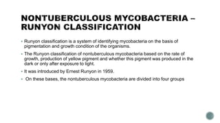
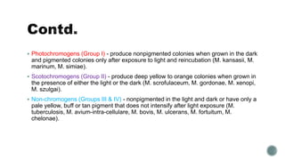
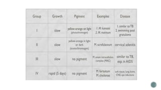
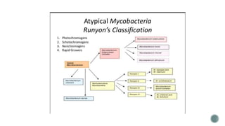
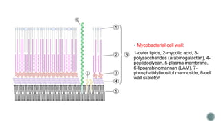
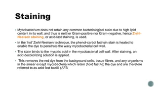
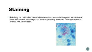
3. The family Mycobacteriaceae consists of a single genus, Mycobacterium.
Mycobacterium sp. are thin, slightly curved to straight, non-spore-forming, nonmotile bacilli,
measuring 0.2 to 6 w x 1.0 to 10.0 pm.
Over 190 species are recognized in this genus.
The Greek prefix myco- means "fungus," alluding to the way mycobacteria have been
observed to grow in a mold-like fashion on the surface of cultures.
It is acid fast and cannot be stained by the Gram stain procedure.
Many mycobacterial species are ubiquitous, and can be found in water, food, soil, and
vegetation.
For example, M. fortuitum has been reported as a commensal on human skin.
M. bovis infection has been acquired by consuming unpasteurized milk
4. Runyon classification is a system of identifying mycobacteria on the basis of
pigmentation and growth condition of the organisms.
The Runyon classification of nontuberculous mycobacteria based on the rate of
growth, production of yellow pigment and whether this pigment was produced in the
dark or only after exposure to light.
It was introduced by Ernest Runyon in 1959.
On these bases, the nontuberculous mycobacteria are divided into four groups
5. Photochromogens (Group I) - produce nonpigmented colonies when grown in the dark
and pigmented colonies only after exposure to light and reincubation (M. kansasii, M.
marinum, M. simiae).
Scotochromogens (Group II) - produce deep yellow to orange colonies when grown in
the presence of either the light or the dark (M. scrofulaceum, M. gordonae, M. xenopi,
M. szulgai).
Non-chromogens (Groups III & IV) - nonpigmented in the light and dark or have only a
pale yellow, buff or tan pigment that does not intensify after light exposure (M.
tuberculosis, M. avium-intra-cellulare, M. bovis, M. ulcerans, M. fortuitum, M.
chelonae).
6.
7.
8. In recent times Runyon’s classification scheme has become less useful because of
finding that pigment production may be temperature dependent.
Furthermore, not all strains of a mycobacterial species may share the same pigment-
producing capability.
An alternate classification scheme is based on the pathogenicity potential of a
species.
This classification system is constantly evolving as the pathogenic potential of various
species comes to be recognized
9. Mycobacteria are aerobic.
They are bacillary in form, they are straight or slightly curved rods between 0.2 and
0.6 µm wide and between 1.0 and 10 µm long.
They are generally nonmotile bacteria, except for the species Mycobacterium
marinum, which has been shown to be motile within macrophages.
They are characteristically acid-fast.
Mycobacteria have an outer membrane.
They possess capsules, and most do not form endospores. M. marinum and perhaps
M. bovis have been shown to sporulate
10. The distinguishing characteristic of all Mycobacterium species is that the cell wall is
thicker than in many other bacteria, being hydrophobic, waxy, and rich in mycolic
acids/mycolates.
The cell wall consists of the hydrophobic mycolate layer and a peptidoglycan layer
held together by a polysaccharide, arabinogalactan.
The cell wall makes a substantial contribution to the hardiness of this genus.
The biosynthetic pathways of cell wall components are potential targets for new drugs
for tuberculosis.
12. Mycobacterium does not retain any common bacteriological stain due to high lipid
content in its wall, and thus is neither Gram-positive nor Gram-negative, hence Ziehl-
Neelsen staining, or acid-fast staining, is used.
In the ‘hot’ Ziehl-Neelsen technique, the phenol-carbol fuchsin stain is heated to
enable the dye to penetrate the waxy mycobacterial cell wall.
The stain binds to the mycolic acid in the mycobacterial cell wall. After staining, an
acid decolorizing solution is applied.
This removes the red dye from the background cells, tissue fibres, and any organisms
in the smear except mycobacteria which retain (hold fast to) the dye and are therefore
referred to as acid fast bacilli (AFB
13. Following decolorization, smear is counterstained with malachite green (or methylene
blue) which stains the background material, providing a contrast color against which
the red AFB can be seen
14. Most Mycobacterium species, including most clinically relevant species, can be cultured in
blood agar.
However, some species grow very slowly due to extremely long reproductive cycles—M.
leprae, may take more than 20 days to proceed through one division cycle (for comparison,
some E. coli strains take only 20 minutes), making laboratory culture a slow process.
A natural division occurs between slowly- and rapidly-growing species.
Mycobacteria that form colonies clearly visible to the naked eye within 7 days on subculture
are termed rapid growers, while those requiring longer periods are termed slow growers.
15. They are aerobes, grow slowly at 37°C.
On Lowenstein-Jensen medium, their growth is more luxuriant (i.e. “eugonic“) than
bovine type (dysgonic) they form thin, white, smooth, slightly moist, wrinkled, granular
and easily broken up growth.
Glycerol favours the growth of human type
16. Pathogenicity:
The most common forms of primary infection is a pulmonary lesion which may be due
to inhalation of organisms in very small particles into the alveoli
Then it is carried by lymphatic drainage to the regional lymph nodes which are
enlarged and followed by caseation and calcification.