Downloaded 175 times
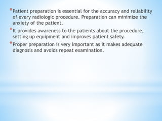
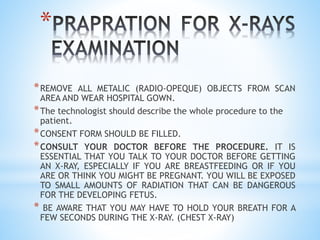


The document provides guidance on patient preparation for various radiological procedures. It emphasizes the importance of preparing the patient to improve accuracy, safety, and minimize anxiety. Key aspects of preparation include fasting, removing metallic objects, emptying bowels and bladder, obtaining consent, and explaining the procedure to relieve anxiety. Proper preparation differs for specific exams and patient populations but aims to provide clear images while reducing risks.


