This document discusses varicoceles, including their definition, etiology, pathophysiology, clinical features, investigations, treatment options, and complications. Varicoceles involve dilated and tortuous veins in the spermatic cord and are a common cause of male infertility. While often asymptomatic, varicoceles can cause pain and negatively impact testicular function and sperm quality. Treatment involves surgical repair of the affected veins to prevent further damage to the testes.
Low sperm count, also known as oligospermia, means having fewer than 15 million sperm per milliliter of semen. It can be caused by medical issues like varicoceles, infections, or medications. Lifestyle factors like smoking, stress, obesity, and environmental exposures may also play a role. Symptoms are typically inability to conceive, though many men with low counts can still father children. Diagnosis involves a semen analysis and may include ultrasounds or hormone testing. Treatment options range from surgery, antibiotics, or assisted reproduction like IVF depending on the underlying cause.
This document discusses hirsutism, excessive hair growth in women. It defines different types such as hypertrichosis, hirsutism, and virilization. Hirsutism specifically refers to male-pattern hair growth in women, such as on the face and abdomen. The causes of hirsutism include excess androgen (male hormone) production from the ovaries or adrenal glands in conditions like polycystic ovarian syndrome. The document provides details on evaluating hirsutism through hormonal testing of testosterone, DHEAS, and 17-hydroxyprogesterone levels. It also discusses the pathophysiology and genetic factors involved in polycystic ovarian syndrome, the most common cause of
Male infertility can be caused by structural or functional abnormalities in sperm, including low sperm count, low motility, and abnormal forms. Oxidative stress from reactive oxygen species is another leading cause, as it can damage sperm DNA and membranes. The body normally defends against oxidative stress through antioxidant enzymes and compounds in seminal plasma. However, oxidative stress overwhelms these defenses in around 25-40% of infertile men. Various antioxidants taken as supplements, either alone or in combination, have been shown to reduce oxidative stress levels, boost sperm parameters, and improve fertility rates. These include CoQ10, lycopene, mixed carotenoids, omega-3 fatty acids, vitamin C, vitamin E, selenium, zinc
Low sperm count | oligospermia | symptoms | management-a medical study martinshaji
this is a common condition observed in men now a days due to unhealthy sedentary lifestyle and also due to some environmental factors ..it is necessary to know some medical facts
please comment
thank you
This document discusses androgen excess in females (hirsutism). It begins by defining hirsutism and virilization. It then covers the biology of hair growth and the roles of androgens and estrogens. It discusses the sources and metabolism of androgens in females. The main causes of hirsutism discussed are physiological puberty/pregnancy/menopause), idiopathic, ovarian (PCOS, tumors), and adrenal (CAH, tumors). The evaluation of hirsutism involves history, exam, and lab tests of hormones like testosterone and DHEA. Key tests aim to identify the underlying cause and assess for conditions like PCOS, tumors, CAH
This document summarizes information about premature ovarian failure (POF). It defines POF as amenorrhea, hypoestrogenism, and elevated gonadotropins in women under age 40. POF prevalence is 1-4% under age 40, increasing to 1% by age 30 and 4% by age 40. Causes include genetic factors, autoimmunity, environmental exposures, infections, and iatrogenic factors. Symptoms include menopausal symptoms and long term risks of osteoporosis and cardiovascular disease. Diagnosis involves lab tests of hormones and imaging. Treatment is hormone replacement therapy. Annual follow up is needed to monitor treatment and screen for other related conditions.
For more Info visit www.healthlibrary.com "Management of PCOS in Unani System of Medicine" by Dr. Shaikh Nikhat held on 11th June 2016.
Management of PCOS in Unani System of Medicine - Unani system have the holistic approach to treat the condition like PCOD / PCOS.
This document discusses the clinical approach and management of hirsutism and virilism. It defines hirsutism as excessive hair growth in androgen-dependent areas, while virilism is a more severe form involving additional masculine features. The clinical approach involves assessing a patient's history and examination findings to determine the severity and underlying cause. Management aims to remove excess hair, suppress androgens, and remove their source. Treatment options depend on the site of androgen overproduction and may include oral contraceptives, antiandrogens, cosmetic treatments like bleaching or laser, and surgery to remove tumors.
The document discusses diseases of the gonads. It begins with a plan to cover topics such as normal sexual differentiation of the gonads, anatomy and physiology of male and female gonads, sexual development in boys and girls, and congenital violations of sexual differentiation. Specific conditions discussed include male hypogonadism, Klinefelter's syndrome, Turner syndrome, hermaphroditism, cryptorchidism, and anorchism. Causes of primary hypogonadism are also reviewed.
Premature ejaculation is a common male sexual dysfunction characterized by the inability to delay ejaculation and can cause distress. It is classified as lifelong or acquired and has prevalence rates ranging from 2-33% depending on age and region. While the exact etiology is unknown, potential contributing factors include anxiety, penile hypersensitivity, serotonin receptor dysfunction, and multifactorial biological and psychosocial influences. Diagnosis involves assessing ejaculation latency, distress levels, and ruling out other conditions. Treatment options include behavioral techniques, pharmacotherapy like SSRIs, and counseling with success rates over 85% depending on commitment to therapy. Prognosis is generally good but relapse can occur without ongoing management.
Invited lecture delivered by Dr Sujoy Dasgupta in a Webinar organized by Sexual medicine Committee of FOGSI (Federation of Obstetric and Gynaecological Societies of India), held in February, 2022
As reproductive clinicians, it is important that the pathological relevance of hyperprolactinemia is established before commencing treatment for this endocrinological disorder.
Most cases of true hyperprolactinemia are associated with amenorrhea or hormone deprivation in premenopausal women and can be managed by dopamine agonist or hormone replacement therapy respectively
This document provides information on erectile dysfunction (ED), including its definition, causes, evaluation, and treatment options. It defines ED as the persistent inability to attain or maintain an erection sufficient for sexual activity. ED can be caused by physical factors like vascular disease or neurological issues, as well as psychological factors like depression or relationship problems. Evaluation involves a medical history, physical exam, and sometimes lab tests or nocturnal penile tumescence testing. Treatment ranges from lifestyle changes and oral medications to devices like pumps or implants. The goal is to help patients achieve erections sufficient for sexual intercourse.
1. The document discusses erectile dysfunction (ED), including its anatomy, physiology, etiology, evaluation, and treatment. It defines ED and describes the neurovascular processes underlying erection.
2. Common organic, psychogenic, and mixed causes of ED are outlined. Evaluation involves history, physical exam, and investigations.
3. Treatment options discussed include lifestyle modifications and first-line oral phosphodiesterase type 5 inhibitors like sildenafil, tadalafil, and vardenafil. Guidelines on their use and dosing are provided.
Urolithiasis is a common disease that is estimated to
produce medical costs of $2.1 billion per year in the United States alone.
Renal colic affects approximately 1.2 million people
each year in USA and accounts for approximately 1% of
all hospital admissions.
Most active emergency departments (EDs) manage
patients with acute renal colic every day.
Erectile dysfunction results from problems affecting the brain, hormones, emotions, nerves, muscles or blood vessels involved in male arousal. It is defined as the inability to achieve or maintain an erection firm enough for sex. Common causes include heart disease, diabetes, smoking, obesity, and certain medications. Evaluation may involve physical exams, blood tests, and questionnaires to check for underlying physical or psychological conditions. Treatment options include oral medications, injections, suppositories, testosterone replacement, and penile prostheses or revascularization surgery if medications are not effective.
This document discusses the causes, evaluation, and treatment of male infertility. It begins by outlining the main causes of male infertility including pretesticular, testicular, post-testicular defects, and idiopathic factors. Evaluation involves a medical history, physical exam, and investigations including standard semen analysis, specialized tests, endocrine testing, and genetic tests. Treatment options are discussed including empiric treatments, therapies for specific diagnoses like varicocele or infections, assisted reproductive techniques like IUI or ICSI, and discussing when such options are appropriate based on resource availability and severity of male factor infertility. The document provides a comprehensive overview of male infertility.
This document appears to be a presentation on LH and its role in human reproduction given by Dr. Sandro Esteves. The presentation covers several key points:
1) LH plays an important role in reproductive cycles through its effects on steroidogenesis, follicular growth, and oocyte maturation.
2) Certain patient groups may benefit from LH supplementation during ovarian stimulation, including poor responders, older women (>35), and those using a GnRH antagonist protocol.
3) The rationale for LH supplementation in older women and poor responders is that it helps restore hormonal balance and the follicular environment by increasing androgen and estrogen production through its effects on the ovaries and granulosa cells.
Ovulation induction - not all fertility treatment is IVF by Dr Aishwarya Parthasarathy, Fertility specialist, A4 Fertility Centre, chennai
#ovulationinduction #FertilityTreamtent #a4fertilitycentre #a4hopsitals #chennai
This document provides information about Dr. Laxmi Shrikhande's credentials and experience in gynecology and fertility. It then summarizes guidelines for assessing and managing polycystic ovary syndrome (PCOS) and infertility. Key recommendations include using letrozole as first-line pharmacological treatment for infertility in PCOS patients, and considering gonadotropins as second-line if letrozole fails. The risks of ovarian hyperstimulation syndrome are also discussed for PCOS patients undergoing fertility treatments like IVF.
Primary amenorrhea is defined as no menstrual periods by age 16. Investigations into primary amenorrhea depend on whether secondary sex characteristics are present or absent. Causes can be physiological, pathological, anatomical, endocrinological, or chromosomal. Common causes include hypothalamic or pituitary disorders, chromosomal abnormalities like Turner's syndrome, congenital adrenal hyperplasia, androgen insensitivity syndrome, or müllerian agenesis. Treatment depends on the underlying cause but may include hormone replacement therapy, vaginal dilation, vaginoplasty, or gonadectomy.
This document provides an overview of male infertility, including its definition, causes, evaluation, and treatment options. It discusses factors that can cause infertility, such as varicocele, genetic disorders, hormonal imbalances, and problems with sperm production or transport. The evaluation of male infertility involves assessing medical history, performing a physical exam, analyzing semen samples, and testing for hormonal and genetic abnormalities if indicated. Treatment depends on the underlying cause but may include surgery, hormone therapy, assisted reproduction techniques like IVF, or empiric supplements for some issues.
1) Infertility is defined as the failure to conceive after 12 months of regular unprotected intercourse. The doctor discusses evaluating male factor infertility through history, physical exam, semen analysis, and tests of sperm function.
2) Semen analysis involves assessing physical parameters like volume, pH, and viscosity. Under the microscope, technicians evaluate sperm count, motility, viability, and morphology. Normal ranges are defined for each parameter.
3) Abnormal results in semen analysis can indicate problems from pre-testicular to post-testicular levels, with various treatable causes like genetic factors, infections, or obstructions. A comprehensive evaluation of male fertility aims to identify underlying issues affecting conception.
Formula for calculating the required dose of iron sucrosedpmo123
This document provides formulas and guidelines for calculating the required dose of iron sucrose for treating severe anemia. It outlines two formulas based on pre-pregnancy weight and hemoglobin percentage deficit. For a patient with a pre-pregnancy weight of 45kg and actual hemoglobin of 6%, the required dose is calculated as 1040mg or 1250mg depending on the formula used. It also provides instructions for administering iron sucrose via intravenous infusion in doses up to 200mg over 30 minutes, up to three times per week. Safety information indicates it has rare minor side effects and allergic reactions occur in 3.3 cases per million per year.
female sexual dysfunction
For More Medicine Free PPT - http://playnever.blogspot.com/
For Health benefits and medicine videos Subscribe youtube channel - https://www.youtube.com/playlist?list=PLKg-H-sMh9G01zEg4YpndngXODW2bq92w
Panel Discussion Problems of MALE INFERTILITY & Management of Oligo Astheno T...Lifecare Centre
This document summarizes a panel discussion on male infertility and the management of oligo astheno teratospermia (OAT). The panel included urologists, IVF experts, and gynaecologists who discussed topics such as the causes of male infertility, recent WHO criteria for semen analysis, what constitutes OAT, specific and idiopathic causes of OAT, how smoking affects fertility, and the steps in evaluating a male for infertility including history, examination, semen analysis, hormone assays, ultrasound, and additional tests or procedures when indicated.
Anorectal malformations (ARMs) occur in 1 in 4,000 to 5,000 births and can range from stenosis to imperforate anus. Cloacal anomalies are even rarer, occurring in 1 in 50,000 births. ARMs are associated with urogenital abnormalities in about 35% of cases as well as spinal defects. Treatment involves surgical repair of the ARM and management of any associated issues like urinary tract defects. Long term management may include bowel management, urinary reconstruction like CIC, and treatment of complications such as incontinence.
Presentation1.pptx, radiological imaging of male infertility.Abdellah Nazeer
This document discusses the role of radiological imaging in evaluating male infertility. It begins by defining male infertility and outlining common causes including anatomic defects, genetic factors, infections, endocrine disorders, varicocele, toxins and other issues. Specific causes are then subcategorized as obstructive (e.g. congenital absence of the vas deferens) or non-obstructive (e.g. varicocele, cryptorchidism). Imaging techniques like ultrasound and MRI are described for evaluating various conditions like varicocele, pituitary disorders, cryptorchidism and obstructions. The conclusion emphasizes that imaging plays a key role in infertility workups by detecting correctable issues and potentially serious underlying disorders.
Male infertility can be caused by abnormalities in sperm morphology, motility, count, or the absence of sperm. Up to 50% of infertility cases involve male factors such as varicocele, undescended testes, infections, genetic issues, environmental toxins, or lifestyle factors. Treatment depends on the underlying cause but may include medications, surgery, assisted reproduction techniques like IVF, or cryopreservation of sperm for future use.
Varicocele is the most common cause of male infertility. It occurs when the veins in the scrotum become enlarged and twisted. The condition is diagnosed through physical examination and Doppler ultrasound. Treatment options aim to disrupt the internal spermatic veins while preserving blood flow, with the most common approaches being inguinal, subinguinal, and microscopic surgery. Patients with higher grade varicoceles or abnormal sperm parameters are more likely to see improved fertility outcomes following treatment. While varicocele repair can improve sperm quality, not all patients will experience improved fertility, and subclinical varicoceles without physical signs do not generally require treatment.
- Anastomotic leakage is a dangerous surgical complication that increases mortality and morbidity. It occurs when there is a defect in the intestinal wall at the site of bowel reconnection that allows internal and external bowel contents to mix.
- Risk factors include patient characteristics like age and comorbidities, neoadjuvant treatments, surgical factors like emergency surgery or surgeon experience, and technical aspects of the anastomosis.
- Preventive measures include temporary stomas, but leakage risk can't be eliminated. Early detection through close postoperative monitoring and imaging is important, as early treatment improves outcomes. Treatment depends on the severity but may include drainage, resection and reanastomosis or stoma formation.
Testicular torsion refers to the torsion of the spermatic cord structures and subsequent loss of the blood supply to the ipsilateral testicle.
Urological emergency; early diagnosis and treatment are vital.
Mainly disease of Neonates, Adolescents.
The rate of testicular viability decreases significantly after 6 hours from onset of symptoms.
Male infertility PPT - Dr P Usha Devi dt 04 Mar.pptslidesharecgr
Male factor infertility can be caused by issues with sperm production, transport, or function. A thorough evaluation of a man's medical history, physical exam, and semen analysis is needed to identify potential causes. Semen analysis involves examining the volume, concentration, mobility, and morphology of sperm and is the first step in evaluating male infertility. Abnormal results on two analyses suggest the presence of male factor infertility and further testing may be needed including hormone levels, genetic testing, or examination under microscope. Identifying the cause is important to determine the best treatment options such as lifestyle changes, medication, surgery, or assisted reproduction.
Varicocele is the abnormal dilatation and tortuosity of the veins within the pampiniform plexus of the spermatic cord. It is usually asymptomatic and found in 15-20% of men. Risk factors include left-sided varicocele which is more common due to anatomical factors. Diagnosis involves physical exam finding of swollen, tortuous veins that increase with straining. Treatment with varicocelectomy is recommended for men with abnormal semen analysis or infertility who are trying to conceive. Complications of surgery are rare but include infection, hydrocele, and recurrence.
Hydronephrosis is the dilation of the renal pelvis and calyces, which can be caused by obstruction of urine flow. It is commonly detected during prenatal ultrasound screening. Common causes in neonates include ureteropelvic junction obstruction, posterior urethral valves, and vesicoureteral reflux. Treatment depends on the severity and includes antibiotics, surgery to repair obstructions, and management of any associated renal issues.
This document provides information about Prune Belly Syndrome from the Department of Urology at Government Royapettah Hospital and Kilpauk Medical College in Chennai, India. It discusses the moderators, history, epidemiology, genetics, clinical findings, urogenital anomalies, extragenitourinary abnormalities, prenatal diagnosis, neonatal presentation, evaluation, and treatment categories of Prune Belly Syndrome. The document contains detailed information over multiple pages and sections.
This document provides an overview of scrotal masses, including relevant anatomy, causes, evaluation, and management. It discusses common etiologies like testicular torsion, epididymitis/orchitis, and varicocele. Testicular torsion is an emergency requiring urgent surgical exploration to preserve testicular function. Epididymitis is usually infectious and treated with antibiotics. Physical exam and ultrasound can help differentiate causes. Painless scrotal masses require evaluation for testicular cancer. Prompt diagnosis and treatment of underlying etiology is important to avoid complications.
The document discusses various perinatal emergencies involving the urinary tract including obstruction, neural tube defects, solid masses, and intersex disorders. It outlines evaluation and treatment approaches for conditions diagnosed during the antenatal period such as bladder exstrophy, prune belly syndrome, and myelodysplasia. The document also covers evaluation and management of scrotal masses, pediatric scrotal tumors, and various intersex disorders including congenital adrenal hyperplasia.
This document discusses several unusual CT manifestations of common abdominal diseases:
1. Solitary rectal ulcer syndrome appears as a solitary linear ulcer on rectosigmoidoscopy in young patients with rectal bleeding.
2. Giant ulcers in the ileum and colon seen on CT in AIDS patients are caused by cytomegalovirus.
3. Ovarian torsion presents on CT as an adnexal mass rotated to the midline and contralateral side with deviation of the uterus and free fluid. The twisted vascular pedicle may also be seen.
4. Chemotherapy-induced pseudocirrhosis appears on CT as diffuse hepatic fibrosis without masses in patients receiving chemotherapy for cancer.
This document provides information on acute scrotum from the Department of Urology at Govt Royapettah Hospital and Kilpauk Medical College in Chennai, India. It discusses the differential diagnoses of acute scrotum including testicular torsion, epididymitis, trauma and tumors. It provides evaluation methods and clinical criteria to differentiate diagnoses. Treatment involves prompt surgical exploration for testicular torsion to preserve viability. Ultrasound Doppler is an important tool to evaluate blood flow and diagnose torsion. Epididymo-orchitis is usually caused by infection and typically has a more gradual onset than torsion. Torsion of appendages like the appendix testis is also discussed.
Congenital genitourinary disorders are birth defects of the kidneys and urinary tract that occur in about 3-6 per 1000 live births. Some common disorders include renal agenesis, hydronephrosis, posterior urethral valves, hypospadias, and Wilm's tumor. Symptoms vary depending on the specific disorder but can include urinary tract infections, blood in the urine, pain, and kidney failure. Treatment involves surgery to correct structural abnormalities, antibiotics to treat infections, and chemotherapy for Wilm's tumor. Nursing care focuses on monitoring the child, preventing infections, providing comfort, and educating parents.
Undescended testis, or cryptorchidism, is a common condition in infants where one or both testes fail to descend into the scrotum. It occurs in approximately 3% of term male infants and is more common in premature infants. Left untreated, undescended testis can lead to infertility and an increased risk of testicular cancer. The standard treatment is orchidopexy surgery to bring the testis into the scrotum, which is usually performed between 6-12 months of age. For intra-abdominal undescended testes, a two-stage surgical approach may be used.
This document provides an overview of the evaluation and investigations for male infertility. It discusses the key components of the medical history and physical exam, including reproductive, sexual, childhood, medical, and family histories. Initial laboratory assessments include semen analysis according to WHO standards and endocrine evaluation if indicated. Imaging tools like ultrasound, Doppler ultrasound, and MRI can identify conditions affecting fertility. Traditional and modern methods are described to further evaluate couples where initial testing is normal but functional defects may still impair fertilization. A postcoital or Sims test examines sperm-mucus interaction ability.
This document provides an overview of common urologic emergencies and their management. It discusses renal colic caused by kidney stones, including pain management with NSAIDs and opioids. It also covers acute urinary retention, priapism, hematuria, and anuria. For each condition, it outlines evaluation, differential diagnosis, and treatment approaches including medical expulsive therapy, ureteral stenting, and surgical procedures.
Role of imaging of diagnosis and management of male infertilityPrasunDas31
MALE INFERTILITY IS ONE OF THE MOST OVERLOOKED AND IGNORED TOPIC.I TRY TO ASSIMILATE ALL PROBABLE IMAGING FEATURE TO CORRECTLY DIAGNOSE IT SO THAT UROSERGEONS CAN PROCEED EASILY.
Resistive index and its applications in UrologyPrateek Laddha
This document discusses the resistive index (RI), a measure of pulsatile blood flow used in urology. It provides background on how the RI is calculated and explores its applications in evaluating conditions like renal obstruction and non-obstructive renal disease. While early studies showed promise for the RI, later research found it lacks specificity. The RI is influenced by vascular compliance and resistance. It may help identify transplant complications but not differentiate causes of dysfunction. The document concludes the RI remains a nonspecific marker of transplant dysfunction.
The document discusses renal vascular anatomy and its implications for surgery. It notes that the renal arteries typically enter the kidney through the hilum and branch into segmental arteries that do not anastomose. It also describes variations such as accessory renal arteries. The segmental arteries further branch within the kidney and an avascular plane exists between anterior and posterior branches. Venous drainage parallels the arterial supply. Imaging like CTA can accurately map the vasculature preoperatively to aid surgery. Understanding variations and collateral circulation is important for procedures like donor nephrectomy.
Primary hyperoxaluria and renal hypercalciuriaPrateek Laddha
This document discusses primary hyperoxaluria and renal hypercalciuria. It defines hyperoxaluria and describes the four main types: primary hyperoxaluria types I and II, enteric hyperoxaluria, dietary hyperoxaluria, and idiopathic/mild hyperoxaluria. It explains oxalate production, absorption, and excretion in the body. For primary hyperoxaluria, it covers the genetic defects, pathophysiology, clinical manifestations, prognosis, treatment including pyridoxine, orthophosphate, magnesium, and sometimes combined liver-kidney transplantation. The document provides details on evaluation, management, and treatment of the different types of hyperoxaluria.
This document provides an overview of the history and techniques for orthotopic neobladder urinary diversion. Some key points:
- Orthotopic diversion was pioneered in the 1950s as an alternative to ureterosigmoidostomy and ileal conduit diversion due to complications of those procedures.
- Patient selection considers oncologic factors like risk of urethral recurrence and tumor stage, as well as patient factors like age, renal function, manual dexterity, and prior treatments.
- Surgical techniques aim to optimize continence by preserving the rhabdosphincter and its innervation during cystectomy. For males the urethra is detached in a retrograde
MRI has several applications in urology, including evaluating the prostate, penis, scrotum, kidneys, and adrenal glands. For the prostate, MRI is useful for detecting and localizing cancer, assessing tumor staging and extent, and guiding biopsies. It provides better soft tissue contrast than other modalities like CT or ultrasound. On T2-weighted MRI sequences, prostate cancer appears as an area of low signal intensity in the peripheral zone compared to the normally high signal prostate tissue. Functional sequences like diffusion-weighted imaging can also identify cancers. MRI is particularly helpful for detecting multifocal tumors and assessing extracapsular extension and seminal vesicle invasion.
The kidneys are located retroperitoneally on either side of the vertebral column. Each kidney receives around 20% of cardiac output through the renal artery. The kidneys filter blood to remove waste and regulate water and electrolyte balance through nephrons, the functional units of the kidney. Filtration occurs through the glomerulus, and most of the filtrate is reabsorbed in the proximal tubule. Remaining fluid is concentrated in the loop of Henle and final urine composition is determined in the distal tubule and collecting duct under hormonal control. The kidneys play key roles in homeostasis by regulating fluid volume and composition, blood pressure, acid-base balance, and red blood cell production.
This document discusses amyloidosis and its effects on the kidneys. It defines amyloidosis as a group of diseases characterized by the deposition of abnormal protein fibrils composed of subunits of normal serum proteins in organs and tissues. There are different types of systemic amyloidosis depending on the precursor protein involved, such as AA, AL, and beta-2 microglobulin amyloidosis which affects those on long-term dialysis. Amyloid deposition in the kidneys is most commonly seen with AL and AA types. Symptoms include proteinuria, edema, and renal dysfunction. Diagnosis involves biopsy of affected tissues with staining techniques to identify amyloid deposits. Treatment aims to eliminate the underlying causes and slow progression of organ damage.
Focussed therapy and imaging in prostate cancerPrateek Laddha
This document discusses focal therapy and imaging techniques for prostate cancer. It provides background on the increasing diagnosis of localized, low-risk prostate cancer and rationale for focal therapy alternatives to radical treatments. It describes improved biopsy strategies using MRI-ultrasound fusion to target small tumors. The document outlines different focal therapy techniques including thermal ablation methods like cryoablation and HIFU, as well as non-thermal methods. It discusses the advantages of focal therapy in preserving function while eliminating cancerous tissue and notes challenges in patient selection and defining clinically significant tumors.
Anatomy of penis and physiology of erectionPrateek Laddha
The document provides information on the anatomy and physiology of the penis and erection. It discusses:
1) The anatomy of the penis including the corpora cavernosa, corpus spongiosum, skin, fascia, tunica albuginea and ligaments.
2) The physiology of erection including relaxation of cavernous smooth muscle allowing blood flow into sinusoids, compression of veins leading to rigidity, and the roles of neurotransmitters, hormones and molecular mechanisms.
3) The phases of erection including flaccid, tumescent, full erection and detumescent phases and changes in blood flow and pressure that occur in each phase.
PGx Analysis in VarSeq: A User’s PerspectiveGolden Helix
Since our release of the PGx capabilities in VarSeq, we’ve had a few months to gather some insights from various use cases. Some users approach PGx workflows by means of array genotyping or what seems to be a growing trend of adding the star allele calling to the existing NGS pipeline for whole genome data. Luckily, both approaches are supported with the VarSeq software platform. The genotyping method being used will also dictate what the scope of the tertiary analysis will be. For example, are your PGx reports a standalone pipeline or would your lab’s goal be to handle a dual-purpose workflow and report on PGx + Diagnostic findings.
The purpose of this webcast is to:
Discuss and demonstrate the approaches with array and NGS genotyping methods for star allele calling to prep for downstream analysis.
Following genotyping, explore alternative tertiary workflow concepts in VarSeq to handle PGx reporting.
Moreover, we will include insights users will need to consider when validating their PGx workflow for all possible star alleles and options you have for automating your PGx analysis for large number of samples. Please join us for a session dedicated to the application of star allele genotyping and subsequent PGx workflows in our VarSeq software.
Are you looking for a long-lasting solution to your missing tooth?
Dental implants are the most common type of method for replacing the missing tooth. Unlike dentures or bridges, implants are surgically placed in the jawbone. In layman’s terms, a dental implant is similar to the natural root of the tooth. It offers a stable foundation for the artificial tooth giving it the look, feel, and function similar to the natural tooth.
Osvaldo Bernardo Muchanga-GASTROINTESTINAL INFECTIONS AND GASTRITIS-2024.pdfOsvaldo Bernardo Muchanga
GASTROINTESTINAL INFECTIONS AND GASTRITIS
Osvaldo Bernardo Muchanga
Gastrointestinal Infections
GASTROINTESTINAL INFECTIONS result from the ingestion of pathogens that cause infections at the level of this tract, generally being transmitted by food, water and hands contaminated by microorganisms such as E. coli, Salmonella, Shigella, Vibrio cholerae, Campylobacter, Staphylococcus, Rotavirus among others that are generally contained in feces, thus configuring a FECAL-ORAL type of transmission.
Among the factors that lead to the occurrence of gastrointestinal infections are the hygienic and sanitary deficiencies that characterize our markets and other places where raw or cooked food is sold, poor environmental sanitation in communities, deficiencies in water treatment (or in the process of its plumbing), risky hygienic-sanitary habits (not washing hands after major and/or minor needs), among others.
These are generally consequences (signs and symptoms) resulting from gastrointestinal infections: diarrhea, vomiting, fever and malaise, among others.
The treatment consists of replacing lost liquids and electrolytes (drinking drinking water and other recommended liquids, including consumption of juicy fruits such as papayas, apples, pears, among others that contain water in their composition).
To prevent this, it is necessary to promote health education, improve the hygienic-sanitary conditions of markets and communities in general as a way of promoting, preserving and prolonging PUBLIC HEALTH.
Gastritis and Gastric Health
Gastric Health is one of the most relevant concerns in human health, with gastrointestinal infections being among the main illnesses that affect humans.
Among gastric problems, we have GASTRITIS AND GASTRIC ULCERS as the main public health problems. Gastritis and gastric ulcers normally result from inflammation and corrosion of the walls of the stomach (gastric mucosa) and are generally associated (caused) by the bacterium Helicobacter pylor, which, according to the literature, this bacterium settles on these walls (of the stomach) and starts to release urease that ends up altering the normal pH of the stomach (acid), which leads to inflammation and corrosion of the mucous membranes and consequent gastritis or ulcers, respectively.
In addition to bacterial infections, gastritis and gastric ulcers are associated with several factors, with emphasis on prolonged fasting, chemical substances including drugs, alcohol, foods with strong seasonings including chilli, which ends up causing inflammation of the stomach walls and/or corrosion. of the same, resulting in the appearance of wounds and consequent gastritis or ulcers, respectively.
Among patients with gastritis and/or ulcers, one of the dilemmas is associated with the foods to consume in order to minimize the sensation of pain and discomfort.
“Psychiatry and the Humanities”: An Innovative Course at the University of Mo...Université de Montréal
“Psychiatry and the Humanities”: An Innovative Course at the University of Montreal Expanding the medical model to embrace the humanities. Link: https://www.psychiatrictimes.com/view/-psychiatry-and-the-humanities-an-innovative-course-at-the-university-of-montreal
STUDIES IN SUPPORT OF SPECIAL POPULATIONS: GERIATRICS E7shruti jagirdar
Unit 4: MRA 103T Regulatory affairs
This guideline is directed principally toward new Molecular Entities that are
likely to have significant use in the elderly, either because the disease intended
to be treated is characteristically a disease of aging ( e.g., Alzheimer's disease) or
because the population to be treated is known to include substantial numbers of
geriatric patients (e.g., hypertension).
Giloy in Ayurveda - Classical Categorization and SynonymsPlanet Ayurveda
Giloy, also known as Guduchi or Amrita in classical Ayurvedic texts, is a revered herb renowned for its myriad health benefits. It is categorized as a Rasayana, meaning it has rejuvenating properties that enhance vitality and longevity. Giloy is celebrated for its ability to boost the immune system, detoxify the body, and promote overall wellness. Its anti-inflammatory, antipyretic, and antioxidant properties make it a staple in managing conditions like fever, diabetes, and stress. The versatility and efficacy of Giloy in supporting health naturally highlight its importance in Ayurveda. At Planet Ayurveda, we provide a comprehensive range of health services and 100% herbal supplements that harness the power of natural ingredients like Giloy. Our products are globally available and affordable, ensuring that everyone can benefit from the ancient wisdom of Ayurveda. If you or your loved ones are dealing with health issues, contact Planet Ayurveda at 01725214040 to book an online video consultation with our professional doctors. Let us help you achieve optimal health and wellness naturally.
These lecture slides, by Dr Sidra Arshad, offer a simplified look into the mechanisms involved in the regulation of respiration:
Learning objectives:
1. Describe the organisation of respiratory center
2. Describe the nervous control of inspiration and respiratory rhythm
3. Describe the functions of the dorsal and respiratory groups of neurons
4. Describe the influences of the Pneumotaxic and Apneustic centers
5. Explain the role of Hering-Breur inflation reflex in regulation of inspiration
6. Explain the role of central chemoreceptors in regulation of respiration
7. Explain the role of peripheral chemoreceptors in regulation of respiration
8. Explain the regulation of respiration during exercise
9. Integrate the respiratory regulatory mechanisms
10. Describe the Cheyne-Stokes breathing
Study Resources:
1. Chapter 42, Guyton and Hall Textbook of Medical Physiology, 14th edition
2. Chapter 36, Ganong’s Review of Medical Physiology, 26th edition
3. Chapter 13, Human Physiology by Lauralee Sherwood, 9th edition
Summer is a time for fun in the sun, but the heat and humidity can also wreak havoc on your skin. From itchy rashes to unwanted pigmentation, several skin conditions become more prevalent during these warmer months.
2. VARICOCELE
Definition
Etiology
Pathophysiology of testicular changes
Clinical features
Investigations
Treatment –
- Expectant treatment
- Indication of intervention
- Treatment options
- Complication of surgery
Complication of untreated varicocele
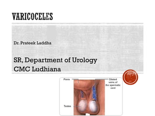
3. DEFINITION
Dilated & tortuous veins of pampaniform plexus of spermatic cord
found in about 15% of male adolescents with a marked left sided
predominance
6. ETIOLOGY
Responsible factors
8-10 cm longer left testicular Vv. increased→
hydrostatic pressure in upright position
Entry of left testicular Vv into renal vein at 900
“Nutcraker phenomenon” due to passage of
left testicular vein between SMA & Aorta
Congenital absence of valve in left vein in
40%
Intrinsic ectasia of plexus due to cremaster
atrophy
Loaded left colon
7. PATHOPHYSIOLOGY OF TESTICULAR
CHANGES
Adverse effects on spermatogenesis –
Reflux of renal and adrenal metabolites
Hyperthermia
Hypoxia
Local testicular hormonal imbalance
Intra testicular hyper perfusion injury
Increased oxidative stress
9. CLINICAL FEATURES (SYMPTOMS)
Asymptomatic - detected during medical
examination or evaluation of infertile male
Constant dragging pain in Testis aggravated by
standing & relieved by lying down
Impaired sperm quality
Cosmetic attention
Swelling in scrotum
Failure of affected testis to grow
10. CLINICAL FEATURES (SIGNS)
Examine in warm room, standing & lying position, with or
without valsulva maneuver
Painless compressible mass with feeling of “Bag of
worms”
Small sized Testis on affected side
11.
12. GRADES OF VARICOCELE
Grade I – Palpable only during valsulva
maneuver
Grade II – Palpable without Valsulva in
standing upright position
Grade III – Visible through scrotal skin
Subclinical – detected during USG
13. INVESTIGATION
Doppler stethoscope (5.3 MHz probe) -audible rush of blood on
valsulva
Colour Doppler –detects Sub Clinical Varicocele also
Ultra sound of abdomen
Semen examination
24. COMPLICATIONS OF TREATMENT
Hydrocele formation – due to ligation of lymphatics
Recurrence
Testicular infarction
Migration of coil to pulmonary artery – usually not fatal
Infection & haemorrhage
26. LET US REVISE
Definition
Etiology
Pathophysiology of testicular changes
Clinical features
Investigations
Treatment
Complication of untreated varicocele
27. dilatation of the pampiniform venous plexus and
the internal spermatic vein
well-recognized cause of decreased testicular
function
very rare < 9 y.o.
~16% of adolescents
~15-20% of all males
40% of infertile males
scrotal varicoceles are the most common cause of poor
sperm production and decreased semen quality
28.
29. The prevalence of varicocele and
associated testicular hypotrophy by age
Age, years Prevalence, %
of varicocele hypotrophic testis
<11 0 0
11–14 6–8 7.3
15–19 11–19 9.3
30. first recognized as a clinical problem in 16th
century
relationship between infertility and
varicocele proposed in late 19th century
thereafter, others reported association with arrest
of sperm secretion and the subsequent restoration
of fertility following repair
enlarged scrotal veins in teenagers
referenced as early as 1885
31. 1950s report of fertility following
varicocele repair in an individual known to
be azoospermic
surgical correction as clinical approach to certain
kinds of male infertility gained support among
American surgeons
Continued research documented recurrent
pattern of low sperm count, poor motility,
and predominance of abnormal sperm forms
(stress pattern of semen)
not specific to varicocele
suggests early evidence of testicular damage
32. 80-90% involve the left testicle
anatomic factors
(1) angle at which left testicular vein enters left renal vein
(2) lack of effective antireflux valves at juncture of testicular
vein and renal vein
(3) increased renal vein pressure due to compression
between the superior mesenteric artery and the aorta (ie,
nutcracker effect)
35-40% of men with palpable left varicocele may
actually have bilateral varicoceles
Recent study by Gat et al ~ 80% of men with a
left clinical varicocele had bilateral varicoceles
revealed by noninvasive radiologic testing
33. Scrotal mass/swelling, symptoms of acute or
chronic scrotal discomfort, differing
testicular sizes without a palpable variocele,
and incidental finding on scrotal US
Grading:
Grade 0 - Subclinical varicocele, Dx by US or
venography
Grade 1 – palpable with Valsalva maneuver
Grade 2 - Easily detected without Valsalva
maneuver
Grade 3 - Detected visually at a distance
34. Most asymptomatic
usually unilateral and almost always left-sided
unilateral right-sided varicocele should prompt
investigation for retroperitoneal process
mass that causes obstruction of the right internal spermatic
vein
Thrombosis/occlusion of the inferior vena cava must be ruled
out in
Situs inversus another etiology of right-sided varicocele
Initial presentation usually occurs during
puberty, with incidence in 13-year-old
adolescent boys equal to that of adult men
35. Multiple investigators have directly correlated the degree of
testicular atrophy with varicocele grade
Steeno et al testis volume reduced by 81% with grade 3 and by
34% with grade 2
No patients with grade 1 had testicular atrophy
36. Unknown how impairment of sperm structure,
function, and production occurs
interference with thermoregulation
other theories include the possible effects of
pressure, oxygen deprivation, heat injury, and
toxins
Despite considerable research, no one theory proved
unquestionably
Regardless, indisputably a significant factor in
decreasing testicular function and in reducing
semen quality in large percentage of men seeking
infertility treatment
37. Histologic studies seminiferous tubule sclerosis, small
vessel degenerative changes, and abnormalities of Leydig,
Sertoli, and germ cells
changes have been documented in patients as young as 12 years
38. Presence of a varicocele does not necessitate
surgical correction
Indications for surgical correction
Relief of significant testicular discomfort or pain not
responsive to routine symptomatic treatment
testicular atrophy (volume difference >20% or > 2cc)
possible contribution to unexplained male infertility
varicocele may cause progressive damage to testes,
resulting in further atrophy and impairment of seminal
parameters
39. The AUA Male Infertility Best Practice Policy
Committee recommends treatment be offered to the
male partner when all the following are present:
varicocele is palpable
couple has documented infertility
female has normal fertility
one or more abnormal semen parameters or sperm function
test results
men who have a palpable varicocele and abnormal
semen analyses findings but are not currently
attempting to conceive should also be offered
varicocele repair
40. No strict criteria necessitate surgical intervention in
adolescents
Each case handled individually
discussion among patient, parents, and physician regarding
risks of intervention and potential impact on future fertility
general guidelines used by some pediatric urologist
include the presence of one or more of the following:
Varicocele associated with decreased ipsilateral testicular
size (20% volume deficit in the involved testis)
Bilateral varicoceles
Symptomatic painful varicocele
Abnormal findings on semen analysis
41. Lipshultz and Corriere (1997)
suggested that varicoceles were associated with
testicular atrophy that was progressive with age
observed that testicular biopsy specimens taken
from prepubertal boys with varicoceles already
revealed histologic abnormalities
Kass and Belman (1987)
first to demonstrate significant increase in
testicular volume after varicocele repair in
adolescents
did not study semen parameters
42. ideal technique is to ligate all of the internal and
external spermatic veins with preservation of
spermatic arteries and lymphatics
internal spermatic artery may be divided with
transperitoneal or retroperitoneal approach
does not usually cause testicular atrophy due to generous
collateral circulation to testicle
3 most common surgical approaches
inguinal
Retroperitoneal
subinguinal
43. Surgical Treatment can be divided into 3 main
categories
Diagnostic procedures
Testis biopsy
Seminal vesicle aspiration
vasography
Procedures to improve sperm production
Varicocelectomy
Procedures to improve sperm delivery
Vasovasostomy
Vasoepididymostomy
44. Varicoceles are now recognized as the most surgically
correctable cause of male infertility.
They are present in 15% of the normal male population.
Up to 40% of patients with male infertility.
Approximately 70% of patients with secondary infertility have been found to have
a varicocele as an underlying cause.
Varicocele repair remains the most cost-effective procedure in
helping a subfertile man establish a pregnancy.
improve spermatogenesis
increase serum testosterone
Vasectomies
½ million performed per year. 75% by urologist
12% of men aged 20 to 39 years in the United States have had a vasectomy
6% will eventually desire a reversal
45. Testicular Biopsy
Azoospermia with normal FSH and normal sized
testicles.
Can be due to obstruction, defect in spermatogenesis, or incomplete
defect
Obstruction vs. spermatogenic failure?
Can also be therapeutic - consider sperm retrieval for IVF/ICSI.
Should be perform on both testes for
nonobstructive azoospermia.
In obstructive azoospermia, should biopsy the
larger testis first.
47. 1. Cord block with 1% lidocaine and
0.25% bupivicaine with 30-ga needle
2.The scrotal skin and tunica vaginalis are
then infiltrated with 2 mL of 1% lidocaine
with a 30-ga needle.
3. A 1- to 2-cm transverse incision is made
to the parietal tunica vaginalis through
the anesthetized region.
4.The tunica vaginalis is then opened
with scissors, and the edges are grasped
and held apart with two small hemostats
or a small self-retaining eyelid retractor.
Lidocaine (2 to 3 mL) is dripped onto the
exposed tunica albuginea to anesthetize
the testicular surface where the biopsy
specimen will be taken.
48. 5.The tunica albuginea is carefully inspected
for the least vascular area for the incision. A 5-
0 Prolene suture is passed at one end of the
proposed site of incision in the testis.
6. A 4- to 5-mm incision is made in the tunica
albuginea by use of a No. 11 scalpel or a
microknife, allowing extrusion of the
seminiferous tubules.
7.With the "no-touch" technique, fine, sharp
iris scissors are used to carefully excise the
extruded tubules.
8.The specimen is then placed in Zenker's,
Bouin's, or buffered glutaraldehyde solution.
The testicular specimen should not be placed
in formalin.
9. “Touch imprint” or wet prep done.Touch
imprint more predictive in the evaluation of
spermatogenesis.
49. 10. If sperm are found and
cryopreservation of testicular
tissue is to be done, additional
testicular tissue can be taken
from the same site and placed
in appropriate medium in
individual Eppendorf tubes for
processing by the andrology
laboratory.
11.The incision is then closed
with the previously placed 5-0
Prolene suture. It is important to
close the tunica vaginalis over
the testis with absorbable
suture, such as 4-0 chromic or
Vicryl.
50. 1. Percutaneous testicular biopsy
can be performed with local
anesthesia in an office-based
setting, and it is generally
associated with less pain and
morbidity than an open
testicular biopsy.
2. A 95% correlation was
described between
percutaneous needle and open
biopsy techniques as long as
sufficient materials are present
for diagnosis.
3. Before the biopsy is performed,
the skin is punctured with a
scalpel to prevent inclusion of
scrotal skin with the specimen.
4. To avoid injury to the
epididymis and the surgeon's
hand, the point of the needle
insertion should be from the
lower pole toward the upper
pole.
52. 15% of the normal male population and in up to 40% of patients with male infertility
World Health Organization reported that varicoceles were found in 25.4% of men with abnormal
semen parameters compared with 11.7% of men with normal semen.
Varicoceles have been associated with impaired semen quality and decreased Leydig cell
function.
However, varicocele repairs have been shown to improve not only spermatogenesis but also
Leydig cell function
most commonly performed surgical procedure in treatment of male infertility.
Grading of Varicocele
I - Palpable only with the Valsalva maneuver
II - Palpable without the Valsalva maneuver
III - Visible through the scrotal skin
Repair of larger varicoceles results in significantly greater improvement in semen quality
than does repair of smaller varicoceles.
On scrotal US – dilated veins > 3.5 mm
Subclinical varicoceles
Diagnosed only on US
Studies have demonstrated that subclinical varicoceles have no impact on fertility and that
repair of subclinical varicoceles does not improve fertility rates.
53. Four indications for treatment in adult men
The couple has known infertility
The female partner has normal fertility or a potentially treatable cause of
infertility
The varicocele is palpable on physical examination, or if it is suspected, the
varicocele is corroborated by ultrasound examination
The male partner has an abnormal semen analysis
In adolescent men
Reduction in ipsilateral testicular size, otherwise observation and /or semen
analysis.
54. Surgical Approaches
Scrotal
No longer used. High failure rate and testicular artery injury risk.
Retroperitoneal
Palomo
High retroperitoneal ligation of the internal spermatic vein above the internal inguinal ring.
A common complication of the retroperitoneal approach is varicocele recurrence or
persistence, estimated to be between 11% and 15%.
The recurrence can be significantly reduced by intentional ligation of the testicular artery.
This is thought to ensure ligation of the periarterial/cremasteric veins and thus to prevent
recurrence.
Laparoscopic
Excessively invasive for what should be a minor outpatient procedure
laparoscopic varicocele repairs have been associated with a recurrence
rate of less than 2% and formation of hydroceles in 5% to 8% of patients
55. Inguinal and subinguinal approach
Preferred approaches
Less morbidity associated with the subinguinal (infrainguinal) approach
than with the laparoscopic and inguinal approach because of the
preservation of the muscle layers and the inguinal canal
However, a greater number of internal spermatic veins and arteries lie
below the external ring, making this procedure technically more
challenging
56. 1. Essentially the same as the
Palomo technique.
2. Establish
pneumoperitoneum using
Veress or Hassan technique.
3. Parietal peritoneum is
incised just lateral to the
spermatic cord.The
testicular artery and veins
are dissected and isolated.
Pulling on the testis can help
identify the vessels.
4. Once the veins are isolated,
they are clipped both
proximally and distally with
titanium endoclips, and
these vessels are then
transected.
57. 1. 3- to 4-cm oblique incision, two
fingerbreadths above the
symphysis pubis and just above the
external ring, is carried laterally
along Langer's lines
2. Incision is carried down to the
external oblique aponeurosis,
which is incised in the direction of
its fibers. Care is taken to identify
and to preserve the ilioinguinal
nerve .
3. The spermatic cord is mobilized
near the pubic tubercle, and a
Penrose drain is passed beneath
the cord.The Penrose drain is used
to elevate the cord and bring it
through the incision.
4. (+/-) microscope/loupes
5. Varicoceles generally appear with
a typical vascular pattern in which
the artery is next to or adherent to
several veins, and there is a
separate isolated vein nearby.
58. 6. Once the dilated veins are isolated, they are
doubly ligated with either 2-0 silk sutures or
small titanium surgical clips.
7.With the microsurgical technique, the
lymphatic channels can be clearly visualized,
and these should be preserved to prevent
postoperative hydrocele formation.
8.The floor of the inguinal canal, near the
external ring, should also be inspected to
identify and ligate any external cremasteric
veins.
9.The cord is placed back into the canal, and
the external oblique fascia is closed with a 3-0
Vicryl suture.The subcutaneous layer is
reapproximated with a 3-0 plain catgut suture,
and the subcuticular layer is closed with a 4-0
Monocryl suture.The incision is infiltrated with
1% lidocaine mixed with an equal amount of
0.5% bupivacaine.
59.
60. Percutaneous Embolization
Cut-down to femoral or internal jugular vein
embolization of the spermatic veins can be accomplished with coils,
balloons, or sclerotherapy
Overall success rate – 68%
Percutaneous varicocele embolization is especially useful in a recurrent or
persistent varicocele, when the anatomy causing the varicocele needs to be
radiographically clarified.
61. Outcomes
studies have shown that repair of varicoceles can retard further damage to
testicular function
overall rate of improvement in semen parameters after varicocelectomy
ranged from 51% to 78%
improve not only semen motility, density, and morphologic features but also
serum FSH and testosterone levels
No difference noted between laparoscopic and open approach, but higher
complications in the lap. Group
Predictors of successful repair
Sperm concentration > 5million/ml or density > 50 million per ejaculate
lack of testicular atrophy
sperm motility of 60% or more
serum FSH values less than 300 ng/mL (normal, 50 to 300 ng/mL)
62.
63. Probability of a live birth after a varicocelectomy was 29.7% versus 25.4% after
IVF-ICSI.
The cost per delivered baby was $26,268 after varicocelectomy compared with
$89,091 with IVF-ICSI.
64. 6% of men who have undergone vasectomy will subsequently request a
vasectomy reversal
Chances for success (patency or pregnancy) based on the personal experience
of the surgeon, the patient's health history, and the results of examination of the
man and the age and reproductive potential of his partner are discussed.
Epididymal obstruction appears, in most instances, to be a time-related
phenomenon
62% of patients who underwent reversal 15 years or more after their
vasectomy required either a unilateral or a bilateral vasoepididymostomy
VE depends on quality of fluid from proximal vas
when the material coming from the proximal vas lumen is thick, pasty,
and devoid of sperm; if the fluid is creamy, containing only debris.
microsurgical vasectomy reversal are superior to results of nonmicrosurgical
techniques
No significant difference if a multilayer anastomosis is performed as opposed to
a modified single-layer technique but the success is physician-dependent.
65. A. Nonlocking needle holder.
B.Suture scissors.
C.Dissecting scissors.
D,E. Very fine pointed and
round-tipped scissors.
F.Round-handled platform
forceps.
G.Curved dilating forceps.
H.Round-handled small knife
blade holder.
I.Microtip bipolar cautery
66. Anesthetic Considerations
1.General vs. local?
2.Preparing the vas for anastomosis
1. Vas grasped through skin
above the vasectomy site.
2. Once the vas is exposed,
injection of a mixture of
0.5% bupivacaine and 1%
lidocaine into the distal
perivasal sheath will
provide sufficient
anesthetic coverage for
the vasal anastomosis to
be performed.
3. Placement of 6-0 Prolene
sutures just into the
muscularis holds the vas
above the incision and
make it easily accessible
for anastomosis.
67. 4.The vas above and below the vasectomy site
should be transected with use of the operating
microscope Once the point of the vas that is to
be cut is chosen, the vasal vessels are secured
with 7-0 Prolene sutures just proximal to the
point of transection. Some experienced
microsurgeons prefer to cut the vas deferens
through the groove of a nerve-holding forceps
to ensure a straight cut.
5. A few drops of fluid from the testicular end
of the vas lumen are placed on a sterile glass
slide and examined by light microscopy.
6. If there are sperm or sperm parts (sperm
heads, sperm with partial tails) in large
numbers or the fluid is clear and copious with
no visible sperm, vasovasostomy is generally
indicated. If the fluid is thick, pasty, and devoid
of sperm or contains only a few sperm heads,
vasoepididymostomy should be considered.
68. 1. The anastomosis is begun by
passing a 9-0 suture through
the muscularis and the
adventitia at the 5- and 7-
o'clock positions .
2. A double-armed 10-0 suture
is passed through the lumen
at the posterior 6-o'clock
position and tied.
3. The next sutures are placed
in the wall of the lumen on
either side of the first.These
sutures are tied after both
are in place.
4. Three to five more sutures
are placed equidistant from
one another to close the
remainder of the lumen but
are left untied until all the
sutures have been placed.
69. 5. Once the anastomosis of
the lumen has been
completed, the 9-0 suture
is again used to bring the
muscularis together. A
suture is placed at the 12-
o'clock position first, then
sequentially around the
cut end of the vas until the
first two sutures are
reached .
6. The adventitia is brought
together over the
muscularis suture line with
interrupted 9-0 sutures to
further enhance the blood
supply at the level of the
anastomosis.
70. 1. A double-armed 10-0 suture
is passed full thickness
through the edge of the
proximal and distal lumen at
the 6-o'clock position.
2. Two more sutures are
placed, full thickness, at the
4- and 8-o'clock positions
and tied.
3. Three more full-thickness
sutures are passed at the 10-,
12-, and 2-o'clock positions
and then tied.
4. The anastomosis is
completed by closing the
muscularis and adventitia to
the opposite side, placing
two 9-0 sutures between
each of the 10-0 full-
thickness sutures.
71. Consider sperm retrieval/cryo during vasovas
8-14% of pts. Use their cryopreserved sperm
Can always do testis biopsy and sperm extraction at a later date.
Post-op Care
Moderate activity for the first week after surgery and to refrain from heavy
exercise and sexual activity for 3 weeks. Examination of the semen occurs at
1 month and every 3 months in the year after surgery. Most patients will
have sperm in their semen within 4 weeks after vasovasostomy.
If sperm are not present by 6 months, the operation is considered a failure.
Repeated surgery or sperm retrieval and IVF-ICSI may be offered.
Complications
Secondary obstruction and consequent azoospermia after initially successful
vasovasostomy have been reported to occur in 3% to 12% of men.
marked decrease in motility and the appearance of sperm heads along with some normal sperm
73. Epididymal Obstruction
Can be idipathic, inflammatory, iatrogenic, congenital.
Time-dependent with vasectomy.
Decision to perform a vasoepididymostomy is based primarily on the quality
of fluid found at the proximal (testicular) vas.
3 microsurgical techniques
Direct end-to-end
End-to-side
End-to-side intussusception
Pre-op Consideration
General/epidural anesthetic
Cryopreserve sperm?
Patient positioning/comfort/safety
74. 1. Testis biopsy to
confirm
spematogenesis.
2. Incision is enlarged
and the testis
delivered out of the
scrotum and
examined. In most
instances, the
epididymis will be
visibly dilated, even
without optical
magnification .
3. Mobilize the distal vas.
4. Confirm vasal patency
with vasography.
75. 1. The epididymal tail can be dissected
free from the inferior aspect of the testis
and the epididymis transected at its
distal end.
2. When the epididymis is cut proximal to
the obstructed area, there will be a
continuous flow of sperm-laden fluid
from one opened epididymal tubule.
3. The lumen of the vas deferens is
anastomosed to the cut, open tubule
exuding sperm.The first step is to
secure the cut end of the abdominal vas
to the epididymal tunic with two 9-0
nylon sutures passed through the edge
of the epididymal tunic and into the
adventitia and muscularis of the vas
deferens at the 5- and 7-o'clock
positions. Four equally spaced double-
armed 10-0 sutures are placed into the
edge of the epididymal tubule, inside
out, and then carried through the vas
lumen, beginning at the 6-o'clock
position.The first suture is tied, but the
sutures at the 3-, 9-, and 12-o'clock
positions are not tied until all are
placed.
76. 4.The muscularis and
adventitia of the vas deferens
are secured to the tunic of
the epididymis with
interrupted 9-0 sutures .
77. 1. The rationale is that there is
far less dissection required,
less troublesome bleeding
from the transected
epididymis, and therefore a
clearer field.
2. Beginning at the level of the
cauda, a 0.5-cm incision is
made in the tunic of the
epididymis, pushing the
tubule toward the tunic
surface.
3. The anterior surface of this
loop is incised along its
longitudinal axis with a
microknife, making an
opening of approximately
0.5 mm.
4. Fluid is examined for
normal-appearing sperm.
78. 5. Once the patent loop is
identified and opened, three
10-0 double-armed sutures are
placed (inside-out) in a
triangular fashion equidistant
from one another
6.The vas deferens is brought
through the uppermost
portion of the tunica vaginalis .
7.Two 9-0 nylon sutures are
used to hold the muscularis
and adventitia of the vas
deferens to the opened
epididymal tunic.
79. 8.The apical suture that was
passed into the epididymal
lumen is now passed into the
lumen of the vas deferens
and secured.
9. Three other sutures are
then placed in between the
previous one and tied
posteriorly to anteriorly.
80. 10.The muscularis and
adventitia of the vas deferens
are approximated to the
epididymal tunic, in a
circumferential fashion, with
eight to ten 9-0 sutures.
81. 1. This technique differs
from the end-to-side
technique in that the
lumen is opened after
the sutures are
positioned in the
epididymal loop.
82. 1. In the two-suture
modification, once the
dilated epididymal loop is
identified and the end of the
vas brought in close to the
epididymal loop, it is
secured to the tunic with a
single 9-0 suture, and two
parallel sutures are passed
into the tubule and left in
position.
83. Post-op Care
Similar to VasoVas
Complications
Infection
Hematoma
DVT
Injury to testicular artery
Results
Very wide variation even with microsurgical techniques.
84. Incision made over external ring at or near the pubic tubercle
obviates the opening of the external oblique aponeurosis
Dilated cremasteric veins ligated
Spermatic cord opened
spermatic veins in pampiniform plexus separated and ligated
any dilated veins that accompany the vas deferens also ligated
Microscopic subinguinal approach
Operating microscope used to dissect out and preserve the
testicular arteries and lymphatic vessels
Some advocate delivering testicle into wound and ligating
external spermatic and gubernacular veins
recurrence rate 0-2%, complication rate 1-5%
85.
86. Incision made over course of inguinal canal
Ligation of cremasteric, deferential, and spermatic veins
performed with arterial preservation
Microscope may be used as well
87. Low abdominal incision above internal ring
High ligation performed of entire spermatic pedicle (Palomo
procedure)
testicular artery–sparing procedure performed by opening the
spermatic fascia to identify and preserve the artery
Laparoscopic-assisted retroperitoneal approach
Artery may be spared
lengthens the procedure
higher recurrence rate (6-15%)
due to inguinal and retroperitoneal collateral veins, failure to ligate fine
periarterial veins when testicular artery preserved
20% incidence of hydroceles at 6 months if lymphatics not
preserved
88. Percutaneous Embolization
Least invasive means of varicocele repair
Internal spermatic vein accessed via cannulation
of femoral vein
balloon and/or coil occlusion of varicocele
failure rate of up to 15%
Antegrade sclerotherapy
success rate is > 90%
hydroceles are not a complication
89. Most methods of varicocelectomy result in
similar short-term results
Open microsurgical inguinal or subinguinal
techniques in adults shown to cause fewer
recurrences and complications
Given that efficacy all techniques is nearly
equivalent, attention must be paid to the
morbidity of the individual procedure and
expertise of the operating surgeon
90. Check patient's semen 3-4 months after surgery if done for
infertility
spermatogenesis requires approximately 72 days
any effects from varicocele repair on semen parameters are
delayed
91. Vasectomy after mass ligation varicocelectomy likely to result
in testicular atrophy
Further supports artery-sparing technique
95. 26y male with 1ry infertility of 3y.
Examination: Bilateral normal testes, Bilateral grade
II varicocele.
Semen x2 Azooepermia, Volume 2-3cc, normal
semen fructose
FSH was normal.
Bilateral inguinal varicocelectomy, testicular biopsy:
Hypospermatogenisis
96. 18 months later one child
Semen:
Volume: 1.5 cc
Conc.: 3 m/cc
Motility: 25%
97. 10-15% general population
40% 1ry infertility
80% 2ry infertility
98. Ambroïse Paré (1500–1590): a clinical problem
Barfield, late 19th century: Relationship to infertility
Lipshultz, 1979: Relationship to testicular atrophy that is
progressive with age
Kass and Belman, 1987:significant increase in testicular volume
after varicocele repair in adolescents
99. Sixty-four infertile male patients with varicocele :
Varicocelectomy 31 cases
No surgery 30 cases
The mean follow-up duration was 76.2 months
The pregnancy rate: (60%) VS (28%)
Int J Urol. 2002 Aug;9(8):455-61.
100. 146 men left varicocelectomy
62 men refused surgery treated with tamoxiphene
Followed up for at least 1 year
Improvement in semen parameters:
83.2% VS 32.3%
Pregnancy within 1 year:
62(46.6%) VS 8 (12.9%) (p<0.001).
Eur Urol. 2001 Mar;39(3):322-5.
101.
102. A meta-analysis was performed to evaluate both
randomized controlled trials and observational studies
using a new scoring system.
Adjust and quantify for various potential sources of bias,
including selection bias, follow-up bias, confounding bias,
information or detection bias, and other types of bias, such
as misclassification
Of 136 studies identified through the electronic and hand
search of references, only 17 studies met our inclusion
criteria
103. Statistically significant improvement in:
Concentration
Motility
Morphology
CONCLUSIONS: Surgical varicocelectomy significantly
improves semen parameters in infertile men with palpable
varicocele and abnormal semen parameters.
Agarwal A, Department of Obstetrics Gynecology, Cleveland
Clinic Urology. 2007 Sep;70(3):532-8
104. 24 pts 63 intrauterine insemination cycles without varicocele
treatment.
34 pts 101 intrauterine insemination cycles following
varicocelectomy.
No statistically significant difference was noted in the mean post-wash
total motile sperm count in the treated and untreated groups.
The pregnancy rate per cycle = 6.3 VS 11.8, p = 0.04
Live birth rate per cycle =1.6 VS 11.8, p = 0.007
Conclusion: A functional factor not measured on routine semen
analysis may affect pregnancy rates in this setting
Daitch JA. J Urol. 2001 May;165(5):1510-3
105. 68 infertile men
Seminal plasma levels of two ROS and six antioxidants on the
day prior to varicocelectomy
Same parameters were measured again 3 and 6 months post-
operatively.
concluded that varicocelectomy reduces ROS levels and
increases antioxidant activity of seminal plasma from infertile
men with varicocele.
Conclusion: Varicocelectomy reduces ROS levels and
increases antioxidant activity of seminal plasma from infertile
men with varicocele.
Mostafa T, Department of Andrology, Faculty of Medicine, Cairo
University Int J Androl. 2001 Oct;24(5):261-5.
106. 286 infertile men
Physical examination, contact thermography, Doppler sonography,
and venography of both testes.
88.8% bilateral
Mean sperm concentration increased from 6.12 +/- 1.02 to 21.3 +/-
1.69 million/mL
mean sperm motility from 16.81 +/- 1.51 to 35.90 +/- 1.41%
mean sperm morphology from 9.75 +/- 0.85 to 16.92 +/- 1.17%.
Pregnancy rate was 43.5%
This may suggest that we should consider varicocele a bilateral
disease
Gat Y. Fertil Steril. 2004 Feb;81(2):424-9.
107. The cost per delivery with ICSI was found to be
$89,091
The cost per delivery after varicocelectomy was
only $26,268
The average published U.S. delivery rate after one
attempt of ICSI was only 28%. whereas a 30%
delivery rate was obtained after varicocelectomy.
CONCLUSIONS: Specific treatment of varicocele-
associated male factor infertility with surgical
varicocelectomy is more cost-effective than primary
treatment with assisted reproduction.
Schlegel PN. Urology. 1997 Jan;49(1):83-90
110. Interestingly, the first study on the importance of
varicocelectomy to male infertility (Tulloch, 1952 ) reported
spontaneous pregnancy after varicocele repair in an
azoospermic man
Tulloch, W.STulloch, W.S Edinb. Med. J. 1952 , 59, 29–34.
112. 13 azo inguinal varicocelectomy
Induction of spermatogenesis was achieved in 3 (23%) patients
Two of them had hypospermatogenesis and one had
maturation arrest at spermatid stage
No pregnancies by natural intercourse
Cakan M. Arch Androl. 2004 May-Jun;50(3):145-50
113. 14 Azo sclerotherapy
7/14 produced sperms
Sperm con 3.1 ± 1.2 × 10⁶/mL
Mean sperm: 2.2 ± 1.9%
mean sperm normal morphology:
7.8 ± 2.2%
2 pregnancies
Poulakis V. Asian J Androl. 2006 Sep;8(5):613-9.
114. 32 men with azoospermia
Improved in 18/32:
sperm concentration in the ejaculate 3.81±1.69 x 10⁶/ml
mean sperm motility: 1.20±3.62%
mean sperm morphology: 8.30±2.64
Nine pregnancies (26%)
Four (12%) unassisted
Five (15%) by ICSI
Gat Y. Human Reproduction 2005 20(4):1013-1017
115. 27 azoospermia microsurgical varicocelectomy
Induction of spermatogenesis was achieved in nine men
(33.3%)
Sperm conc 1.2 x 10(6)/mL to 8.9 x 10(6)/mL
Motility 24% to 75.7%,
One patient with maturation arrest established pregnancy
Five relapsed into azoospermia 6 months after the recovery of
spermatogenesis
Pasqualotto FF, Fertil Steril. 2006 Mar;85(3):635-9.Pasqualotto FF, Fertil Steril. 2006 Mar;85(3):635-9.
Is the Effect Durable?
116. 17 azo microsur
Spermatozoa in the ejacultae 47% (8/17)
Only 35% (6/17) of them had motile sperm
Mean time for appearance of spermatozoa in the ejaculates was 5
months (3 to 9 months).
Esteves SC. Int Braz J Urol. 2005 Nov-Dec;31(6):541-8.
117.
118.
119. 33 men with infertility & varicocele
7 has coexisting genetic infertility:
Abnormal karyotype in 4
Y chromosome microdeletion in 3
26 No defect
Same semen parameters
All had varicocelectomy
54% VS 0% improvement
CONCLUSIONS: From this early experience, men with
varicocele and genetic lesions appear to have a poorer
response to varicocele repair than men without coexisting
genetic lesions.
120. What are the sonographic findings that could predict the outcome of
varicocele repair in the treatment of male infertility?
107 patients with varicocele.
CONCLUSIONS: The best preoperative sonographic parameters of
success of varicocele repair are:
The presence of normal-sized testes
Clinically palpable veins
Bilateral varicocele
Donkol RH. J Ultrasound Med. 2007 May;26(5):593-9.
121. grade 1--small (22 patients)
grade 2--medium (44)
grade 3--large (20)
Sperm count, per cent motility, per cent tapered
forms were measured preoperatively and
postoperatively.
Conclusion: infertile men with a large varicocele
have poorer preoperative semen quality but repair
of the large varicocele in those men results in
greater improvement than repair of a small or
medium sized varicocele.
Goldstein M.J Urol. 1993 Apr;149(4):769-71
123. Preoperative FSH levels between men who did
(14.8 ± 3.1 IU/L) and did not (19.4 ± 3.8 IU/L) show
improvement in semen parameters after sclerotherapy were
not significantly different
Czplick M.Czplick M. Arch Androl. 1979;3(1):51-5
124. Germinal Aplasia
Maturation arrest at spermatocyte stage
Hypospermatogenisis
Maturation arrest at spermatid stage
125. 13 Azoospermic patients
Age
Preoperative sex hormones
Unilaterl VS Bilateral
Varicocele grade
Hypospermatogenesis and late maturation arrest
Arch Androl. 2004 May-Jun;50(3):145-50
No association
127. subclinical in 73 patients
Clinical in 66 patients, based on palpation in addition to
ultrasonography.
Conclusion: ligation of varicoceles detected using Doppler
ultrasonography, whether palpable or not, results in an
increase in sperm concentration and motility.
Pierik FH, Rotterdam, The Netherlands. Int J Androl. 1998 Oct;21(5):256-
60.
128. 76 underwent varicocele repair
Improvement: Clinical VS subclinical:67% VS 41%
But: Equal number were worse postoperatively and,
thus, mean sperm count was unchanged for the
group with subclinical varicocele
Conclusion: The results of our study suggest that
subclinical varicocelectomy is of questionable
benefit.
Jarow JP North Carolina, USA. J Urol. 1996 Apr;155(4):1287-90
129. 350 patients:
Ejaculated sperm
Epididymal
Testicular
CONCLUSION: The fertilizing ability of sperm in ICSI is highest with
normal ejaculated semen and lowest with sperm extracted from a
testicular biopsy in non-obstructive azoospermia.
Aboulghar M. Fertil Steril. 1997 Jul;68(1):108-11Aboulghar M. Fertil Steril. 1997 Jul;68(1):108-11
130. Varicocele may cause any variation of severity in spermogram including
azoospermia.
The treatment of varicocele may significantly improve spermatogenesis and
renew sperm production.
Adequate treatment may spare the need for TESE as preparation for ICSI in
>30% of azoospermic patients.
Since achievement of pregnancy in IVF units is higher when spermatogenesis is
better, the treatment of varicocele is an effective medical adjunct for IVF units
prior to the treatment.
In men with spermatogenic failure, freshly ejaculated sperm are easier to use,
and fertilization ability in ICSI is higher with normal semen than with sperm
retrieved by TESE