Management of cacrinoma cervix: Techniques of radiotherapy (2D conventional, 3D Conformal radiotherapy (3DCRT) and IMRT with a review of various contouring guidelines.
1) Patellofemoral pain syndrome is a common condition caused by multiple factors affecting the patella, hip, trunk, and foot.
2) Theories for its pathogenesis include maltracking of the patella, weakness or timing issues of the vastus medialis muscle, altered hip and trunk muscle function, and pronated foot mechanics.
3) Treatment approaches aim to address these various local and proximal factors and may include taping, foot orthoses, and physiotherapy exercises. Short-term benefits have been seen from foot orthoses but combined physiotherapy may be most effective.
The document discusses the evolution of breast cancer treatment from radical mastectomies to more conservative local treatments like lumpectomy and sentinel node biopsy, as well as the use of accelerated partial breast irradiation techniques like intraoperative radiotherapy (ELIOT) to further reduce treatment times and exposure to radiation. It presents data from trials at the European Institute of Oncology demonstrating the safety and effectiveness of ELIOT compared to conventional external beam radiotherapy for early-stage breast cancer.
This document discusses the use of intensity-modulated radiation therapy (IMRT) for treating cervix cancer. It notes that IMRT is rarely used currently but could help reduce dose to normal tissues and potentially replace brachytherapy. The document outlines the need for accurate target volume definition using imaging like MRI and CT. It also describes the inverse planning process for IMRT and challenges like organ motion. While IMRT may help spare organs at risk, issues like increased leakage, integral dose and treatment time must be considered.
This chapter discusses total body irradiation (TBI) techniques and equipment. TBI uses megavoltage photon beams to destroy a patient's bone marrow and tumor cells prior to a bone marrow transplant. Two common TBI techniques are described: bilateral TBI where the patient is seated and treated from the left and right sides, and anteroposterior/posterioanterior TBI where the patient stands upright. Compensators and beam spoilers are used to improve dose uniformity. Dosimetry considerations and equations for calculating dose are also provided.
Ligation-assisted endoscopic enucleation for the diagnosis and resection of s...Enrique Moreno Gonzalez
Ligation-assisted endoscopic enucleation (EE-L) was developed for the pathological
diagnosis and resection of small gastrointestinal tumors originating from the muscularis
propria. The technique combines endoscopic band ligation and endoscopic enucleation. The aim of this study was to evaluate the efficacy and safety of EE-L in the diagnosis and
resection of gastrointestinal tumors originating from the muscularis propria.
This document summarizes a study that evaluated the use of magnetic resonance (MR) imaging to predict the depth and extent of bowel wall infiltration in patients with deep infiltrating endometriosis (DIE) of the bowel. MR images of 28 patients who underwent segmental bowel resection were analyzed and findings were correlated with histopathology. The study found that MR imaging has high sensitivity, specificity, and accuracy for diagnosing DIE infiltrating the muscular layer of the bowel wall. Characteristic MR imaging findings for muscular layer infiltration included a "fan shaped" configuration and hypointensity on T2- and T1-weighted images. MR imaging was less accurate for diagnosing infiltration of the submucosal and mucosal
This document discusses the integration of intensity-modulated radiation therapy (IMRT) and brachytherapy in the treatment of cervical cancer. It notes that while IMRT and brachytherapy were once seen as competing approaches, they can be integrated to provide comprehensive adaptive radiation treatment. Several studies demonstrate the tumor shrinkage observed during both external beam radiation and brachytherapy, allowing opportunities for treatment adaptation. Overcoming technical challenges like deformable image registration will be needed to enable daily adaptive IMRT integrated with adaptive brachytherapy planning based on imaging. This has the potential to further improve outcomes for cervical cancer patients.
Radiotherapy planning in carcinoma cervix dr rekhaDr Rekha Arya
This document discusses radiotherapy planning and techniques for treating carcinoma of the cervix. It describes conformal radiotherapy which aims to deliver a high dose to the target volume while minimizing dose to surrounding tissues. Intensity-modulated radiotherapy (IMRT) is recommended to reduce toxicities to organs near the cervix such as the small bowel, rectum, and bladder. The workflow of conformal radiotherapy involves patient positioning using devices, immobilization, image acquisition with CT and/or MRI, treatment planning using defined target volumes and organs at risk, and dose prescription according to ICRU guidelines.
This document discusses image-guided radiotherapy (IGRT) for gynecologic malignancies. It begins by defining IGRT and noting that its definition is not standardized. It then discusses various imaging modalities that can be used for IGRT, including CT, MRI, PET, and ultrasound. In particular, it explores how these advanced imaging techniques can help with target delineation and treatment delivery verification. It also examines different IGRT approaches like planar, volumetric, and adaptive IGRT. Adaptive IGRT holds promise for adjusting treatment plans based on tumor response over the course of therapy but faces technical challenges that require further study.
The document summarizes findings from a pooled analysis of 1200 patients treated with intraoperative radiotherapy (IORT) using electrons to deliver a 10 Gy boost to the tumor bed, followed by whole breast irradiation. Key findings included:
1. With a median follow-up of 59.6 months, the in-breast tumor control rate was 99.3% and 8 in-breast recurrences were observed.
2. Disease-free survival at 7 years was 87.7% and disease-specific survival was 94.5%.
3. True local recurrences accounted for 3 of the 8 in-breast recurrences, while the other 5 were out-of-quadrant recurrences.
This document discusses prostate motion and its impact on image-guided radiotherapy for prostate cancer. It finds that the rectum is a major source of interfractional prostate variation. Strategies like rectal emptying can help reduce shifts. Daily imaging allows for reduced planning target volume margins and decreased rectal toxicity despite dose escalation. However, optimal clinical target volume to planning target volume expansions remain unclear due to factors like extracapsular extension and residual errors. Different image guidance methods each have benefits and limitations for margin reduction and dose escalation in prostate cancer radiotherapy.
This document provides an overview of various imaging techniques used in orthopaedics, including their principles, applications, advantages, and limitations. It discusses plain film radiography, computed tomography, magnetic resonance imaging, ultrasound, radionuclide imaging techniques like bone scintigraphy, and emerging techniques like SPECT and PET imaging. The key information provided includes the physical principles behind each technique, their clinical utility in evaluating bone fractures, tumors, infections and other musculoskeletal abnormalities, and factors like radiation exposure, soft tissue contrast, and cost.
Current methods for contouring obese patients in the extended field of view (eFOV) of CT scans can be inaccurate and increase radiation exposure. Objects in the eFOV can be geometrically distorted by up to 15% and multiple scans increase unnecessary radiation. X-Line allows for more accurate contouring in the eFOV by placing radiopaque lines on the patient's skin that create landmarks visible on CT images, enabling the contour to be delineated by connecting the dots. This single-scan method improves contouring accuracy in the distorted eFOV and reduces additional radiation from repeat scans.
Pettine et al treatment of discogenic back pain with autologous bmc inje...Jason Attaman
This study evaluated the safety and effectiveness of treating discogenic back pain by injecting autologous bone marrow concentrate (BMC) directly into damaged discs. Twenty-six patients received injections of their own BMC into one or two painful discs. At two years follow-up, most patients experienced significant reductions in pain and disability, with 81% avoiding back surgery. No complications occurred from the injections. The results provide preliminary evidence that BMC injections may be a safe and effective non-surgical treatment for discogenic back pain.
Posttreatment imaging in head and neck cancer can help evaluate tumor response and recurrence but is complicated by changes from surgery, radiation, and reconstruction techniques. The summary describes:
1) Tumor recurrence typically appears as an infiltrative enhancing mass within 2 years, most commonly at surgical margins. Differentiating recurrence from fibrosis can be difficult.
2) Common surgery complications include fluid collections, fistulas, and flap necrosis appearing as fluid collections or thromboses. Late radiation effects involve surrounding tissues like skin thickening, bone necrosis, and vascular damage.
3) Imaging findings of recurrence, complications, and treatment effects can overlap, requiring correlation with clinical history and prior imaging for accurate interpretation.
Importance of Planning CT Simulation(3D) in Radiothrapy/Radiation oncology.Saikat Roy
CT simulation is an important part of the radiotherapy planning process. It allows for 3D visualization of patient anatomy using CT scans. Various immobilization devices are used to accurately position the patient for treatment planning and delivery. The summary describes the key steps in CT simulation including patient setup using immobilization devices, obtaining CT images with appropriate parameters, and noting important details in the patient record for their specific diagnosis and treatment area. CT simulation provides critical 3D information to optimize radiotherapy treatment planning.
TREATMENT OF TGN WITH CYBERKNIFE FRAMELESS RADIOSURGERY SYSTEMSubrata Roy
Trigeminal neuralgia (TN or TGN) is a long-term pain disorder that affects the trigeminal nerve, the nerve responsible for sensation in the face, and motor functions such as biting and chewing. It is a form of neuropathic pain. There are two main types: typical and atypical trigeminal neuralgia. The typical form results in episodes of severe, sudden, shock-like pain in one side of the face that lasts for seconds to a few minutes. Groups of these episodes can occur over a few hours. The atypical form results in a constant burning pain that is less severe. Episodes may be triggered by any touch to the face. Both forms may occur in the same person. It is regarded to be one of the most painful disorders known to medicine and often results in depression.
Virtual Navigator Real-Time Ultrasound Fusion Imaging with Positron Emission ...rosopeplaton
Enzo Di Mauro, Marco Solbiati, Stefano De Beni, Leonardo Forzoni, Sara D’Onofrio, Luigi Solbiati
Real-time fusion imaging technologies are
increasingly being used among interventional radiologists,
mostly Computed Tomography (CT) or Magnetic Resonance
Imaging (MRI) dataset, fused with Ultrasound (US) imaging. In
addition, fusion of Positron Emission Tomography (PET) and
CT is increasingly diffused in clinical practice, due to the wide
availability of PET scanners and the capability to make either a
direct (acquisitions performed within the same system) or an
indirect (procedure performed on an external workstation,
merging the two different sets of acquired data) fusion with CT
data. The present work describes the feasibility of real-time
fusion imaging directly between PET data and US imaging,
with CT scans being used only for PET-US fusion registration.
Data on multimodality registration precision and clinical
applications are presented as well.
"INCIDENCE OF INCIDENTAL FINDINGS ON MRI SPINE AND PATIENT BENEFITS : SEE BE...Earthjournal Publisher
This document discusses a study of 300 patients undergoing MRI of the spine. The researchers found that around 50% of patients had incidental extra-spinal findings. The most common findings were renal cysts, ovarian/uterine cysts, and abnormalities of the kidneys and genitourinary system. Other frequent findings included lesions in the lungs, chest, liver, and brain. Reporting these incidental findings provides valuable clinical information for managing patients. Detecting conditions like kidney tumors early through incidental findings can help avoid long-term health risks. The study concludes it is important for radiologists to include and discuss any significant incidental extra-spinal findings in their reports.
The document provides information about the CyberKnife radiosurgery system. It describes the key components of the system including the linear accelerator, robotic manipulator, imaging system, tracking methods, and treatment workflow. The CyberKnife can accurately deliver radiation to tumors anywhere in the body using image-guidance and robotic mobility to track and correct for tumor movement during treatment without needing immobilization frames.
Foundations of Diagnostic Imaging for Physical TherapistDana Tew
Here are my recommendations for imaging based on the provided cases:
Case 1:
1st option: MRI of the left knee
2nd option: CT of the left knee
The patient has a long history of left knee pain that has progressively worsened. An MRI would be best to evaluate for internal derangements like a meniscal tear or cartilage damage. A CT could also help evaluate bony abnormalities.
Case 2:
1st option: CT head
2nd option: MRI brain
The acute onset of symptoms suggests a cerebrovascular accident (stroke). A non-contrast CT would be fastest to rule out hemorrhage. An MRI with contrast would provide more detail on the location and extent of injury
This document provides information on evaluating and managing chest wall masses. It discusses:
1) Performing a thorough history, physical exam, and imaging to determine if the mass is primary or secondary.
2) Obtaining a biopsy for masses ≥3cm or if resection would be extensive to determine if the mass is benign or malignant.
3) For benign primary masses, complete resection is usually curative. For malignant primary masses, radical resection with reconstruction is required.
Epidural adhesiolysis has been accepted as a treatment for post laminectomy syndrome, failed back syndrome, & radicular syndromes.
The efficacy of caudal approach epidural adhesiolysis depends on the proper diagnosis, patient’s condition, and better techinuqe.
The combined use of long term patient education for neural flossing exercises & the inclusion of the facet-delayed treatment in the algorithm further improves patient outcome.
Additional studies are underway to further refine the technique & indications.
Eficiența iontoforezei cu_acid_acetic_și_a_ultrasunetului_în_tratamentul_pint...Georgeta Alexandru
This case report documents the treatment of a 29-year-old woman with a calcaneal spur on her left heel using acetic acid iontophoresis followed by ultrasound therapy. The patient underwent 20 minutes of iontophoresis with 5% acetic acid solution applied to the heel spur area, followed by 8 minutes of pulsed ultrasound daily for 6 weeks. Additionally, she performed stretching and strengthening exercises. Radiographic imaging after treatment showed an 85% reduction in the size of the heel spur, and the patient's pain decreased 100% according to a pain scale. This case demonstrates that a therapeutic program of acetic acid iontophoresis and ultrasound may be effective for reducing the size of calcaneal spurs
IGRT is a radiation therapy process that uses imaging to ensure accurate patient positioning and alignment. Frequent imaging during treatment, such as with CBCT, allows corrections to be made for set-up errors and organ motion. This improves the precision of radiation delivery to the target volume while reducing doses to healthy tissues, leading to better treatment outcomes and fewer side effects.
Bone metastases occur when cancer spreads from its original site, such as the breast, prostate, or lungs, to the bone. Common sites for bone metastases include the spine, pelvis, and ribs. Bone metastases can be either osteoblastic, causing abnormal bone growth, or lytic, creating holes in the bone. Imaging tests like MRI, bone scans, and PET scans can detect bone metastases. Radiation therapy is effective at reducing pain from bone metastases and can provide lasting symptom relief for many patients.
This document discusses the management of bone metastases. It begins by explaining how tumor cells interact with bone cells, disrupting normal bone metabolism and increasing osteoclast activity. This leads to skeletal complications over several years for cancers like myeloma, breast, and prostate. Common sites of bone metastases are then outlined. Treatment options discussed include systemic therapies like bisphosphonates and denosumab which target osteoclasts and RANKL, as well as local therapies like surgery, radiation, vertebroplasty, and kyphoplasty. Denosumab is positioned as an alternative to zoledronic acid, with potential advantages of subcutaneous dosing and reduced risks of osteonecrosis of the jaw and renal toxicity. Guidelines recommend
Pathophysiology of Metastatic Bone Disease and the Role of Bisphosphonatesshabeel pn
This document summarizes metastatic bone disease and the role of bisphosphonates. It discusses how bisphosphonates like pamidronate and zoledronic acid inhibit osteoclast activity to prevent skeletal complications from bone metastases. Clinical trials showed bisphosphonates reduced skeletal complications, bone pain, and hypercalcemia compared to placebo in cancers like breast cancer and multiple myeloma. Zoledronic acid was found to be more potent than pamidronate in suppressing bone turnover based on markers and time to first skeletal event.
Bone scintigraphy uses radiolabeled phosphonates injected intravenously to evaluate bone formation. It produces whole body images of tracer distribution in the skeleton. Increased uptake indicates elevated osteoblastic activity such as might occur with fractures, tumors, or metastases. The scan has high sensitivity but low specificity for bone abnormalities, so findings must be interpreted in clinical context. It is useful for detecting skeletal involvement by cancer or other bone diseases.
1. The document discusses the classification, diagnosis, and imaging appearances of various bone tumors and tumor-like lesions.
2. Key bone tumors discussed include osteosarcoma, giant cell tumor, and bone cyst. Osteosarcoma is classified based on X-ray signs into osteosclerotic, osteolytic, or mixed types.
3. Imaging findings help determine if a bone lesion is benign or malignant based on characteristics such as boundaries, destruction pattern, periosteal reaction, and surrounding soft tissues.
m.gaia studio is a leading mobile games studio in São Paulo, Brazil that has received venture capital funding and international awards. They focus on creating engaging games for players and have had success getting games featured on Xbox Live Indie Games and in the Brazilian press. The company is led by CEO Túlio Soria and has a team of 9 people with experience in programming, art, and game development. Their current games include Jetpack Mouse and Escape Video.
This document discusses the target audience and how to attract them for a media product focused on Brit Pop music. The target audience is male students aged 18-20 with an interest in Brit Pop music and local bands. The ideal reader is described as liking this type of music, socializing with friends, going to pubs and local music venues. The product would attract this audience through its cover featuring an attractive model, bold red and white design that stands out, cover lines about Gallagher's and new acts, and a stereotypical MOD tag line.
The document provides tips for giving a successful speech. It recommends picking an interesting topic you are knowledgeable about, practicing your speech out loud multiple times, and getting to know your audience. The tips also suggest arriving early to a speaking event to familiarize yourself with the room, relaxing your nerves by smiling and counting to three before speaking, visualizing yourself giving a confident speech, and focusing on engaging the audience with your message rather than your own anxieties. The overall message is that preparation, practice, knowing your topic and audience, and concentrating on the content of your speech rather than your nerves are keys to effective speaking.
The document contains summaries of several camps and outdoor programs located in the Bay Area, including those focused on outdoor leadership, wildlife education, computer programming, horseback riding, music, and general summer activities for youth. The camps aim to inspire responsibility, build character, form lasting friendships, and provide challenging and meaningful summer adventures for kids and teens through their educational programs.
Застосування геоінформаційних технологій Olena Ursu
Застосування геоінформаційних технологій «Панорама» та баз даних ДЗЗ при веденні інформаційних систем забезпечення містобудівної діяльності. Презентація ТОВ "ГІСІНФО" під час конференції Smart City, Вінниця, 26-27 липня 2012 року
This document provides a collection of clinical case studies for self-learning in clinical medicine. It includes 13 case studies with relevant patient histories, examination findings, test results and radiological images. Learners are asked to examine the information and materials provided to make diagnoses and identify any abnormalities. The goal is to enhance clinical reasoning skills through active engagement with real medical cases.
MR Guided Focused Ultrasound (MRgFUS) Overview of Present and Future Clinical...u.surgery
The document provides an overview of MR Guided Focused Ultrasound (MRgFUS) and its present and future clinical research studies for various applications. It discusses how MRgFUS works using focused ultrasound to ablate tissue only at the focal point with MR guidance and thermometry for precise targeting and treatment monitoring. Current research studies described include pain palliation of bone metastases, breast cancer, and prostate cancer treatments. Preliminary results for bone metastases and breast cancer indicate reduced pain and high tumor necrosis. Future areas of research discussed include brain treatments and expanding applications to other organs.
This document provides information about Magnetic Resonance Therapy (MRT) treatments using MBST Medical devices. It discusses the company history and international use of over 700 devices. MRT non-invasively treats conditions like arthritis, osteoporosis, and muscle/tendon injuries through cell regeneration. Several clinical studies demonstrate its effectiveness, showing improvements like increased cartilage volume and reduced pain. The document also outlines MRT treatment principles, available MBST device models, pricing, and the treatment process. It encourages questions and provides contact information.
This document provides information on the evaluation and treatment of metastatic bone disease and spinal cord compression. It discusses:
1. Common sites of bone metastases from various primary cancers. Imaging tools to evaluate bone metastases like x-rays, bone scans, CT, PET, and MRI scans are described.
2. A multi-disciplinary treatment approach is recommended, including medical treatment, surgery, radiotherapy, radionuclides, chemotherapy, and hormonal therapy.
3. Details are provided on conventional and advanced radiation therapy techniques for treating bone metastases and spinal cord compression, including stereotactic radiosurgery. Overall pain relief rates, time to pain relief, and the benefits of combining surgery and radiation therapy are
MANAGEMENT OF VERTEBRAL BODY METASTATIC TUMOURS.pptxAlangsungyu Ajem
Management of vertebral body metastatic tumors typically involves a multidisciplinary approach. Spinal metastases are common, with up to 70% of cancer patients developing spinal metastases. Modern treatments include surgery, radiation therapy such as stereotactic body radiotherapy, and medical therapies like chemotherapy and hormone therapy. The goals of treatment are palliation through pain relief and preservation of neurological function and quality of life. Surgical indications include spinal instability, pain resistant to other therapies, and neurological deficits. Outcomes are often improved when surgery is combined with radiation.
This document summarizes guidelines for palliative radiotherapy for bone metastases. It finds that single fraction or short fractionated regimens of 8 Gy, 20 Gy in 4 fractions, 24 Gy in 6 fractions or 30 Gy in 10 fractions provide effective pain relief with minimal side effects. Bisphosphonates or surgery do not obviate the need for radiotherapy but may be used in combination. Stereotactic body radiotherapy and radiopharmaceuticals may benefit select patients but require further study.
This document describes a study evaluating the efficacy of using a dynamic hip screw (DHS) drill under image intensifier guidance to ablate osteoid osteoma of the lower extremities in children and adolescents. Eighteen patients between ages 4-16 were treated with this minimally invasive technique, with 16 achieving initial success from one drilling. Complications included one tibial fracture and two skin abrasions. At follow-up, all patients were classified as having a good clinical response. The combination of intraoperative localization using image intensifier guidance and ablation with a DHS drill provided an efficient, safe, and curative procedure with minimal bone loss for treating osteoid osteoma.
It is an oncologic emergency. This slides contains a brief discussion on mechanism of spinal cord compression , common malignancies presenting with spinal cord compression , approach to a patient with cord compression like features and management this catastrophic situation.
Bone is commonly affected by metastasis. Radiation therapy is effective for relieving bone pain from metastases. Shorter fractionation schedules like single 8 Gy fractions provide pain relief but have higher retreatment rates compared to longer schedules like 30 Gy in 10 fractions. Newer techniques like SBRT and hemibody irradiation also effectively palliate bone pain with acceptable toxicity. Bisphosphonates combined with radiation can further improve pain relief and increase bone density. Surgery to stabilize fractures is recommended for high risk or impending fractures to allow early mobility.
The document discusses the historical perspectives and principles of limb salvage surgery for malignant bone sarcomas. It notes that before the 1970s, amputation was standard treatment but survival rates were low. Developments in chemotherapy in the 1970s-80s improved outcomes. Neoadjuvant chemotherapy provided pre-op tumor downstaging while the patient awaited surgery. Modern imaging allows better staging and understanding of tumor extent. With multimodal treatment including chemotherapy and improved surgical techniques, 80-85% of patients can now be treated with limb-preserving wide resection. Limb salvage provides similar survival benefits to amputation.
The management of painful bone metastases requires multidisciplinary care, with external beam radiation therapy (EBRT) providing relief that is effective and time efficient.
In our study the efficacy of external beam irradiation in the palliation of bone metastasis-related symptoms is confirmed by this study, even with short treatments and single-dose administrations. This is important for both patient expectations and the necessity for improved resource allocation with reference to the territorial distribution and waiting lists of radiotherapy centers. The issue of their efficacy in combination with antiblastic drugs (Bisphosphonates drugs such as Zoledronic acid) and/or external beam irradiation(EBRT) remains open and will be clarified only with further randomized clinical trials.
This case report describes a 14-year-old female patient with a history of osteosarcoma in the left femur who later developed a metachronous osteosarcoma in the right femur. She initially received chemotherapy for the first osteosarcoma but later experienced pain in the right knee and imaging showed a tumor in the right distal femur. She ultimately developed metastases in the lung and died. While it is difficult to distinguish primary from metastatic osteosarcoma histologically, the features in this case were consistent with a second primary tumor in the right femur rather than a metastasis, as it appeared more than 10 months after the initial diagnosis. Metachronous osteosarcomas developing more than
Neck pain is an aching, burning, stabbing, shooting, or cramping pain. PCI is well known Neck Pain Treatment Clinic in Mumbai. Visit http://goo.gl/pqRBvJ & Get Relief from Neck Pain
1) Stereotactic radiosurgery (SRS), stereotactic radiotherapy (SRT), and stereotactic body radiotherapy (SBRT) use focused radiation beams and precise targeting to deliver high doses of radiation to small, well-defined tumors with minimal damage to surrounding healthy tissue.
2) Studies have found stereo treatments improve survival rates for certain brain tumors compared to traditional treatments and offer an effective non-invasive option for inoperable lung tumors.
3) The advantages of stereo treatments include targeting small tumors with high radiation doses using fewer treatment sessions, resulting in reduced side effects and improved quality of life for patients.
Controversias en cancer de pulmón oligometastásico (tratamiento sistémico vs ...Mauricio Lema
This document discusses controversies around treating oligometastatic non-small cell lung cancer (NSCLC) with systemic therapy versus surgery. It reviews evidence that local ablative therapies like surgery and stereotactic body radiotherapy (SBRT) may cure a minority of patients with oligometastatic NSCLC, especially those with few metastases, longer disease-free intervals, and complete resection of metastases. However, the evidence is limited and does not allow for clear recommendations. The decision to use local ablative therapies depends on a case-by-case clinical judgment weighing factors like patient health, number/location of metastases, and whether ablation can render all visible disease.
This document provides an overview of palliative radiation therapy for cancer patients. It discusses the fundamentals of how radiation works and advances that have allowed it to more effectively treat cancer. It then focuses on how palliative radiation can effectively relieve symptoms from bone metastases, lung cancer, bleeding, and other cancers in 1-3 fractions rather than longer courses of treatment. Studies show short fractionation schedules provide pain relief comparable to longer schedules with fewer side effects and greater convenience. The document provides guidance on discussing palliative radiation options with radiation oncologists to help simplify the process for hospice patients.
Stereotactic body radiotherapy (SBRT) delivers high-dose radiation to tumors in a small number of fractions using high precision. For prostate SBRT, the target and organs at risk are contoured on planning CT. A dose of 35-38Gy in 5 fractions is used as primary treatment for low risk prostate cancer. Rigid image guidance and intrafraction monitoring are important to minimize setup errors. ExacTrac X-ray positioning co-registers X-rays with digitally reconstructed radiographs and corrects for rotational and translational deviations, achieving sub-millimeter accuracy. This allows safe dose escalation for prostate SBRT.
Background:
Osteoarthritis (OA) is one of the most common joint disorders in the elderly
Webermedical intra-articular laser ( WEL) therapy makes it possible to irradiate directly in the tissue.Infrared laser(IR) has dose-dependent anti-inflammatory effect on OA . Thermal radiofrequency (RF) of the knee joint articular nerve branches were targeted to address the entire nociception and stiffness in OA3.This study aimed to investigate the effects of WEL and TRF on nonspecific knee joint pain.
Case report:
50 patients suffering from non-specific knee pain for more than 3 months with no response to conservative treatments ,were enrolled in the study with Knee pain rated 44 mm or greater on the pain visual analogue scale (VAS) and 4 or greater on the Numeric rating scale ( NRS).Procedures performed from Feb. to Oct. 2014 Cairo, Egypt. Assessment of pain was done at baseline then weekly post procedure for 1 month then at 1-month follow-up visits for 6 months.
Patients of the 1st group received WEL Blue and IR, using Ultrasound guided imaging, 2 WEL needles were advanced successively in the retro-patellar recess. The procedure includes 3 sessions with 1 week interval. Patients of the 2nd group received RF with temperature 80°C for 90 sec.,2 cycles, using fluoroscopy guided imaging ,3 of the 5 genicular nerves were targeted.
CT-Guided Percutaneous Radiofrequency Thermal Ablation of Osteoid Osteoma-Cri...CrimsonPublishersOPROJ
CT-Guided Percutaneous Radiofrequency Thermal Ablation of Osteoid Osteoma by Pedro Manuel Serrano* in Crimson Publishers: Orthopedic Research and Reviews Journal
INSIGHTEC: Education as a powerful marketing strategyINSIGHTEC Ltd
INSIGHTEC, founded in 1999, develops and distributes the Exablate platform which provides non-invasive treatments for a variety of oncology and gynecology indications. The Exablate platform uses revolutionary MRgFUS technology which combines high intensity focused ultrasound guided by Magnetic Resonance Imaging (MRI).
TURNING A REVOLUTIONARY MEDICAL CONCEPT INTO REALITY: THE INSIGHTEC STORYINSIGHTEC Ltd
A fascinating lecture given to students at the Kellogg School of Management, by Kobi Vortman, PhD., INSIGHTEC's Founder and Vice Chairman of the board. It deals with the business & technology challenges of discovering the next medical advancement, that will change the face of medicine as we know it today.
MRI guided ultrasound, by definition, requires MRI equipment for the treatment. It’s a CAPEX based investment in a treatment option that can treat a variety and growing number of indications. Therefore, different medical centers choose to approach and structure their MRgFUS capabilities differently. We see three leading Focused Ultrasound (FUS) models emerging, each presenting different structural, economical and clinical implications, each with its own advantages and disadvantages. Let’s take a closer look at each of these models.
INSIGHTEC develops and distributes the Exablate platform which provides non-invasive treatments for a variety of oncology and gynecology indications. The Exablate platform uses revolutionary MRgFUS technology which combines high intensity focused ultrasound guided by Magnetic Resonance Imaging (MRI). Focused ultrasound waves, guided by magnetic resonance imaging are used to safely ablate targeted tissue, ensuring a high rate of effectiveness with minimal side effects.
INSIGHTEC develops the world’s foremost
MR-guided Focused Ultrasound treatment
platform for a variety of neurosurgery, oncology and gynecology indications.
Treatments are totally non invasive, safe and effective, and are performed in ambulatory setting.
MRgFUS in Locally Non-Advanced Prostate CancerINSIGHTEC Ltd
MR guided Focused Ultrasound in Locally Non-Advanced Prostate Cancer
MRgFUS
Sapienza University of Rome
Dpt of Radiological Sciences
MR guided Focused Ultrasound Therapy 2011
September 22-23 2011
Rome
MRgFUS for uterine fibroids- The New GenerationINSIGHTEC Ltd
The document summarizes the development of an enhanced MRgFUS system for treating uterine fibroids. The new system aims to increase treatment efficacy and safety while allowing for a wider selection of patients. Key enhancements include a fifth transducer position for beam optimization, beam shaping capabilities to overcome anatomical obstacles, and an automated 3D treatment planner. Preliminary results from a multi-center study show the new system achieved high non-perfused volume ratios of 67.8% on average with no serious adverse events reported. The system demonstrates potential for improving treatment of difficult fibroid cases.
TEST BANK For Community and Public Health Nursing: Evidence for Practice, 3rd...Donc Test
TEST BANK For Community and Public Health Nursing: Evidence for Practice, 3rd Edition by DeMarco, Walsh, Verified Chapters 1 - 25, Complete Newest Version TEST BANK For Community and Public Health Nursing: Evidence for Practice, 3rd Edition by DeMarco, Walsh, Verified Chapters 1 - 25, Complete Newest Version TEST BANK For Community and Public Health Nursing: Evidence for Practice, 3rd Edition by DeMarco, Walsh, Verified Chapters 1 - 25, Complete Newest Version Test Bank For Community and Public Health Nursing: Evidence for Practice 3rd Edition Pdf Chapters Download Test Bank For Community and Public Health Nursing: Evidence for Practice 3rd Edition Pdf Download Stuvia Test Bank For Community and Public Health Nursing: Evidence for Practice 3rd Edition Study Guide Test Bank For Community and Public Health Nursing: Evidence for Practice 3rd Edition Ebook Download Stuvia Test Bank For Community and Public Health Nursing: Evidence for Practice 3rd Edition Questions and Answers Quizlet Test Bank For Community and Public Health Nursing: Evidence for Practice 3rd Edition Studocu Test Bank For Community and Public Health Nursing: Evidence for Practice 3rd Edition Quizlet Test Bank For Community and Public Health Nursing: Evidence for Practice 3rd Edition Stuvia Community and Public Health Nursing: Evidence for Practice 3rd Edition Pdf Chapters Download Community and Public Health Nursing: Evidence for Practice 3rd Edition Pdf Download Course Hero Community and Public Health Nursing: Evidence for Practice 3rd Edition Answers Quizlet Community and Public Health Nursing: Evidence for Practice 3rd Edition Ebook Download Course hero Community and Public Health Nursing: Evidence for Practice 3rd Edition Questions and Answers Community and Public Health Nursing: Evidence for Practice 3rd Edition Studocu Community and Public Health Nursing: Evidence for Practice 3rd Edition Quizlet Community and Public Health Nursing: Evidence for Practice 3rd Edition Stuvia Community and Public Health Nursing: Evidence for Practice 3rd Edition Test Bank Pdf Chapters Download Community and Public Health Nursing: Evidence for Practice 3rd Edition Test Bank Pdf Download Stuvia Community and Public Health Nursing: Evidence for Practice 3rd Edition Test Bank Study Guide Questions and Answers Community and Public Health Nursing: Evidence for Practice 3rd Edition Test Bank Ebook Download Stuvia Community and Public Health Nursing: Evidence for Practice 3rd Edition Test Bank Questions Quizlet Community and Public Health Nursing: Evidence for Practice 3rd Edition Test Bank Studocu Community and Public Health Nursing: Evidence for Practice 3rd Edition Test Bank Quizlet Community and Public Health Nursing: Evidence for Practice 3rd Edition Test Bank Stuvia
One health condition that is becoming more common day by day is diabetes.
According to research conducted by the National Family Health Survey of India, diabetic cases show a projection which might increase to 10.4% by 2030.
Muktapishti is a traditional Ayurvedic preparation made from Shoditha Mukta (Purified Pearl), is believed to help regulate thyroid function and reduce symptoms of hyperthyroidism due to its cooling and balancing properties. Clinical evidence on its efficacy remains limited, necessitating further research to validate its therapeutic benefits.
share - Lions, tigers, AI and health misinformation, oh my!.pptxTina Purnat
• Pitfalls and pivots needed to use AI effectively in public health
• Evidence-based strategies to address health misinformation effectively
• Building trust with communities online and offline
• Equipping health professionals to address questions, concerns and health misinformation
• Assessing risk and mitigating harm from adverse health narratives in communities, health workforce and health system
Histololgy of Female Reproductive System.pptxAyeshaZaid1
Dive into an in-depth exploration of the histological structure of female reproductive system with this comprehensive lecture. Presented by Dr. Ayesha Irfan, Assistant Professor of Anatomy, this presentation covers the Gross anatomy and functional histology of the female reproductive organs. Ideal for students, educators, and anyone interested in medical science, this lecture provides clear explanations, detailed diagrams, and valuable insights into female reproductive system. Enhance your knowledge and understanding of this essential aspect of human biology.
Local Advanced Lung Cancer: Artificial Intelligence, Synergetics, Complex Sys...Oleg Kshivets
Overall life span (LS) was 1671.7±1721.6 days and cumulative 5YS reached 62.4%, 10 years – 50.4%, 20 years – 44.6%. 94 LCP lived more than 5 years without cancer (LS=2958.6±1723.6 days), 22 – more than 10 years (LS=5571±1841.8 days). 67 LCP died because of LC (LS=471.9±344 days). AT significantly improved 5YS (68% vs. 53.7%) (P=0.028 by log-rank test). Cox modeling displayed that 5YS of LCP significantly depended on: N0-N12, T3-4, blood cell circuit, cell ratio factors (ratio between cancer cells-CC and blood cells subpopulations), LC cell dynamics, recalcification time, heparin tolerance, prothrombin index, protein, AT, procedure type (P=0.000-0.031). Neural networks, genetic algorithm selection and bootstrap simulation revealed relationships between 5YS and N0-12 (rank=1), thrombocytes/CC (rank=2), segmented neutrophils/CC (3), eosinophils/CC (4), erythrocytes/CC (5), healthy cells/CC (6), lymphocytes/CC (7), stick neutrophils/CC (8), leucocytes/CC (9), monocytes/CC (10). Correct prediction of 5YS was 100% by neural networks computing (error=0.000; area under ROC curve=1.0).
Our backs are like superheroes, holding us up and helping us move around. But sometimes, even superheroes can get hurt. That’s where slip discs come in.
Adhd Medication Shortage Uk - trinexpharmacy.comreignlana06
The UK is currently facing a Adhd Medication Shortage Uk, which has left many patients and their families grappling with uncertainty and frustration. ADHD, or Attention Deficit Hyperactivity Disorder, is a chronic condition that requires consistent medication to manage effectively. This shortage has highlighted the critical role these medications play in the daily lives of those affected by ADHD. Contact : +1 (747) 209 – 3649 E-mail : sales@trinexpharmacy.com
- Video recording of this lecture in English language: https://youtu.be/kqbnxVAZs-0
- Video recording of this lecture in Arabic language: https://youtu.be/SINlygW1Mpc
- Link to download the book free: https://nephrotube.blogspot.com/p/nephrotube-nephrology-books.html
- Link to NephroTube website: www.NephroTube.com
- Link to NephroTube social media accounts: https://nephrotube.blogspot.com/p/join-nephrotube-on-social-media.html
These lecture slides, by Dr Sidra Arshad, offer a quick overview of the physiological basis of a normal electrocardiogram.
Learning objectives:
1. Define an electrocardiogram (ECG) and electrocardiography
2. Describe how dipoles generated by the heart produce the waveforms of the ECG
3. Describe the components of a normal electrocardiogram of a typical bipolar lead (limb II)
4. Differentiate between intervals and segments
5. Enlist some common indications for obtaining an ECG
6. Describe the flow of current around the heart during the cardiac cycle
7. Discuss the placement and polarity of the leads of electrocardiograph
8. Describe the normal electrocardiograms recorded from the limb leads and explain the physiological basis of the different records that are obtained
9. Define mean electrical vector (axis) of the heart and give the normal range
10. Define the mean QRS vector
11. Describe the axes of leads (hexagonal reference system)
12. Comprehend the vectorial analysis of the normal ECG
13. Determine the mean electrical axis of the ventricular QRS and appreciate the mean axis deviation
14. Explain the concepts of current of injury, J point, and their significance
Study Resources:
1. Chapter 11, Guyton and Hall Textbook of Medical Physiology, 14th edition
2. Chapter 9, Human Physiology - From Cells to Systems, Lauralee Sherwood, 9th edition
3. Chapter 29, Ganong’s Review of Medical Physiology, 26th edition
4. Electrocardiogram, StatPearls - https://www.ncbi.nlm.nih.gov/books/NBK549803/
5. ECG in Medical Practice by ABM Abdullah, 4th edition
6. Chapter 3, Cardiology Explained, https://www.ncbi.nlm.nih.gov/books/NBK2214/
7. ECG Basics, http://www.nataliescasebook.com/tag/e-c-g-basics
Osteoporosis - Definition , Evaluation and Management .pdfJim Jacob Roy
Osteoporosis is an increasing cause of morbidity among the elderly.
In this document , a brief outline of osteoporosis is given , including the risk factors of osteoporosis fractures , the indications for testing bone mineral density and the management of osteoporosis
Part II - Body Grief: Losing parts of ourselves and our identity before, duri...
MRgFUS for Bone Metastases
1. MRgFUS for Bone Metastases Rome, September 2011 update and future trends Raphael Pfeffer Sheba Medical Center, Israel Leading the world with innovative MR guided focused ultrasound therapy
2.
3.
4. Importance of MR Guidance and Control Planning : 3D Imaging for precise tumor targeting Beam path visualization for controlled treatment MR thermometry for real-time temperature feedback Post treatment contrast imaging for precise treatment validation CLOSED LOOP THERAPY Modify parameters based on thermal feedback
5. Real time MR thermometry and tissue ablation MR thermometry demonstrates clear correlation with tissue ablation and sharp edges of sonication Thermal dose threshold (above 240 minutes at 43° C) Pathology of same sonication Temperature map of a sonication 3 1 2 3 1 2 1 2 3 2 Temperature rise at margin 3 Temperature rise in untreated area, 7mm from center 1 Temperature rise at center of sonication spot
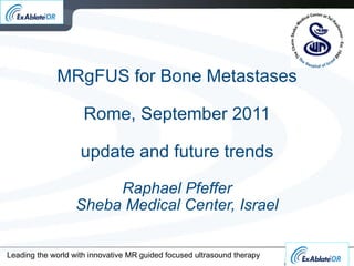
10. Leading the world with innovative MR guided focused ultrasound therapy Summary of Three Studies
11.
12. MRgFUS bone metastasis treatment overview Left : Screening MR Axial T1w with contrast, showing enhancement of the bone metastasis in the left iliac bone (marked with orange dashed circle); Middle : MR Axial T2w planning image with ultrasound beam overlay (blue); Right : Post treatment MR axial T1w with contrast, showing non-enhancement in the treated area (marked with orange dashed circle).
17. Leading the world with innovative MR guided focused ultrasound therapy Ribs: A- macro pathology showing lesion B- CT images showing new bone formation. Note: At 3M new bone formation & thickening of the cortical layer in the area of treatment MRgFUS and tumor control Interaction of Focused Ultrasound with bone A B
18. Tumor Control – preliminary clinical data Leading the world with innovative MR guided focused ultrasound therapy Immediately Post treatment T1w Contrast Enhanced subtraction Pre-Treatment T1w Contrast enhanced *courtesy of Sheba Medical Center Dr Yael Inbar
19. BM 016 Prospective, randomized study MRgFUS vs External Beam RadioTherapy Goal : Pain control local tumor ablation within the bone Endpoints : 1. Pain assessment 2. Follow up MR Imaging
31. Utilization of MRI in Radiation Oncology MRI Simulator in radiotherapy suite Why not adapt MRI Simulator for MRgFUS? increased use of MRgFUS for oncological indications CLOSED LOOP Increased uptake of MRI simulators
32.
Editor's Notes
The bone periosteom is a thin membrane covering the bone. The pain sensation from the bone originates from the periosteum For a successful treatment it is important to be able to sedate the patient (usually with Morphine) so he / she can tolerate the treatment. The high absorption of the bone allows faster treatments using wide beam approach, thus heating the area where the acoustic beam intersects with the bone, (right diagram). There is no significant heating on the other side of the bone since most of the energy is being absorbed by the bone. Prescribed energy is about half of energy usually prescribed in UF treatment. Therefore, treatment is less likely to cause complications (e.g., skin burn). Treatment is also faster because cooling time required in lower levels of energy is significantly shorter.
The bone periosteum is a thin membrane covering the bone. The pain sensation from the bone originates from the periosteum For a successful treatment it is important to be able to sedate the patient (usually with Morphine) so he / she can tolerate the treatment. The high absorption of the bone allows faster treatments using wide beam approach, thus heating the area where the acoustic beam intersects with the bone, (right diagram). There is no significant heating on the other side of the bone since most of the energy is being absorbed by the bone. Prescribed energy is about half of energy usually prescribed in UF treatment. Therefore, treatment is less likely to cause complications (e.g., skin burn). Treatment is also faster because cooling time required in lower levels of energy is significantly shorter.
This closed loop therapy paradigm of MR together with focused ultrasound allows the ability to get immediate feedback, react to that feedback, and know immediately what the outcome is. All this provides the physician with much more information than traditional surgical procedures. This ensures efficacy and safety of the procedure. Magnetic Resonance together with focused ultrasound provides precise visualization of the tumor, other organs, and the beam path for exact targeting and to ensure that there are no unwanted structures or organs in the beam path. It also allows for real time thermal imaging to evaluate if the beam actually reached the target and if so, if it reached coagulation temperatures. If it doesn’t reach the right temperature, then the physician has the ability to change the parameters.
An efficacy VAS analysis was conducted on 25 patients who both had full treatment and reached the 3-month follow-up visit. The mean baseline pain score before treatment was 5.9 (range: 3.5-8.5), mean pain score 3 days after treatment was 3.8 (range: 0-8.5), and by the three month post treatment follow up it had dropped to 1.8 (range: 0-8). A reduction of two points on 0-10 pain scale at three months proved to be statistically significant (p-value<0.003). 72% of patients (18/25) had a significant reduction in pain (> 2 points) at the 3-month follow up. Of these 50% (9/18) reported a pain score of zero. 24% had no response and 1 patient (4%) experienced worsened pain levels. It is significant to point out that 52% of patients reported substantial pain relief as early as at the 3 days post treatment 12 out of 25 patients used opioid analgesics and we were able to collect medication data on 10 of these patients. 66% of these decreased the use of analgesic medication , and 22% had increased their medication dosage. Most of the patients taking non-opioid analgesics (13/25) also reported a drop in medication usage. When combining both the medication and VAS score, 36% of patient had partial response and 36% had complete response according to the working group criteria for treatment outcome.
The illustration shows the usage of wide beam approach on a pelvic tumor. Difficult to see here but patient is not on back but somewhat tilted Details in the next slides
The bone periosteom is a thin membrane covering the bone. The pain sensation from the bone originates from the periosteum For a successful treatment it is important to be able to sedate the patient (usually with Morphine) so he / she can tolerate the treatment. The high absorption of the bone allows faster treatments using wide beam approach, thus heating the area where the acoustic beam intersects with the bone, (right diagram). There is no significant heating on the other side of the bone since most of the energy is being absorbed by the bone. Prescribed energy is about half of energy usually prescribed in UF treatment. Therefore, treatment is less likely to cause complications (e.g., skin burn). Treatment is also faster because cooling time required in lower levels of energy is significantly shorter.
One patient can be treated for more than one painful lesion
This closed loop therapy paradigm of MR together with focused ultrasound allows the ability to get immediate feedback, react to that feedback, and know immediately what the outcome is. All this provides the physician with much more information than traditional surgical procedures. This ensures efficacy and safety of the procedure. Magnetic Resonance together with focused ultrasound provides precise visualization of the tumor, other organs, and the beam path for exact targeting and to ensure that there are no unwanted structures or organs in the beam path. It also allows for real time thermal imaging to evaluate if the beam actually reached the target and if so, if it reached coagulation temperatures. If it doesn’t reach the right temperature, then the physician has the ability to change the parameters.