TREATMENT OF TGN WITH CYBERKNIFE FRAMELESS RADIOSURGERY SYSTEM

Trigeminal neuralgia (TN or TGN) is a long-term pain disorder that affects the trigeminal nerve, the nerve responsible for sensation in the face, and motor functions such as biting and chewing. It is a form of neuropathic pain. There are two main types: typical and atypical trigeminal neuralgia. The typical form results in episodes of severe, sudden, shock-like pain in one side of the face that lasts for seconds to a few minutes. Groups of these episodes can occur over a few hours. The atypical form results in a constant burning pain that is less severe. Episodes may be triggered by any touch to the face. Both forms may occur in the same person. It is regarded to be one of the most painful disorders known to medicine and often results in depression.

Recommended
More Related Content
What's hot
What's hot (20)
Similar to TREATMENT OF TGN WITH CYBERKNIFE FRAMELESS RADIOSURGERY SYSTEM
Similar to TREATMENT OF TGN WITH CYBERKNIFE FRAMELESS RADIOSURGERY SYSTEM (20)
More from Subrata Roy
More from Subrata Roy (13)
Recently uploaded
Recently uploaded (20)
TREATMENT OF TGN WITH CYBERKNIFE FRAMELESS RADIOSURGERY SYSTEM
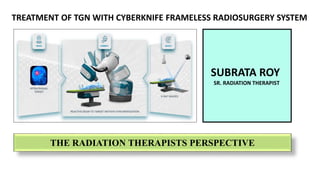
- 1. THE RADIATION THERAPISTS PERSPECTIVE SUBRATA ROY SR. RADIATION THERAPIST HCG CANCER CENTRE MUMBAI SUBRATA ROY SR. RADIATION THERAPIST TREATMENT OF TGN WITH CYBERKNIFE FRAMELESS RADIOSURGERY SYSTEM
- 2. Table of Presentation What is TGN History Behind Trigeminal Neuralgia Classifications of TGN Types of TGN General Characteristics of TGN Treatment strategies for Trigeminal Neuralgia Cyberknife Treatment Preparation for TGN Treatment Delivery (Therapists Role & Observations) Efficiency Of Cyberknife System In View of Treating Small Field Target Results and outcome Analysis. Conclusions
- 3. What Is TGN Trigeminal neuralgia or tic douloureux is a neuropathic disorder of trigeminal nerve that causes episodes of intense pain in eyes, lips, scalp, forehead and jaws. It has been labeled as Suicide disease due to insignificant number of people taking their own life because they are unable to have their pain controlled by medication or surgery. Synonyms: Tic Doulourex Trifacial Neuralgia Fothergills disease
- 4. TGN is 5th Cranial Nerve (CN V) It is largest of the cranial nerves Trigeminal (tri- three , geminus-twinned) Responsible for sensation in the face and motor functions The patients typically presents with severe episodic lancinating, shock like pain sensation in face TGN is most chronic pain condition of the trigeminal nerve Incidence - 4-5/1 lac population
- 5. History of Trigeminal Neuralgia Aretaeus of Cappadocia –At the end of first century -1st clinical description of TN 1677 John Locke, gave the first full description with its treatment. 1756 Nicolaus Andre - tic douloureux 1773 John Fothergill in published detailed description of TN
- 6. Classifications of Trigeminal Neuralgia TGN TYPICAL TGN ATYPICAL TGN PRIMARY /IDIOPATHIC SECONDARY Typical idiopathic TN due to vascular compression with morphologic changes of the trigeminal nerve root Diagnosis of secondary TN relies on the demonstration of a major neurologic disease that causes the neuralgia. A tumor at the cerebellopontine angle or MS causes TN in 15% of patients TN with continuous pain TN type 2 or atypical, idiopathic TN. (Danger!!!)
- 7. • V1- Supraorbital ridge affected side • V2 –Skin of upper Lip,Ala,Cheeks & Upper gums. • V3-Lower lip, teeth or gums of lower jaw Trigger Zones & Trigger Points
- 8. Washing the FACE Brushing Teeth Shaving Vibrations from Walking Going Out In Cold Wind Trigger Factors Eating ,drinking water
- 9. Types Of TGN Typical Trigeminal Neuralgia Atypical Trigeminal Neuralgia Pre- Trigeminal Neuralgia Multiple Sclerosis Related Trigeminal Neuralgia Secondary Or Tumour Related Trigeminal Neuralgia Trigeminal Neuropathy Or Post- Traumatic Trigeminal Neuralgia Failed Trigeminal Neuralgia
- 10. General Characteristics Of TGN Incidence: 8 : 1,00,000 Age: 5th – 6th decade of life Sex: Female > male ; 1.6 > 1.0 Affliction for side: Right > left Division of trigeminal nerve involvement: V3>V2>V1
- 11. Treatment strategies for Trigeminal Neuralgia First line treatment option: Medical management (Pharmacotherapy) Treatment options for medically refractory TGN: • Surgical: microsurgical vascular decompression (MVD) or ablative procedures like rhizotomy • Minimal-invasive percutaneous retro gasserian rhizotomies (PRR) • Nerve block • Gycerol injection • Stereotactic Radiosurgery
- 12. Stereotactic Radiosurgery Options Cyberknife Gamma Knife X-Knife (LINAC BASED)
- 13. There is a Treatment Option that can be done without going under the knife that called as Cyberknife Thanks to modern technology, Now Patients can have some surgeries without being cut open!
- 14. Introducing Cyberknife Frameless Radiosurgery System
- 15. Functional Neurosurgery requires the less possible invasive treatment Frameless radiosurgery reduces as much as possible the invasively of a surgical approach to trigeminal neuralgia The non-isocentric approach introduces a series of new and unexplored issues for treatment with a non-isocentric technique, the dose is not the only parameter to consider actually, Volume of the treated nerve and the dose received by the surrounding structures must be taken into consideration What Makes It Distinct Cyberknife In order to better understand about Cyberknife, lets take a look at the following Highlights about this…
- 17. Cyberknife Treatment:- Cyberknife Frameless Radio surgical System Offering highly Precise ,Non-Surgical Treatment for TGN.
- 18. Cyberknife Treatment Preparation for TGN The Cyberknife is made up of a radiation delivery device (Linac) attached to a robotic arm. Cyberknife is capable to treat tumors anywhere in the body. There is no need for a patient to be still or head frames to hold them in place because the Cyberknife automatically adjusts to the movement of the patient or tumor. The Cyberknife is Pain-free, no anesthesia is required Treatment Preparation in Cyberknife Usually Completed in Following Steps :- U frame Thermoplastic Mould Preparation Acquiring CT Scan with Predefined Standard Cyberknife 6D Skull TGN Protocol Obtaining MRI Series Treatment Planning Using Cyberknife Treatment Planning System
- 19. Radiation Therapists Played an Immense Role In the Treatment Preparation & Treatment delivery Of Trigeminal Neuralgia, While they are involving in Following Steps to made this Delivery Successfully:- U Frame Thermoplastic Mould Preparation (Immobilization Part) Acquiring CT Scan with Predefined Standard Cyberknife 6D Skull TGN Protocol. U Frame Thermoplastic Mould Preparation (Immobilization Part) U Frame Thermoplastic Mask is Having a thickness of 1.25 mm & giving high quality rigidness which is very essential while we are delivering high dose of radiation to the pinpoint Target 0.01 c.c While making the U frame Mask Needs to be very confirmative on the selection of proper Head Rest Which will directly reflating on the Patient Comfort and as well as the reproducibility and minimizing the setup error in Sub mm range.
- 20. • All TGN Patients Head Mask, Proper Nasal Impression Should be given by therapist which will create impact on the Setup accuracy as per the Standard. • In Case Of Female Patients Long Hair will always gives a bigger challenge to the therapists to reproduce the setup on daily basis . • Immobilization with Hair Out Or Hair In Should be mentioned on the Patient Setup Information page and same protocol needs to be fixed on institutional basis to improve the Bench mark of Good Patient setup
- 21. Acquiring CT Scan with Predefined Standard Cyberknife 6d Skull TGN Protocol All patient immobilization devices that will be used during patient treatment must be used during CT scanning. Include the CT table top, U Frame Base Plate, Head Rest, index bar, and any pads used. Field of View (FOV): Include the entire circumference of the skull. Use the smallest FOV that encompasses the anatomy for the best pixel resolution. Include part of the headrest and extend 1 cm anterior past the tip of the nose & as much anatomy as possible. The CT scan is used to create Digitally Reconstructed Radiographs (DRR) images. DRR images must have a 0 – 1 cm gap anterior and superior to the patient skull. AP and lateral scout images help ensure there are approximately 0 – 1 cm gaps anterior and superior to the skull.
- 22. Things to Keep in Mind by Therapists While Performing CT Simulation Guidelines For Patient Orientation as Per Cyberknife Protocol Patient Simulation Orientation Must be Checked Before Performing
- 23. Treatment Planning Using Cyberknife Treatment Planning System • RT planning CT and MRI (T1 ,T2) images fused with deformable image registration software • Trigeminal nerve is delineated till it enters in meckel’s cave • Choice of target – Anterior (Cisternal) OR Posterior (Dosral root entry zone - REZ) decided depending neurovascular conflict location • OARs Contouring - Brainstem, Temporal Lobe, Blood vessel, Cochlea, VII-VIII nerve complex Target Delineation Fusion & Contouring
- 24. Treatment Plan Preparation Dose Volume Histogram
- 25. TGN Dose Prescription & Treatment Planning :- Sharp fall off of Dose around the small Field Target Plan Approval Criteria's: • Target Coverage • Sharp Dose Fall Off • 95%, 50%,30% Dose • Dose Heterogeneity • Brain Stem and Other OAR Dose Limits A dose of 80-85Gy max dose prescribed to the Dorsal root entry zone and 20% IDL touching the brainstem.
- 26. • Prescription dose 80 Gy to 85 Gy at Isocenter (Maximum Point Dose) • More than 50Gy (58% ISDL) to encompass TGN • Maximum Brainstem Point dose 25Gy (30% ISDL) or 0.03cc 18Gy • Vestibular cochlea nerve max dose < 6Gy • Temporal Lobe Max Dose 30 Gy Institutional Plan Evaluation Criteria
- 27. Dose Distribution & Target Coverage ( CT Plane )
- 28. Dose Distribution & Target Coverage on 3D FIESTA seq MRI
- 29. Dose Distribution & Target Coverage
- 30. Treatment Delivery (Therapists Role & Observations) Treatment Delivery Console The treatment delivery computer is the main computer used for delivering and monitoring patient treatment. It includes a flat panel monitor. The monitor, keyboard, and mouse are located in the Control Room. The treatment delivery computer is usually located in the Equipment Room. 6D Skull Tracking System The 6D Skull Tracking system enables tracking of intracranial targets without the need for stereotactic frames. 6D Skull Tracking mode works by computing the offset between Live X-ray images and reference DRR images by identifying and matching skeletal features of the skull. Tracking the target relies on the fixed relationship between the target volume and these skeletal features.
- 31. Left and Right Screens of the Delivery Phase The Delivery phase for 6D Skull Tracking mode
- 32. Non-Isocentric Beam Delivery Highly collimated beams Non-convergent beams Superior conformality while Maximizing homogeneity Non-Coplanar Beam Delivery Automatically minimizes entrance/exit beam interactions. No patient or Linac re-positioning required.
- 33. 6D Skull tracking system used for intra-cranial lesions up to C2,Bony anatomy of the skull is used as reference for tracking This tracking feature allows direct and non-invasive tracking of Intracranial lesions Target tracking and motion compensation are accomplished by identifying and tracking rigid Skull anatomy image intensity and brightness gradients between the DRR and LIVE images The naming of this method as 6D because the corrections are made for the 3 translational motions and 3 rotational motions
- 34. All the Offset Correction values should be Constantly below 0.3mm during the entire course of Treatment in all direction including the rotational Correction ,if the Planning done on Normal Skull Path Image Interval Should be kept as low as Possible (Usually 30/min) ( To Observe and made corrections for intrafraction Movement) LONGITUDINAL LATERAL VERTICAL ROLL PITCH YAW
- 35. Efficiency of CK in view of Treating Small Field Target Among the advanced Technologies available, Cyberknife Radiosurgery System is a more recent image-guided frameless robotic system, which is designed for SRS and stereotactic radiation therapy (SRT) The associated 6D skull tracking system enables highly conformal intracranial high-dose tumour targeting, with sub-mili-meter precision in beam delivery allowing for steep dose gradients achievable around the contoured tumour. An additional benefit is the frameless nature of the system which makes it easy to opt for hypofractionation regimens to minimize toxicity Single fraction SRS can be used to treat small to moderate sized OGMs (< 10 cm3) that are not spatially associated with the optic apparatus while larger OGMs (> 10 cm3) can be treated using fractionated planning to achieve normal tissue sparing and preserve cranial nerve function.
- 36. Cyberknife is a safe, effective, minimally invasive and potentially cost-effective treatment modality for patients with medically intractable TN or those who are ineligible or refuse open surgery. Our results demonstrate that a CKRS treatment is associated with good outcomes in the majority of patients with sustained relief of TN pain in most responding to therapy AS THIS STUDY SHOWS
- 37. Results & Outcome Analysis Pain relief after Cyberknife Radiosurgery A large proportion of patients (42.9%) reported pain relief within 1 month following CKRS treatment. 19 % of patients reported relief within 6 months of treatment and another 19% reported relief no relief of symptoms. In 14.3% relief was experienced within 3 months from treatment. The median time to recurrence of severe pain was 19 months (mean 27.8 months; range 1–129 months). Treatment related complications The majority of the patients (18 or 72%) did not experience any new bothersome post-treatment facial numbness 2 patients developed new somewhat bothersome facial numbness No patients developed any new very bothersome facial numbness Indeed 2 patients reported improvement in her facial pain from very bothersome facial numbness to no facial numbness.
- 38. Sex (M vs. F) Age (<60 vs. >60 years) Side (Right vs. Left) Pain type (TN1 vs. TN2) Target length (<5 vs. 5 to 6 mm) Target volume (<30 mm3 vs. >30 mm3) Target dose (<58 versus >58 Gy) Factors affecting outcome:-
- 39. Conclusions Safe handling and Operation of the Cyberknife System requires careful attention familiarity with emergency procedures & constant Close View on CCTV while Robot moves around the patient Careless operation of the Cyberknife System can damage the system, its components or other property; cause poor performance; or lead to serious bodily injury and possibly death. Therapists who operates the Cyberknife System must read, understand, and be thoroughly familiar with the information in the manual. Trigeminal Neuralgia has been an enigma to physicians for a long course of time. There have been various advances in the understanding of the pathogenesis of the disease per se and the treatment modalities.
- 40. THANK YOU FOR KIND ATTENTION