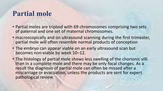
GESTATIONAL
TROPHOPLASTIC DISEASE
DR.ATHRAA J. ALENEZE
terminology
• In latin "hydatid" means "drop of water” "mole" means "spot”.
• H. mole is a pregnancy characterized by vesicular swelling of placental villi
and usually the absence of an intact fetus.
• the terminal extremities of the chorionic villiare converted intotransparent vesicles withclear,
viscidcontents. These vary in size fromminute bodies a few millimeters in diameter to cystic
structures the size of hazel-nuts, and hang in clusters fromthe villous stems, to which they are
connectedby thinpedicles, giving to theexternal surface of thechoriona grape-like
appearance.
J. Whitridge Williams (1903)
Introduction
1. Gestational Trophoblastic diseases- heterogeneous
group of interrelated lesions that arise from abnormal
proliferation of placental trophoblast.
2. GTN-subset of malignancies that have varying
propensities for local invasion & metastasis
3. GTN-rare human tumors, cured even in the presence of
widespread dissemination
WHO Classification GTD
Premalignant Diseases
Complete Hydatidiform Mole ( C M )
Partial Hydatidiform Mole ( P M )
Atypical placental site nodules
Malignant Diseases (Gestational Trophoblastic Neoplasia)
Nonmetastatic
• Invasive Mole
• Placental site trophoblastic tumor ( PSTT )
• epithelioid trophoblastic tumor (ETT)
Metastatic
• Gestational Choriocarcinoma
Pathogenesis
• Paternal genes have more control over placental growth, while maternal genes
have more control over fetal growth. Thus, with excess paternal genes, there is
excessive placental or trophoblastic proliferation.
• Complete mole is recognized by the presence of characteristic grapelike structures,
which represent swollen chorionic villi, and the absence of a viable fetus, The
chorionic villi are diffusely hydropic and enveloped by hyperplastic and atypical
trophoblast
• Complete moles are usually diploid, the most common type being 46XX; in most
cases, a haploid sperm divides within an ovum without a nucleus.
• partial moles usually have recognizable embryonic and fetal tissues, with focal
hydropic swelling of the chorionic villi and focal trophoblastic hyperplasia.
• Partial moles are generally triploid, for example 69XXY; they result most often from
Di spermic fertilization of normal ova.
• When a fetus is present it often has the features of triploidy, including growth
retardation and multiple congenital malformations
Epidemiology
• An ethnic predisposition is seen with hydatidiform mole, which has increased
prevalence in Asians, Hispanics, and American Indians
• The incidence in the United States and Europe has been relatively constant at 1 to
2 per 1000 deliveries
• The strongest risk factors are age and a prior hydatidiform mole.
• Women at both extremes of reproductive age(below 15yr and above 45yr) are
most vulnerable. Specifically, adolescents and women aged 36 to 40 years have a
twofold risk, but those older than 40 have an almost tenfold risk
• With a prior complete mole, the risk of another mole is 0.9%, and with a previous
partial mole, the rate is 0.3%. After two prior complete moles, approximately 20%
of women have a third mole
• partial molar pregnancies are slightly more common than complete moles, with an
approximate ratio of 60 : 40 in the UK series.
• Nutritional factors- low socio-economic status, carotene & animal fat soluble
vitamin deficiency
Partial mole
• Partial moles are triploid with 69 chromosomes comprising two sets
of paternal and one set of maternal chromosomes.
• macroscopically and on ultrasound scanning during the first trimester,
partial mole will often resemble normal products of conception
• The embryo can appear viable on an early ultrasound scan but
becomes non‐viable by week 10–12.
• The histology of partial mole shows less swelling of the chorionic villi
than in a complete mole and there may be only focal changes. As a
result the diagnosis of partial mole can often be missed after a
miscarriage or evacuation, unless the products are sent for expert
pathological review.
Partial mole
• The clinical presentation of partial mole is most frequently via a failed
pregnancy rather than irregular bleeding or by detection on routine
ultrasound.
• U/S exam shows the following:
• Large placenta with cystic spaces within it, and an empty gestational sac or sac
containing amorphous echoes, or growth restricted fetus
• increase in the ratio of transverse/AP diameter of gestational sac to a ratio of >1.5
• The obstetric management is by suction evacuation and histological review.
• All patients with partial mole should be followed up by registration and serial
HCG measurement
• Fortunately, partial mole rarely transforms into malignant disease, and there
is an overall risk of 0.5–1% of patients requiring chemotherapy after a partial
mole
Complete mole
• In complete moles the genetic material is totally male in origin
• The clinical diagnosis of complete mole most often occurs as a result of
first‐trimester bleeding or an abnormal ultrasound. There is no fetal material
and the histology shows the characteristic edematous villous stroma.
• he textbook ‘bunch of grapes’ appearance is only seen in the second
trimester and as most cases are diagnosed earlier
• Obstetric management is by suction evacuation followed by registration and
serial hCG measurement.
• Complete molar pregnancies have an appreciable risk of proceeding to
invasive disease, with approximately 15% of patients with complete mole
requiring chemotherapy
Familial recurrent hydatidiform mole syndrome
• Occasionally we see patients who have repetitive complete molar pregnancies and
who have failed to conceive a normal baby
• Patients who have had three consecutive complete moles should be genetically
tested to determine whether they have FRHM syndrome where the DNA is
biparental in origin rather than androgenetic
• mutations in two genes have been associated with this autosomal recessive
condition, NLRP7 and KHD3CL
• It is member of CATERPILLAR family involved in inflammation & Apoptosis in genetic
CM
• Unfortunately, the risk of subsequent malignancy requiring chemotherapy is the
same for these patients as for those with androgenetic complete moles. Therefore
recommending repeated rounds of attempted natural conception does not seem
appropriate.
Twin pregnancies
Histological appearances of complete (a) and partial (b) mole. (a) 46XX, paternal. Haploid sperm fertilizes
empty ovum and undergoes cytogenesis; diploid sperm fertilizes empty ovum. (b) Triploid: 69,XXY 69,XXX or
69,XYY. Defective zona pellucidum; Di spermic fertilization of haploid ovum
Sonograms of hydatidiform moles. A. Sagittal view of a uterus with a complete hydatidiform mole. The characteristic
“snowstorm” appearance is due to an echogenic uterine mass, marked by calipers, that has numerous anechoic cystic
spaces. Notably, a fetus and amnionic sac are absent. B. In this image of a partial hydatidiform mole, the fetus is seen
above a multicystic placenta.
Twin pregnancies
• The risk of having a twin pregnancy comprising a complete mole and healthy
co‐twin is estimated to be 1–5 per 100 000
• The risk of malignancy was not increased by allowing such pregnancies to
continue
• Nearly 40% of them resulted in a healthy take‐home baby with most of the
rest undergoing spontaneous loss from presumed continued molar growth
• The mode of delivery has been a mixture of normal vaginal and caesarean
section depending on local assessment of what was considered the safest
option
• After appropriate counselling, it seems reasonable to allow women to
continue such pregnancies but under close monitoring to ensure that potential
complications are detected early and dealt with promptly
Atypical placental site nodules
• Following a normal pregnancy, placental remnants in the uterine wall
usually spontaneously regress and disappear. Occasionally, they may
persist and form placental site nodules
• These may present with irregular bleeding that often results in
dilation and curettage.
• Some placental site nodules may have atypical histopathological
features and, if so, can in 10–15% of cases be associated with or
subsequently develop into either a PSTT or ETT
• If atypical placental site nodule is confirmed, then these patients are
registered and seen centrally for monitoring
CLINICAL FEATURES
Complete Mole
• Vaginal bleeding
• Uterus is larger than gestational age
• Hyperemesis Gravidarum
• PET 10-15%
• hyperthyroidism 7%
• Theca lutein cysts
• Beta h CG levels >100 000
• Passage of grapes like vesicle
Clinical features
PARTIAL MOLE
• Uterus is often not enlarged more than POG
• More often presents as Missed or incomplete
abortion
• Pre evacuation HCG levels are not more than
100,000IU/ L
• Macroscopic : villous swelling is less intense
• Embryo is present
Management of Molar Pregnancy
• Suction curettage is the method of choice for evacuation of complete as well as
partial mole
• Medical evacuation of complete molar pregnancy should be avoided as far as
possible because of theoretical risk of embolization & dissemination of
trophoblastic tissue through the venous system
• In case of partial mole ,medical evacuation can be used when the size of the fetus
prevent the use of suction curettage
• Cervix can be prepared immediately prior to evacuation
• Due to lack of fetal part , suction catheter size 12mm is sufficient to evacuate
• . It is recommended that, when necessary, oxytocic therapy is only commenced
once evacuation is complete.
• If there is significant haemorrhage prior to or during evacuation and some degree
of control is needed, oxytocic agent may be used according to clinical judgement. It
is also suggested that prostaglandin analogues should be reserved for cases for
which oxytocic therapy is ineffective
Management of Molar Pregnancy
• Second uterine evacuation
• There is NO clinical indication for the routine use of second uterine
evacuation.
• It may be useful for symptom control in selected patients with heavy
bleeding or curative if the recurrent molar tissue is confined to the
uterine cavity
Management of Molar Pregnancy
• Hysterectomy may serve as an option in the following cases of H.mole:
• Elderly multiparous women (age > 40 yrs 0 who don’t wish to become pregnant in
the future
• Those women with H.mole desiring sterilization
• Sever infection or uncontrolled bleeding
• Patient with non metastatic persistent disease who completed her family
• If theca lutein cysts are present they must be left as it is, or they can be
aspirated to reduce their size , with ovarian conservation
• Since hysterectomy does not prevent metastatic disease, follow up with β-
HCG level is essential even after surgery
Management of Molar Pregnancy
• Serial assay of serum β-HCG level should be carried out in 2 wkly basis ,
until 3 negative level are obtained
• Average time for first normal HCG post evacuation is7 weeks for partial
moles and 9 weeks for complete moles.
• Once β-hCG is undetectable, this is confirmed with monthly
determinations for another 6 months
• Anti-D Prophylaxis is given to Rh negative women because fetal tissues
with a partial mole may include red cells with D-antigen
• Those with suspected complete mole are similarly treated because a
definitive diagnosis of complete versus partial mole may not be confirmed
until histological evaluation of the evacuated products.
Management of Molar Pregnancy
• If hCG has reverted to normal within 56 days (8wks)of the pregnancy event
then follow up will be for 6 months from the date of uterine evacuation.
• If hCG has not reverted to normal within 56 days of the pregnancy event
then follow-up will be for 6 months from normalization of the hCG level.
• Follow up of partial molar pregnancy is stopped, once the HCG levels has
normalized on two samples, at least 4 wk. apart
• Women who have not received chemotherapy, do not require measurement
of there β –HCG levels after any subsequent pregnancy event
Management of Molar Pregnancy
• Women with GTD should be advised to use barrier methods of
contraception until hCG levels revert to normal.
• Once hCG level have normalized, the combined oral contraceptive
pill may be used
• Intrauterine contraceptive devices should not be used until hCG
levels are normal to reduce the risk of uterine perforation
• Women should be advised not to conceive until their follow-up is
completed
Gestational trophoblastic Neoplasia
Nonmetastatic
Invasive Mole
• Clinical features
• Clinical diagnosis by persistence or rising titers of β-HCG in the weeks after
molar evacuation & USG
• Persistent bleeding p/v
• Lower abdominal pain due to invasion in myometrium, vulva, vagina or intra
abdominal metastasis
• It may spread to adjacent pelvic structures bladder , rectum; hematuria,
bleeding P/ R
• Pulmonary metastasis
• Mostly due to initial diagnosis of CM is missed & not on β-HCG follow up
• Invasive mole nearly always arises from a complete mole
Placental Site Trophoblastic Tumor (PSTT)
and epithelioid trophoblastic tumor(ETT)
• rare slow growing tumor, and is the rarest form of GTD, accounting for 0.2%
of all registered cases of GTD
• Little or no HCG is produced ( Free βhCG fragment )
• Microscopically: In the normal placenta it is distinct from villous trophoblast
& infiltrates the decidua, myometrium, & spiral arteries.
• Pathologically, ETT is distinguished from PSTT by its smaller cells, its smaller,
more monomorphic cells and by its nested, nodular, well‐circumscribed
growth pattern unlike the sheet‐ like infiltrative pattern seen with PSTT
• Mainly from intermediate trophoblast derived from cytotrophoblast
Origin of PSTT. (A) PSTT originates from extravillous trophoblasts, and then acquires the abilities of proliferation and migration.
Afterwards, those cells migrate away from placenta and invade the decidual artery and uterine spiral artery to remodel the
blood vessels which in turn provide nutrition for the embryo. The disruption of this well-regulated invasion process may lead to
PSTT. (B) During delivery, placenta detaches from decidual, leaving small nodules and form placental site nodules in the
myometrium. In the process of reabsorbing, some adverse trigger or stimuli may cause atypical mitosis and result in neoplasm.
• PSTT/ETT most commonly follows a normal pregnancy but can also occur
after a non‐molar abortion or a molar pregnancy.
• in PSTT the average interval between the prior pregnancy and
presentation is 3.4years. The most frequent presentations are
amenorrhoea or abnormal bleeding.
• lung and liver is the most common sites of distant spread
• Spread is late local Infiltration & metastasis is through lymphatic
Metastatic tumor
CHORIOCARCINOMA
Clinical features
• Occurs mainly following any form of pregnancy, mainly after complete mole, but can
also arise after any other type of pregnancy including a partial mole, a non‐molar
abortion, miscarriage, ectopic or term pregnancy
• Clinical features of bleeding p/v , lower abdominal pain, or in 1/3 of cases no pelvic
symptoms but symptoms of distant metastasis lungs , brain ,liver, skin, cauda equina
& the heart may present
• Highly malignant , appears as soft purple largely hemorrhagic mass
• Microscopic: implanting blastocyst with central cores of mononuclear
cytotrophoblast surrounded by rim of multinucleated syncytiotrophoblast & distinct
absence of chorionic villi extensive areas of necrosis & haemorrhage &frequent
evidence of tumor in the sinuses
• The hypervascularity & absence of connective tissue support are the reason for its
highly malignant behavior
• DIAGNOSIS IS BY β -HCG
Investigations
• Quantitative beta hCG
• X Ray Chest
• Pelvic Doppler USG
• Abdominal doppler USG to rule out liver &
renal metastasis
• CT chest , abdomen
• MRI brain
• Beta h CG in cerebrospinal fluid
• PET scan
• Genetic studies can reflect its origin from a
complete or partial mole or a normal
diploid conception combined with a range
of gross abnormalities without any specific
characteristic patterns
FIGO REQUIREMENT FOR MAKING DIAGNOSIS OF
GTN
• 1. Four values or more of plateau hCG over at least 3 weeks: days 1, 7,
14 and 21.
• 2. A rise of hCG of 10% or greater for 3 values or more over at least 2
weeks: days 1, 7, and 14.
• 3. Histologic diagnosis of choriocarcinoma.
• 4. Persistence of hCG beyond 6 months after mole evacuation.
Metastatic disease
Low Risk GTN
• FIGO score 6 or less.
• Drugs schedules:
• single agent chemotherapy Most commonly used regimen.
• Methotrexate: 1 mg/kg 1M on days 1, 3, 5, 7
• Folinic acid 0.1 mg/kg 1M on days, 2, 4, 6 and 8.
• Side Effects: Stomatitis, conjunctivitis, abdominal and chest pain.
• Actinomycin- D:(1.25mg/m2), i.v. repeated every 2 weeks (primary therapy in case of abnormal
liver function)
• Bleeding usually responds well to strict bed rest and less than 1% of low‐risk patients
require emergency interventions such as embolization, vaginal packing or
hysterectomy.
• Treatment is continued until normalization of the serum hCG level (0–4 IU/L) and then
a further three cycles (6weeks) to ensure eradication of any residual disease that is
below the level of serological detection
Treatment and human chorionic gonadotrophin (hCG) graph of a low‐risk patient with a gestational
trophoblast tumor, successfully treated with methotrexate (MTX) and folinic acid (FA) chemotherapy.
Role of hysterectomy in non metastatic
disease i.e. Stage-I
• Choice (older patient, localized disease, family complete).
• Excessive uterine bleeding (before or during treatment).
• Chemo resistant (localized) uterine tumor.
• Placental site trophoblastic tumor stage 1
Follow up in Low risk GTN
• Weekly β-HCG titer until normal for 3 consecutive weeks.
• Monthly β-HCG level until normal for 12 consecutive months.
• Effective contraception during the follow up
• FIGO recommends additional 3 courses of chemotherapy after initial
negative β-HCG.
High Risk GTN
• Stage I, II, III With FIGO score 7 or greater or Stage IV
• Primary intensive combination chemotherapy
• Regimes given :
• MAC.
• Modified Bagshawe (CHAMOCA)
• EMA-CO
• EMA-EP
• TE/TP (paclitaxel)
MAC-III Regime
• Mtx 1 mg/kg IM/1V Days 1, 3, 5, 7
• Folinic acid 0.1 mg/kg IM Days 2, 4, 6, 8
• Actinomycin-D 12 g/kg IV Days 1-5
• Cyclophosphamide 3 mg/kg IV Days 1-5
• To be repeated every 15 days if toxicity permits
EMA-CO Regime
• Etoposide, methotrexate, actinomycin D, alternating weekly with
cyclophosphamide and oncovin.
• Day 1
• Actinomycin D 500 micrograms IV push IV.
• Etoposide 100 mg/m2 over 30-50 min.
• Methotrexate 100 mg/m2 IV infusion over 1 hr and then
• Methotrexate 200 mg/m2 IV infusion over 12 hrs
• Day 2
• Actinomycin D 500 micrograms IV push
• new IV Etoposide 100 mg/m2 over 30-50 min.
• Folinic acid 15 mg IV push Q 6 hrs for 8 doses beginning 24 hrs after methotrexate bolus.
• Day 8
• Vincristine (Oncovin) 1 mg/m2 IV
• Cyclophosphamide 600 mg/m2 IV
• SIDE EFFECT:- Myelosuppression ,Mucositis , Neuropathy ,Reversible alopecia
Treatment and human chorionic gonadotrophin (hCG) graph of a low‐risk patient with a gestational trophoblast
tumor, initially treated with methotrexate (MTX) and folinic acid (FA) chemotherapy, changing to EMA‐CO
treatment in response to an hCG plateau.
EMA-EP regime
• In patient resistant to EMA-CO
• On day 8
• Etoposide- 10 mg/m2 iv.
• Cisplatin- 80 mg/m2 iv
• treatment – until 3 consecutive weekly titers normal.
• 2-4 cycles given further after initial normal β-HCG.
Follow up OF High risk GTN
• Weekly until β-HCG normal for 3 consecutive weeks.
• Monthly β-HCG for 24 consecutive months.
• Contraception in follow up period.
• Salvage of EMA/CO failures
• Of the high‐risk patients treated with EMA/CO, approximately 18–20% develop
resistance
• In this situation, the EP/EMA or TE/TP regimens may be used, which incorporate
cisplatin and additional etoposide into the combination along with paclitaxel in the
TE/TP regimen.
• salvage about 80% of patients failing EMA/CO if combined with surgry
• Ultra‐high‐risk disease
• high‐risk patients at greatest risk of death
• Death from :fatal haemorrhage, metabolic upset from tumor lysis, very
advanced disease and from multiresistant disease.
• Factors associated with deaths included
• a FIGO score above 12,
• very advanced lung disease
• liver with or without brain metastases
• an interval from the last‐known and presumed causative pregnancy greater than 2.8
years
GTD.pptx

GTD.pptx

  • 1.
  • 2.
    terminology • In latin"hydatid" means "drop of water” "mole" means "spot”. • H. mole is a pregnancy characterized by vesicular swelling of placental villi and usually the absence of an intact fetus. • the terminal extremities of the chorionic villiare converted intotransparent vesicles withclear, viscidcontents. These vary in size fromminute bodies a few millimeters in diameter to cystic structures the size of hazel-nuts, and hang in clusters fromthe villous stems, to which they are connectedby thinpedicles, giving to theexternal surface of thechoriona grape-like appearance. J. Whitridge Williams (1903)
  • 3.
    Introduction 1. Gestational Trophoblasticdiseases- heterogeneous group of interrelated lesions that arise from abnormal proliferation of placental trophoblast. 2. GTN-subset of malignancies that have varying propensities for local invasion & metastasis 3. GTN-rare human tumors, cured even in the presence of widespread dissemination
  • 5.
    WHO Classification GTD PremalignantDiseases Complete Hydatidiform Mole ( C M ) Partial Hydatidiform Mole ( P M ) Atypical placental site nodules Malignant Diseases (Gestational Trophoblastic Neoplasia) Nonmetastatic • Invasive Mole • Placental site trophoblastic tumor ( PSTT ) • epithelioid trophoblastic tumor (ETT) Metastatic • Gestational Choriocarcinoma
  • 7.
    Pathogenesis • Paternal geneshave more control over placental growth, while maternal genes have more control over fetal growth. Thus, with excess paternal genes, there is excessive placental or trophoblastic proliferation. • Complete mole is recognized by the presence of characteristic grapelike structures, which represent swollen chorionic villi, and the absence of a viable fetus, The chorionic villi are diffusely hydropic and enveloped by hyperplastic and atypical trophoblast • Complete moles are usually diploid, the most common type being 46XX; in most cases, a haploid sperm divides within an ovum without a nucleus. • partial moles usually have recognizable embryonic and fetal tissues, with focal hydropic swelling of the chorionic villi and focal trophoblastic hyperplasia. • Partial moles are generally triploid, for example 69XXY; they result most often from Di spermic fertilization of normal ova. • When a fetus is present it often has the features of triploidy, including growth retardation and multiple congenital malformations
  • 9.
    Epidemiology • An ethnicpredisposition is seen with hydatidiform mole, which has increased prevalence in Asians, Hispanics, and American Indians • The incidence in the United States and Europe has been relatively constant at 1 to 2 per 1000 deliveries • The strongest risk factors are age and a prior hydatidiform mole. • Women at both extremes of reproductive age(below 15yr and above 45yr) are most vulnerable. Specifically, adolescents and women aged 36 to 40 years have a twofold risk, but those older than 40 have an almost tenfold risk • With a prior complete mole, the risk of another mole is 0.9%, and with a previous partial mole, the rate is 0.3%. After two prior complete moles, approximately 20% of women have a third mole • partial molar pregnancies are slightly more common than complete moles, with an approximate ratio of 60 : 40 in the UK series. • Nutritional factors- low socio-economic status, carotene & animal fat soluble vitamin deficiency
  • 10.
    Partial mole • Partialmoles are triploid with 69 chromosomes comprising two sets of paternal and one set of maternal chromosomes. • macroscopically and on ultrasound scanning during the first trimester, partial mole will often resemble normal products of conception • The embryo can appear viable on an early ultrasound scan but becomes non‐viable by week 10–12. • The histology of partial mole shows less swelling of the chorionic villi than in a complete mole and there may be only focal changes. As a result the diagnosis of partial mole can often be missed after a miscarriage or evacuation, unless the products are sent for expert pathological review.
  • 11.
    Partial mole • Theclinical presentation of partial mole is most frequently via a failed pregnancy rather than irregular bleeding or by detection on routine ultrasound. • U/S exam shows the following: • Large placenta with cystic spaces within it, and an empty gestational sac or sac containing amorphous echoes, or growth restricted fetus • increase in the ratio of transverse/AP diameter of gestational sac to a ratio of >1.5 • The obstetric management is by suction evacuation and histological review. • All patients with partial mole should be followed up by registration and serial HCG measurement • Fortunately, partial mole rarely transforms into malignant disease, and there is an overall risk of 0.5–1% of patients requiring chemotherapy after a partial mole
  • 12.
    Complete mole • Incomplete moles the genetic material is totally male in origin • The clinical diagnosis of complete mole most often occurs as a result of first‐trimester bleeding or an abnormal ultrasound. There is no fetal material and the histology shows the characteristic edematous villous stroma. • he textbook ‘bunch of grapes’ appearance is only seen in the second trimester and as most cases are diagnosed earlier • Obstetric management is by suction evacuation followed by registration and serial hCG measurement. • Complete molar pregnancies have an appreciable risk of proceeding to invasive disease, with approximately 15% of patients with complete mole requiring chemotherapy
  • 13.
    Familial recurrent hydatidiformmole syndrome • Occasionally we see patients who have repetitive complete molar pregnancies and who have failed to conceive a normal baby • Patients who have had three consecutive complete moles should be genetically tested to determine whether they have FRHM syndrome where the DNA is biparental in origin rather than androgenetic • mutations in two genes have been associated with this autosomal recessive condition, NLRP7 and KHD3CL • It is member of CATERPILLAR family involved in inflammation & Apoptosis in genetic CM • Unfortunately, the risk of subsequent malignancy requiring chemotherapy is the same for these patients as for those with androgenetic complete moles. Therefore recommending repeated rounds of attempted natural conception does not seem appropriate.
  • 15.
    Twin pregnancies Histological appearancesof complete (a) and partial (b) mole. (a) 46XX, paternal. Haploid sperm fertilizes empty ovum and undergoes cytogenesis; diploid sperm fertilizes empty ovum. (b) Triploid: 69,XXY 69,XXX or 69,XYY. Defective zona pellucidum; Di spermic fertilization of haploid ovum
  • 16.
    Sonograms of hydatidiformmoles. A. Sagittal view of a uterus with a complete hydatidiform mole. The characteristic “snowstorm” appearance is due to an echogenic uterine mass, marked by calipers, that has numerous anechoic cystic spaces. Notably, a fetus and amnionic sac are absent. B. In this image of a partial hydatidiform mole, the fetus is seen above a multicystic placenta.
  • 18.
    Twin pregnancies • Therisk of having a twin pregnancy comprising a complete mole and healthy co‐twin is estimated to be 1–5 per 100 000 • The risk of malignancy was not increased by allowing such pregnancies to continue • Nearly 40% of them resulted in a healthy take‐home baby with most of the rest undergoing spontaneous loss from presumed continued molar growth • The mode of delivery has been a mixture of normal vaginal and caesarean section depending on local assessment of what was considered the safest option • After appropriate counselling, it seems reasonable to allow women to continue such pregnancies but under close monitoring to ensure that potential complications are detected early and dealt with promptly
  • 19.
    Atypical placental sitenodules • Following a normal pregnancy, placental remnants in the uterine wall usually spontaneously regress and disappear. Occasionally, they may persist and form placental site nodules • These may present with irregular bleeding that often results in dilation and curettage. • Some placental site nodules may have atypical histopathological features and, if so, can in 10–15% of cases be associated with or subsequently develop into either a PSTT or ETT • If atypical placental site nodule is confirmed, then these patients are registered and seen centrally for monitoring
  • 20.
    CLINICAL FEATURES Complete Mole •Vaginal bleeding • Uterus is larger than gestational age • Hyperemesis Gravidarum • PET 10-15% • hyperthyroidism 7% • Theca lutein cysts • Beta h CG levels >100 000 • Passage of grapes like vesicle
  • 21.
    Clinical features PARTIAL MOLE •Uterus is often not enlarged more than POG • More often presents as Missed or incomplete abortion • Pre evacuation HCG levels are not more than 100,000IU/ L • Macroscopic : villous swelling is less intense • Embryo is present
  • 22.
    Management of MolarPregnancy • Suction curettage is the method of choice for evacuation of complete as well as partial mole • Medical evacuation of complete molar pregnancy should be avoided as far as possible because of theoretical risk of embolization & dissemination of trophoblastic tissue through the venous system • In case of partial mole ,medical evacuation can be used when the size of the fetus prevent the use of suction curettage • Cervix can be prepared immediately prior to evacuation • Due to lack of fetal part , suction catheter size 12mm is sufficient to evacuate • . It is recommended that, when necessary, oxytocic therapy is only commenced once evacuation is complete. • If there is significant haemorrhage prior to or during evacuation and some degree of control is needed, oxytocic agent may be used according to clinical judgement. It is also suggested that prostaglandin analogues should be reserved for cases for which oxytocic therapy is ineffective
  • 23.
    Management of MolarPregnancy • Second uterine evacuation • There is NO clinical indication for the routine use of second uterine evacuation. • It may be useful for symptom control in selected patients with heavy bleeding or curative if the recurrent molar tissue is confined to the uterine cavity
  • 24.
    Management of MolarPregnancy • Hysterectomy may serve as an option in the following cases of H.mole: • Elderly multiparous women (age > 40 yrs 0 who don’t wish to become pregnant in the future • Those women with H.mole desiring sterilization • Sever infection or uncontrolled bleeding • Patient with non metastatic persistent disease who completed her family • If theca lutein cysts are present they must be left as it is, or they can be aspirated to reduce their size , with ovarian conservation • Since hysterectomy does not prevent metastatic disease, follow up with β- HCG level is essential even after surgery
  • 25.
    Management of MolarPregnancy • Serial assay of serum β-HCG level should be carried out in 2 wkly basis , until 3 negative level are obtained • Average time for first normal HCG post evacuation is7 weeks for partial moles and 9 weeks for complete moles. • Once β-hCG is undetectable, this is confirmed with monthly determinations for another 6 months • Anti-D Prophylaxis is given to Rh negative women because fetal tissues with a partial mole may include red cells with D-antigen • Those with suspected complete mole are similarly treated because a definitive diagnosis of complete versus partial mole may not be confirmed until histological evaluation of the evacuated products.
  • 26.
    Management of MolarPregnancy • If hCG has reverted to normal within 56 days (8wks)of the pregnancy event then follow up will be for 6 months from the date of uterine evacuation. • If hCG has not reverted to normal within 56 days of the pregnancy event then follow-up will be for 6 months from normalization of the hCG level. • Follow up of partial molar pregnancy is stopped, once the HCG levels has normalized on two samples, at least 4 wk. apart • Women who have not received chemotherapy, do not require measurement of there β –HCG levels after any subsequent pregnancy event
  • 27.
    Management of MolarPregnancy • Women with GTD should be advised to use barrier methods of contraception until hCG levels revert to normal. • Once hCG level have normalized, the combined oral contraceptive pill may be used • Intrauterine contraceptive devices should not be used until hCG levels are normal to reduce the risk of uterine perforation • Women should be advised not to conceive until their follow-up is completed
  • 28.
  • 29.
    Invasive Mole • Clinicalfeatures • Clinical diagnosis by persistence or rising titers of β-HCG in the weeks after molar evacuation & USG • Persistent bleeding p/v • Lower abdominal pain due to invasion in myometrium, vulva, vagina or intra abdominal metastasis • It may spread to adjacent pelvic structures bladder , rectum; hematuria, bleeding P/ R • Pulmonary metastasis • Mostly due to initial diagnosis of CM is missed & not on β-HCG follow up • Invasive mole nearly always arises from a complete mole
  • 31.
    Placental Site TrophoblasticTumor (PSTT) and epithelioid trophoblastic tumor(ETT) • rare slow growing tumor, and is the rarest form of GTD, accounting for 0.2% of all registered cases of GTD • Little or no HCG is produced ( Free βhCG fragment ) • Microscopically: In the normal placenta it is distinct from villous trophoblast & infiltrates the decidua, myometrium, & spiral arteries. • Pathologically, ETT is distinguished from PSTT by its smaller cells, its smaller, more monomorphic cells and by its nested, nodular, well‐circumscribed growth pattern unlike the sheet‐ like infiltrative pattern seen with PSTT • Mainly from intermediate trophoblast derived from cytotrophoblast
  • 32.
    Origin of PSTT.(A) PSTT originates from extravillous trophoblasts, and then acquires the abilities of proliferation and migration. Afterwards, those cells migrate away from placenta and invade the decidual artery and uterine spiral artery to remodel the blood vessels which in turn provide nutrition for the embryo. The disruption of this well-regulated invasion process may lead to PSTT. (B) During delivery, placenta detaches from decidual, leaving small nodules and form placental site nodules in the myometrium. In the process of reabsorbing, some adverse trigger or stimuli may cause atypical mitosis and result in neoplasm.
  • 33.
    • PSTT/ETT mostcommonly follows a normal pregnancy but can also occur after a non‐molar abortion or a molar pregnancy. • in PSTT the average interval between the prior pregnancy and presentation is 3.4years. The most frequent presentations are amenorrhoea or abnormal bleeding. • lung and liver is the most common sites of distant spread • Spread is late local Infiltration & metastasis is through lymphatic
  • 34.
  • 35.
    CHORIOCARCINOMA Clinical features • Occursmainly following any form of pregnancy, mainly after complete mole, but can also arise after any other type of pregnancy including a partial mole, a non‐molar abortion, miscarriage, ectopic or term pregnancy • Clinical features of bleeding p/v , lower abdominal pain, or in 1/3 of cases no pelvic symptoms but symptoms of distant metastasis lungs , brain ,liver, skin, cauda equina & the heart may present • Highly malignant , appears as soft purple largely hemorrhagic mass • Microscopic: implanting blastocyst with central cores of mononuclear cytotrophoblast surrounded by rim of multinucleated syncytiotrophoblast & distinct absence of chorionic villi extensive areas of necrosis & haemorrhage &frequent evidence of tumor in the sinuses • The hypervascularity & absence of connective tissue support are the reason for its highly malignant behavior • DIAGNOSIS IS BY β -HCG
  • 36.
    Investigations • Quantitative betahCG • X Ray Chest • Pelvic Doppler USG • Abdominal doppler USG to rule out liver & renal metastasis • CT chest , abdomen • MRI brain • Beta h CG in cerebrospinal fluid • PET scan • Genetic studies can reflect its origin from a complete or partial mole or a normal diploid conception combined with a range of gross abnormalities without any specific characteristic patterns
  • 37.
    FIGO REQUIREMENT FORMAKING DIAGNOSIS OF GTN • 1. Four values or more of plateau hCG over at least 3 weeks: days 1, 7, 14 and 21. • 2. A rise of hCG of 10% or greater for 3 values or more over at least 2 weeks: days 1, 7, and 14. • 3. Histologic diagnosis of choriocarcinoma. • 4. Persistence of hCG beyond 6 months after mole evacuation.
  • 38.
  • 41.
    Low Risk GTN •FIGO score 6 or less. • Drugs schedules: • single agent chemotherapy Most commonly used regimen. • Methotrexate: 1 mg/kg 1M on days 1, 3, 5, 7 • Folinic acid 0.1 mg/kg 1M on days, 2, 4, 6 and 8. • Side Effects: Stomatitis, conjunctivitis, abdominal and chest pain. • Actinomycin- D:(1.25mg/m2), i.v. repeated every 2 weeks (primary therapy in case of abnormal liver function) • Bleeding usually responds well to strict bed rest and less than 1% of low‐risk patients require emergency interventions such as embolization, vaginal packing or hysterectomy. • Treatment is continued until normalization of the serum hCG level (0–4 IU/L) and then a further three cycles (6weeks) to ensure eradication of any residual disease that is below the level of serological detection
  • 42.
    Treatment and humanchorionic gonadotrophin (hCG) graph of a low‐risk patient with a gestational trophoblast tumor, successfully treated with methotrexate (MTX) and folinic acid (FA) chemotherapy.
  • 43.
    Role of hysterectomyin non metastatic disease i.e. Stage-I • Choice (older patient, localized disease, family complete). • Excessive uterine bleeding (before or during treatment). • Chemo resistant (localized) uterine tumor. • Placental site trophoblastic tumor stage 1
  • 44.
    Follow up inLow risk GTN • Weekly β-HCG titer until normal for 3 consecutive weeks. • Monthly β-HCG level until normal for 12 consecutive months. • Effective contraception during the follow up • FIGO recommends additional 3 courses of chemotherapy after initial negative β-HCG.
  • 45.
    High Risk GTN •Stage I, II, III With FIGO score 7 or greater or Stage IV • Primary intensive combination chemotherapy • Regimes given : • MAC. • Modified Bagshawe (CHAMOCA) • EMA-CO • EMA-EP • TE/TP (paclitaxel)
  • 46.
    MAC-III Regime • Mtx1 mg/kg IM/1V Days 1, 3, 5, 7 • Folinic acid 0.1 mg/kg IM Days 2, 4, 6, 8 • Actinomycin-D 12 g/kg IV Days 1-5 • Cyclophosphamide 3 mg/kg IV Days 1-5 • To be repeated every 15 days if toxicity permits
  • 47.
    EMA-CO Regime • Etoposide,methotrexate, actinomycin D, alternating weekly with cyclophosphamide and oncovin. • Day 1 • Actinomycin D 500 micrograms IV push IV. • Etoposide 100 mg/m2 over 30-50 min. • Methotrexate 100 mg/m2 IV infusion over 1 hr and then • Methotrexate 200 mg/m2 IV infusion over 12 hrs • Day 2 • Actinomycin D 500 micrograms IV push • new IV Etoposide 100 mg/m2 over 30-50 min. • Folinic acid 15 mg IV push Q 6 hrs for 8 doses beginning 24 hrs after methotrexate bolus. • Day 8 • Vincristine (Oncovin) 1 mg/m2 IV • Cyclophosphamide 600 mg/m2 IV • SIDE EFFECT:- Myelosuppression ,Mucositis , Neuropathy ,Reversible alopecia
  • 48.
    Treatment and humanchorionic gonadotrophin (hCG) graph of a low‐risk patient with a gestational trophoblast tumor, initially treated with methotrexate (MTX) and folinic acid (FA) chemotherapy, changing to EMA‐CO treatment in response to an hCG plateau.
  • 49.
    EMA-EP regime • Inpatient resistant to EMA-CO • On day 8 • Etoposide- 10 mg/m2 iv. • Cisplatin- 80 mg/m2 iv • treatment – until 3 consecutive weekly titers normal. • 2-4 cycles given further after initial normal β-HCG.
  • 50.
    Follow up OFHigh risk GTN • Weekly until β-HCG normal for 3 consecutive weeks. • Monthly β-HCG for 24 consecutive months. • Contraception in follow up period.
  • 51.
    • Salvage ofEMA/CO failures • Of the high‐risk patients treated with EMA/CO, approximately 18–20% develop resistance • In this situation, the EP/EMA or TE/TP regimens may be used, which incorporate cisplatin and additional etoposide into the combination along with paclitaxel in the TE/TP regimen. • salvage about 80% of patients failing EMA/CO if combined with surgry • Ultra‐high‐risk disease • high‐risk patients at greatest risk of death • Death from :fatal haemorrhage, metabolic upset from tumor lysis, very advanced disease and from multiresistant disease. • Factors associated with deaths included • a FIGO score above 12, • very advanced lung disease • liver with or without brain metastases • an interval from the last‐known and presumed causative pregnancy greater than 2.8 years

Editor's Notes

  • #5 Presomite embryo and the trophoblast at the end of the third week. Tertiary and secondary stem villi give the trophoblast a characteristic radial appearance. Intervillous spaces, which are found throughout the trophoblast, are lined with syncytium. Cytotrophoblastic cells surround the trophoblast entirely and are in direct contact with the endometrium. The embryo is suspended in the chorionic cavity by means of the connecting stalk.
  • #10 the change in overall risk due to an increased incidence of complete molar pregnancies. In the 18–40 age group, complete moles make up about 40% of all molar pregnancies, but in the 45+ age group they account for over 90% of cases
  • #14 As these genes are normally expressed in both the egg and the genital tract/uterus, the question then arises as to whether the recurrent moles are due to a defective egg or a problem with uterine implantation. To help resolve this, one affected patient agreed to try egg donation from an unaffected relative and this resulted in a normal pregnancy
  • #19 serious and potentially life‐ threatening complications for the mother, including haemorrhage, uterine perforation, pre‐eclampsia, toxaemia and malignant change in the mole requiring chemotherapy
  • #21 “Snowstorm” appearance of complete mole on ultrasound examination. The arrow points to intrauterine tissue. Sonographic image of an ovary with theca-lutein cysts in a woman with a hydatidiform mole.
  • #26 If hCG has reverted to normal within 56 days(8wks) of the pregnancy event then follow up will be for 6 months from the date of uterine evacuation. If hCG has not reverted to normal within 56 days of the pregnancy event then follow-up will be for 6 months from normalisation of the hCG level.