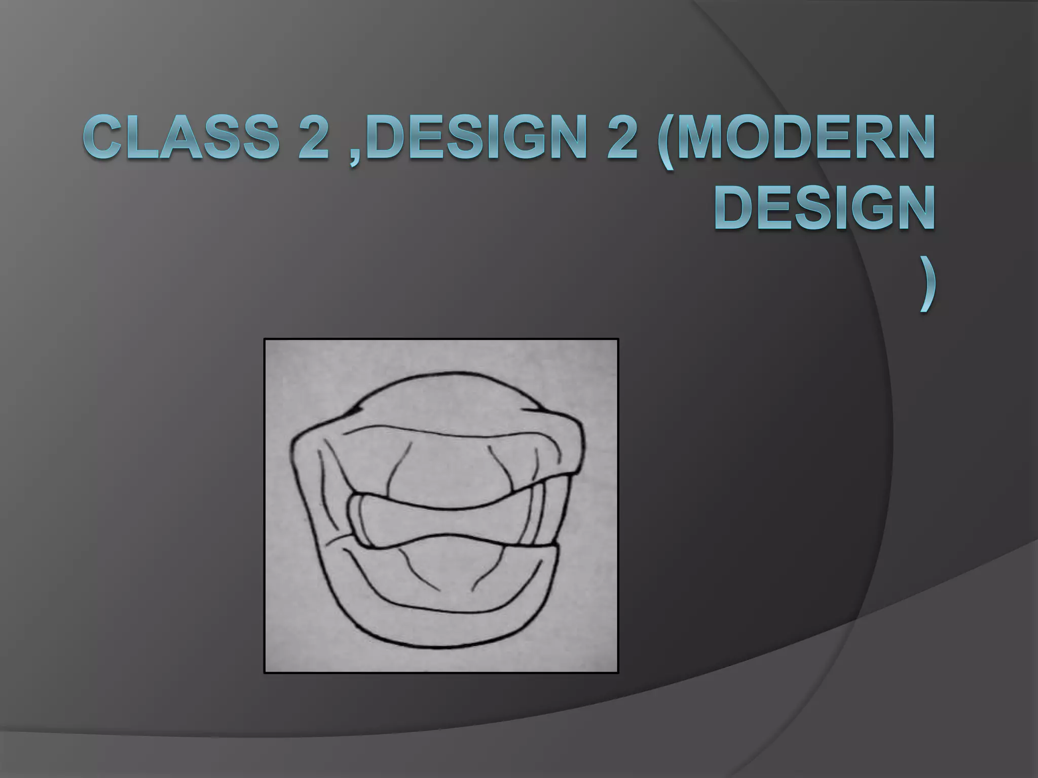
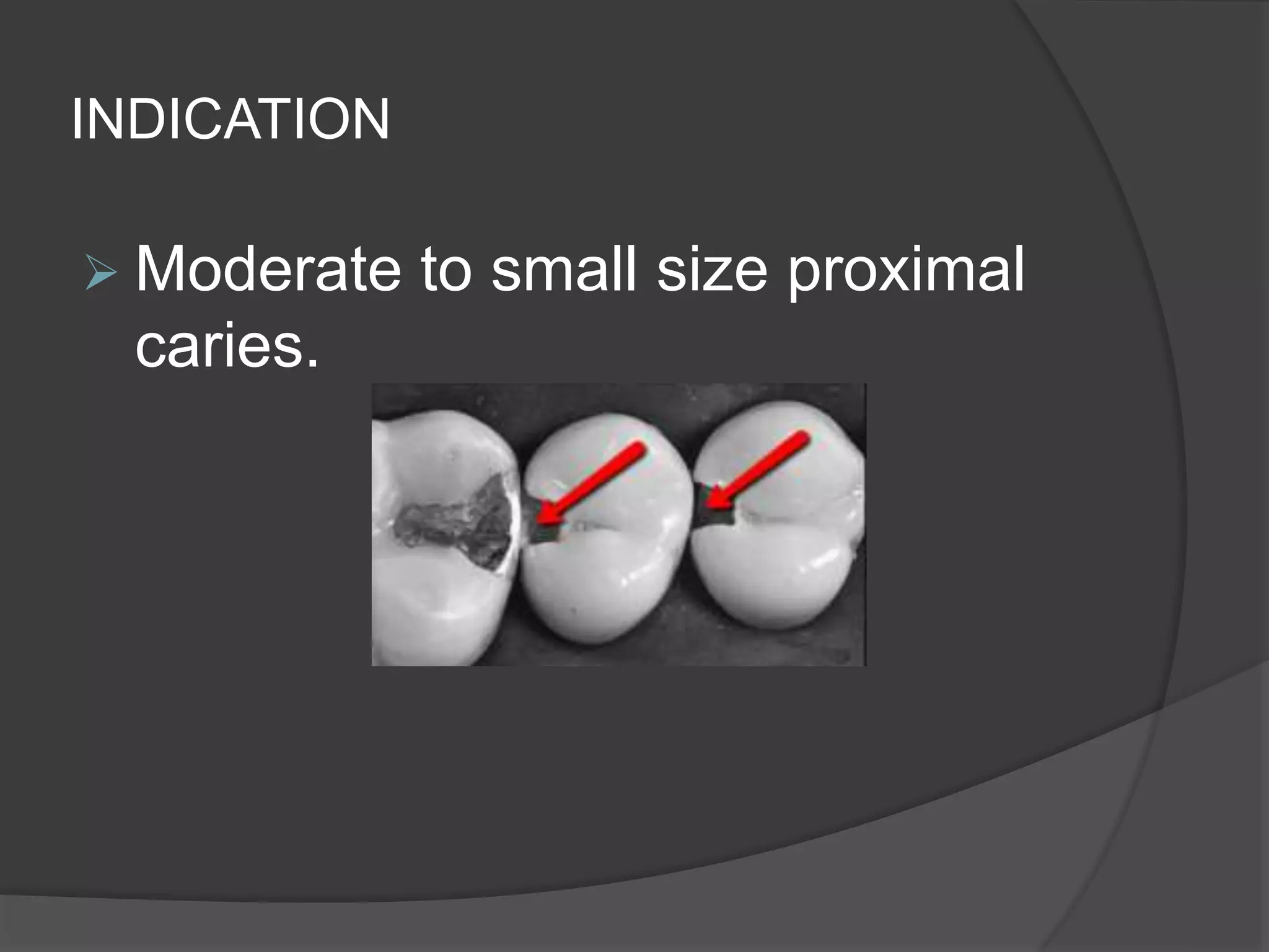
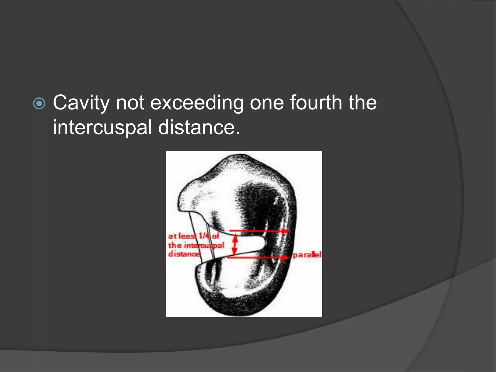
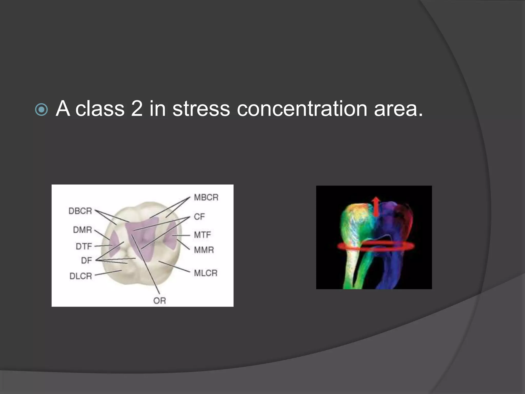
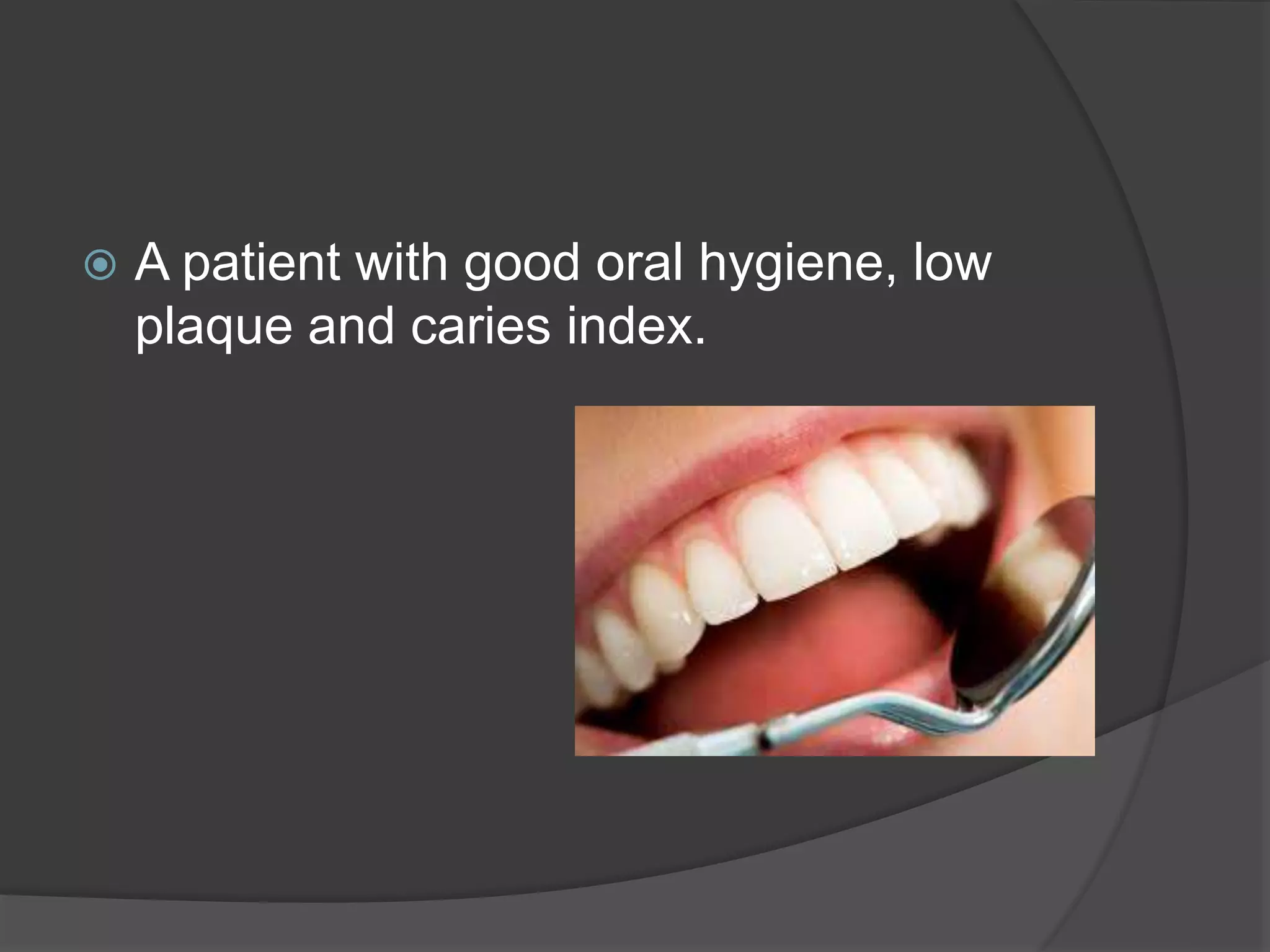
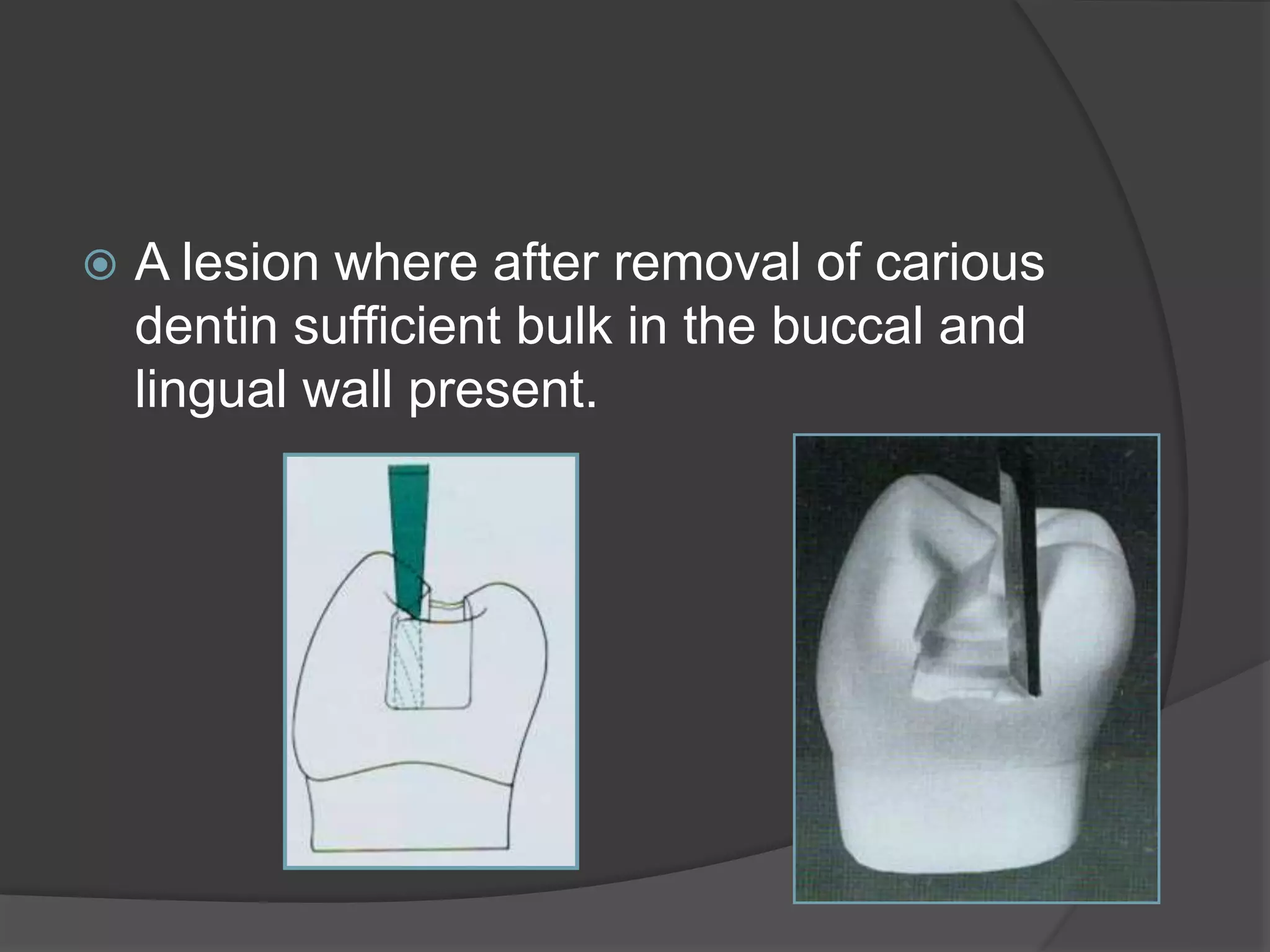
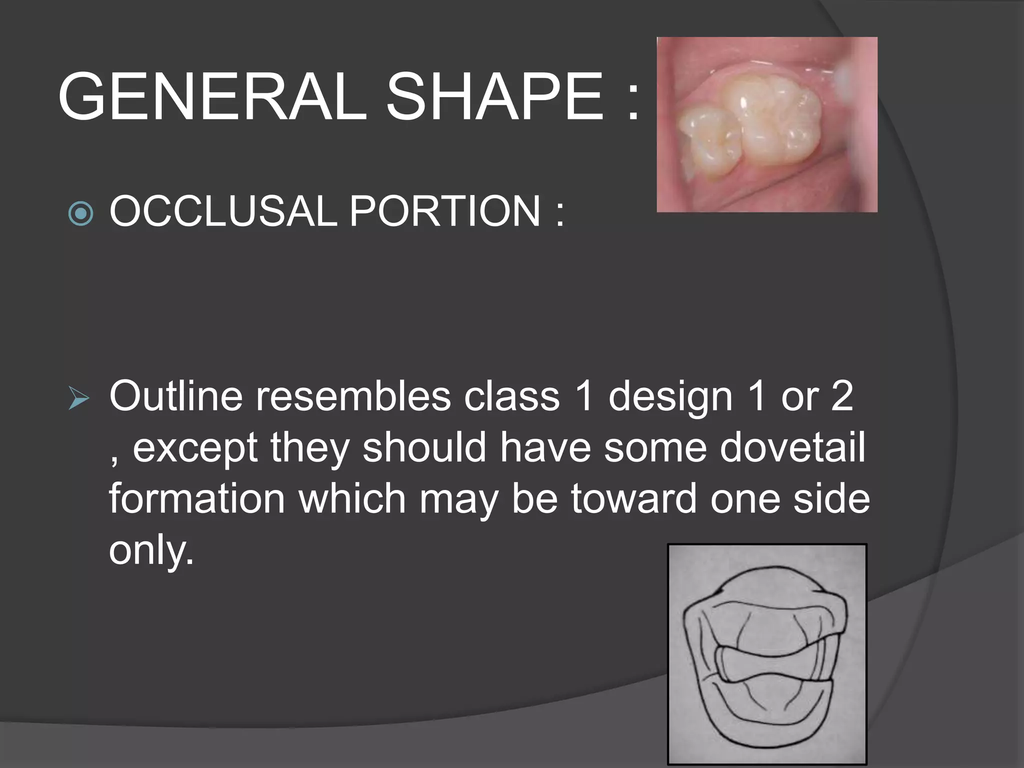
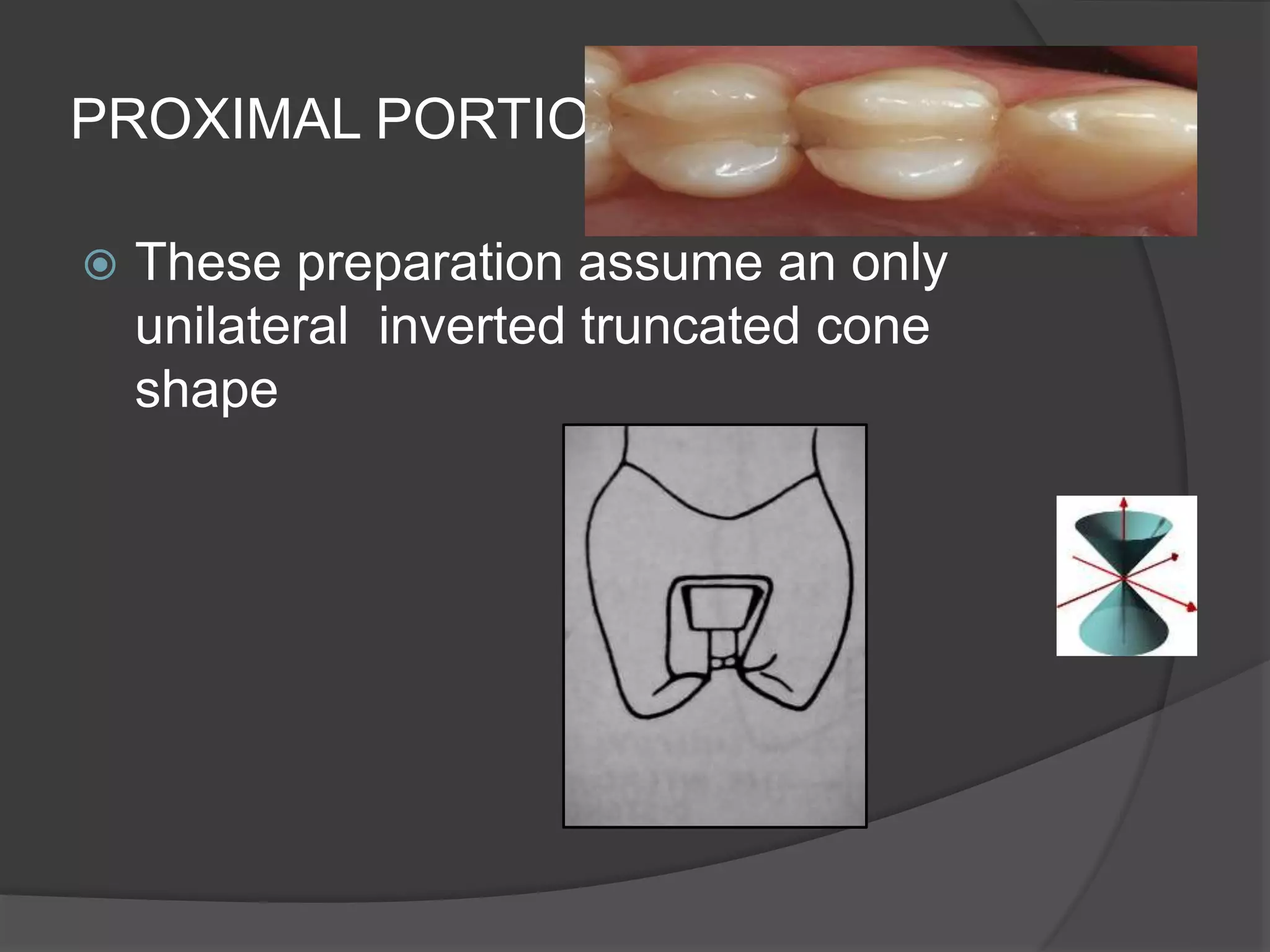
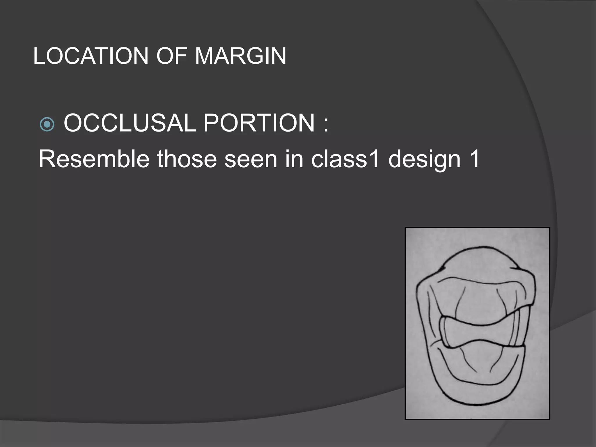
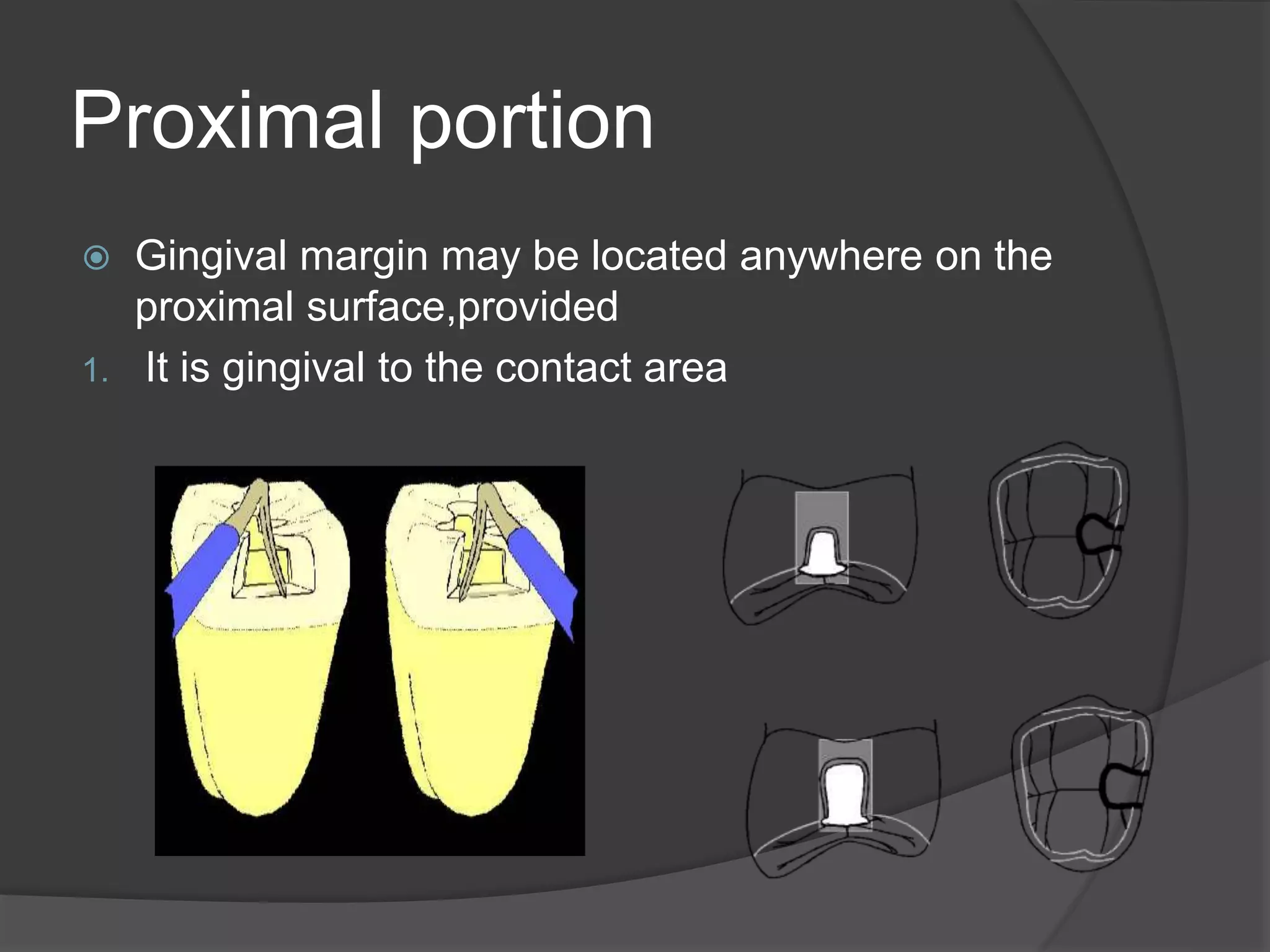
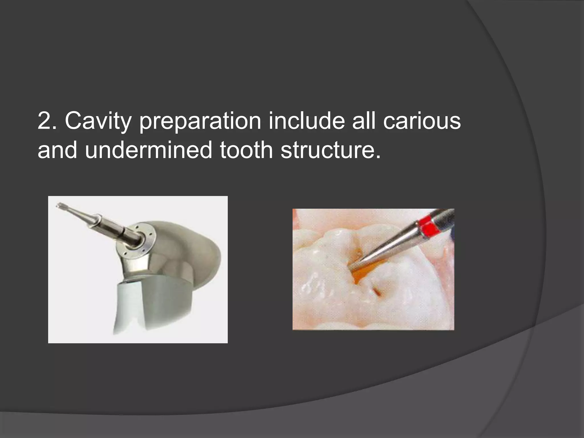
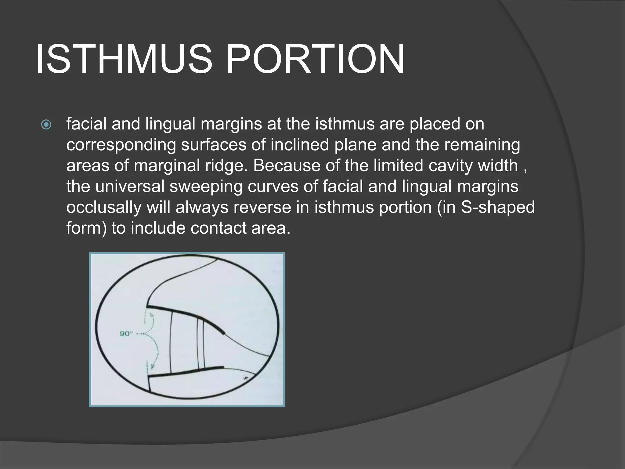
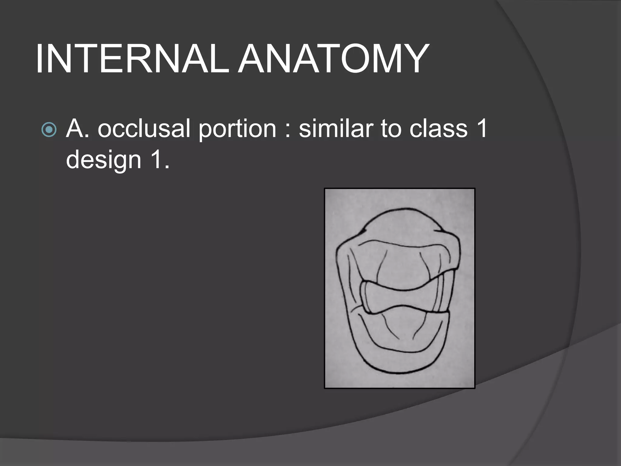
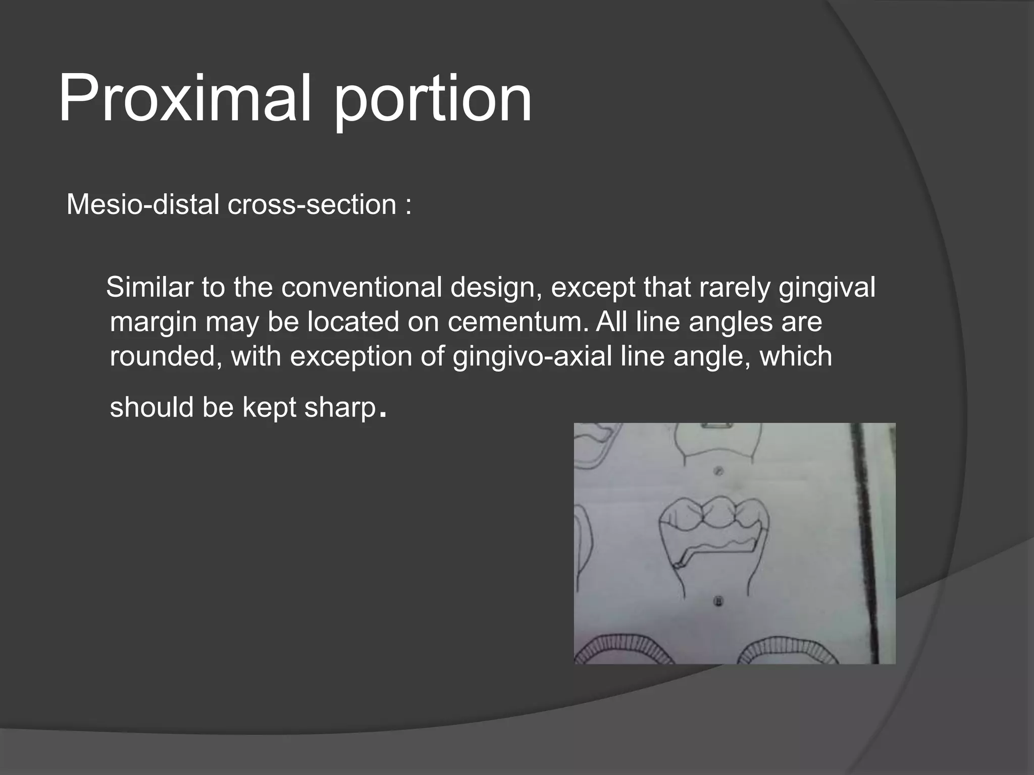
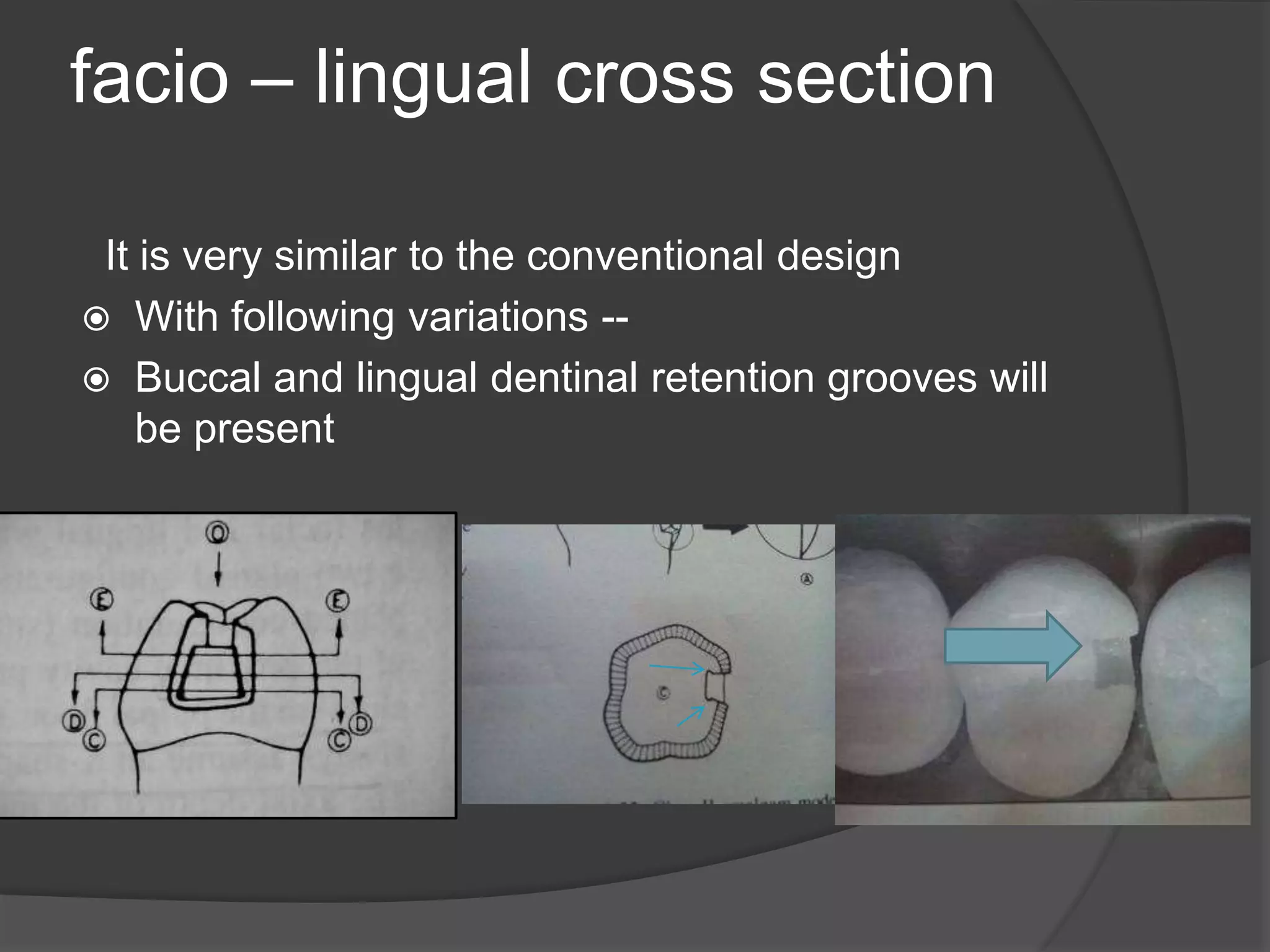
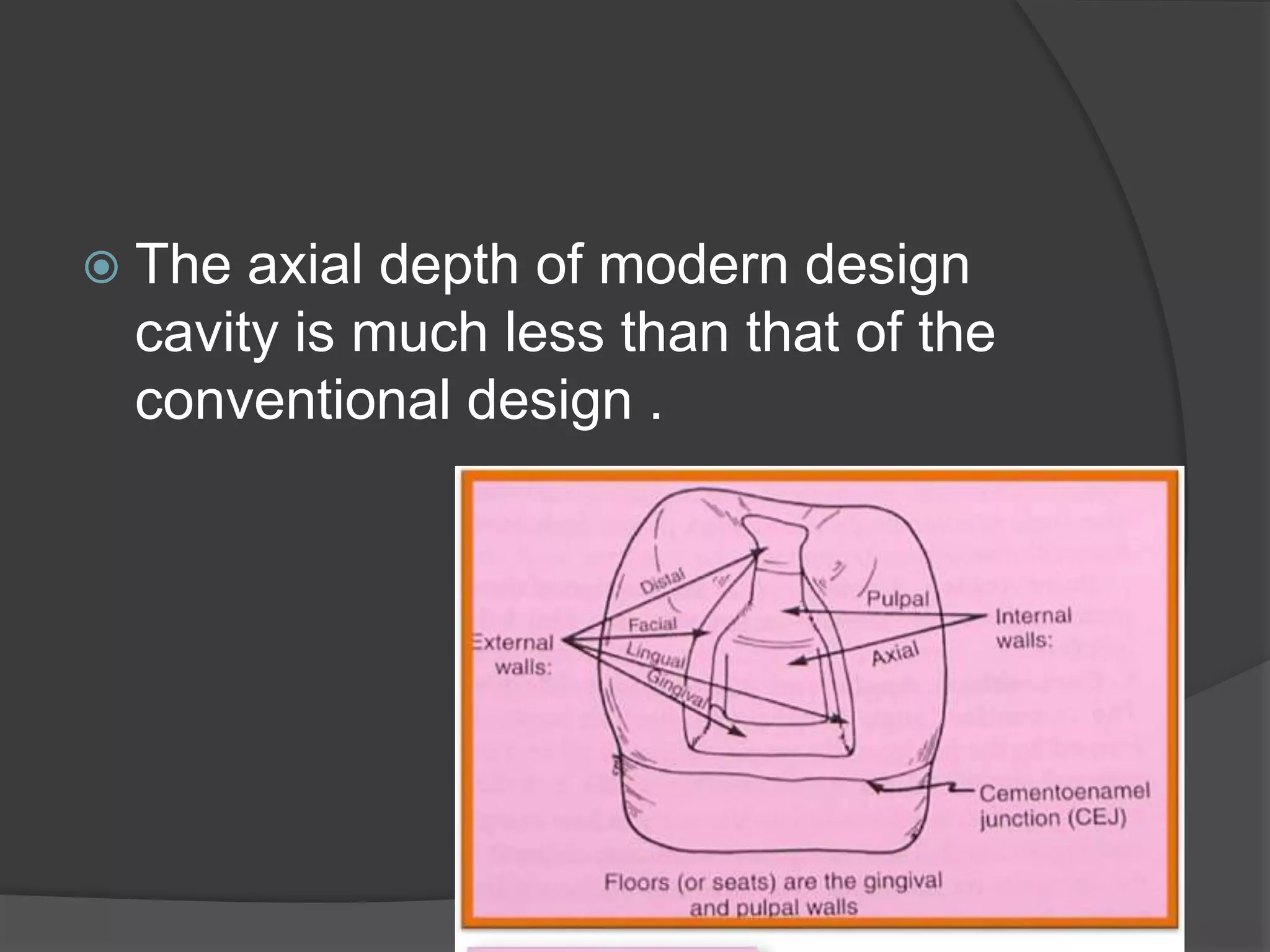
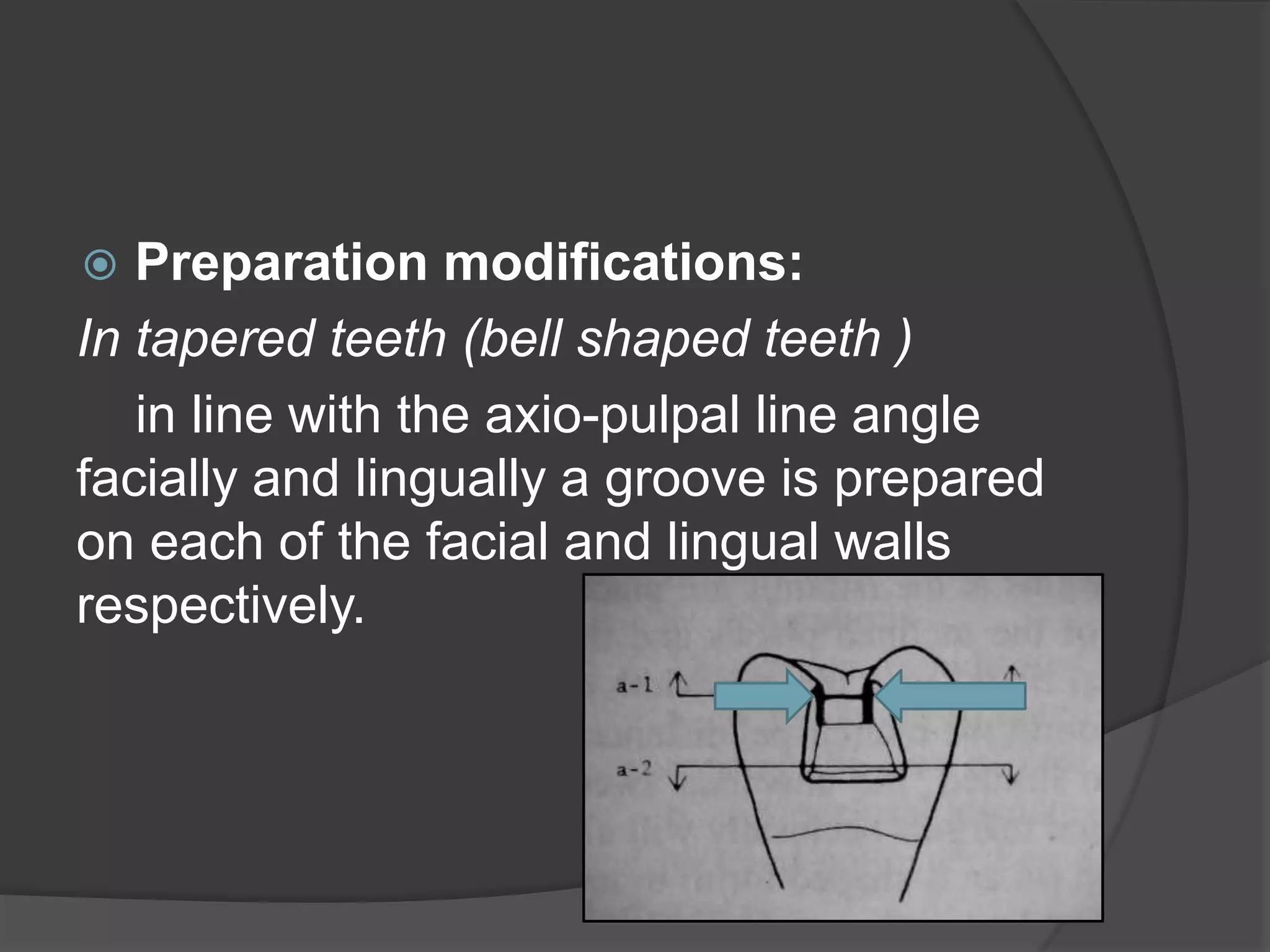
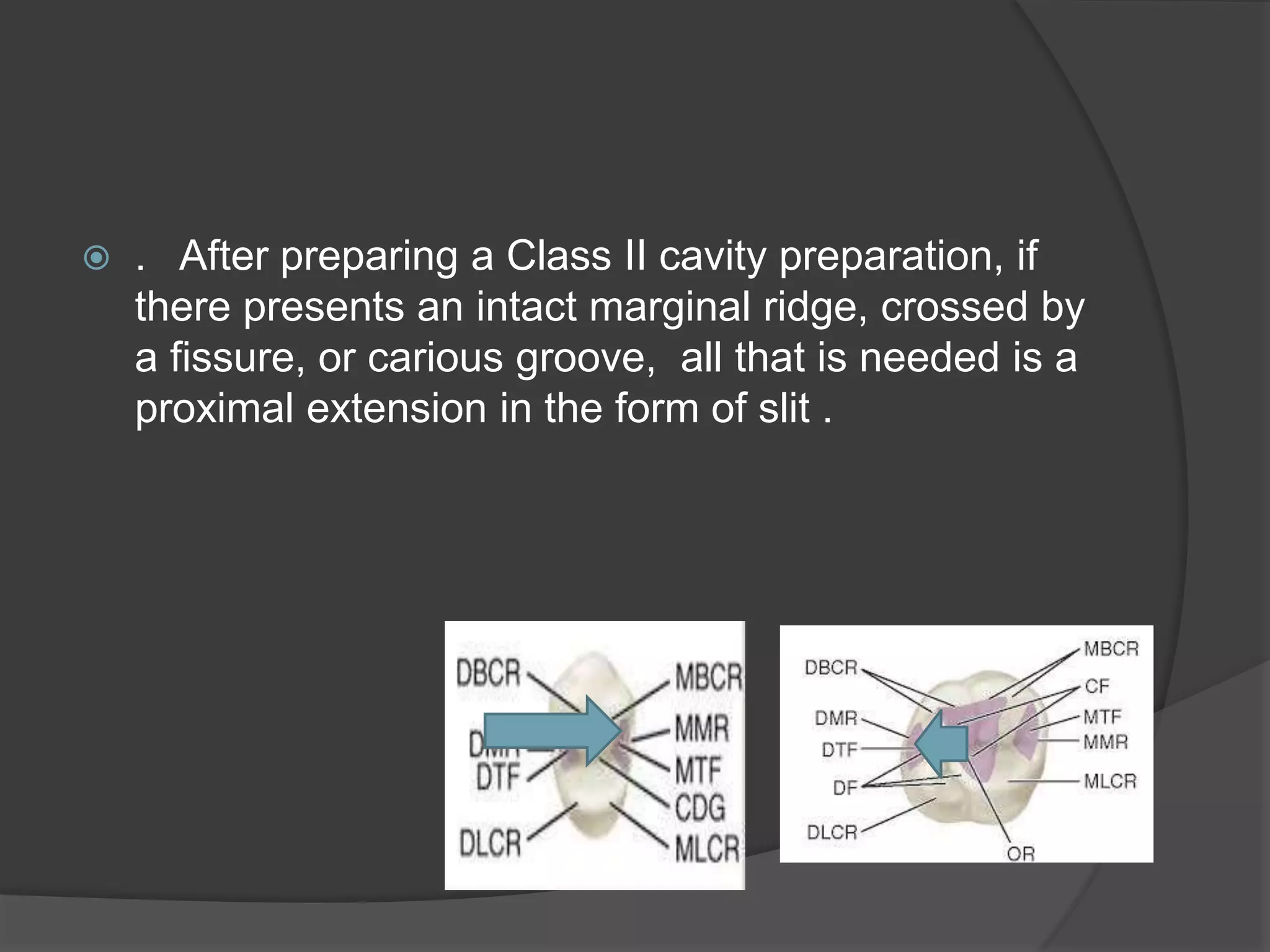
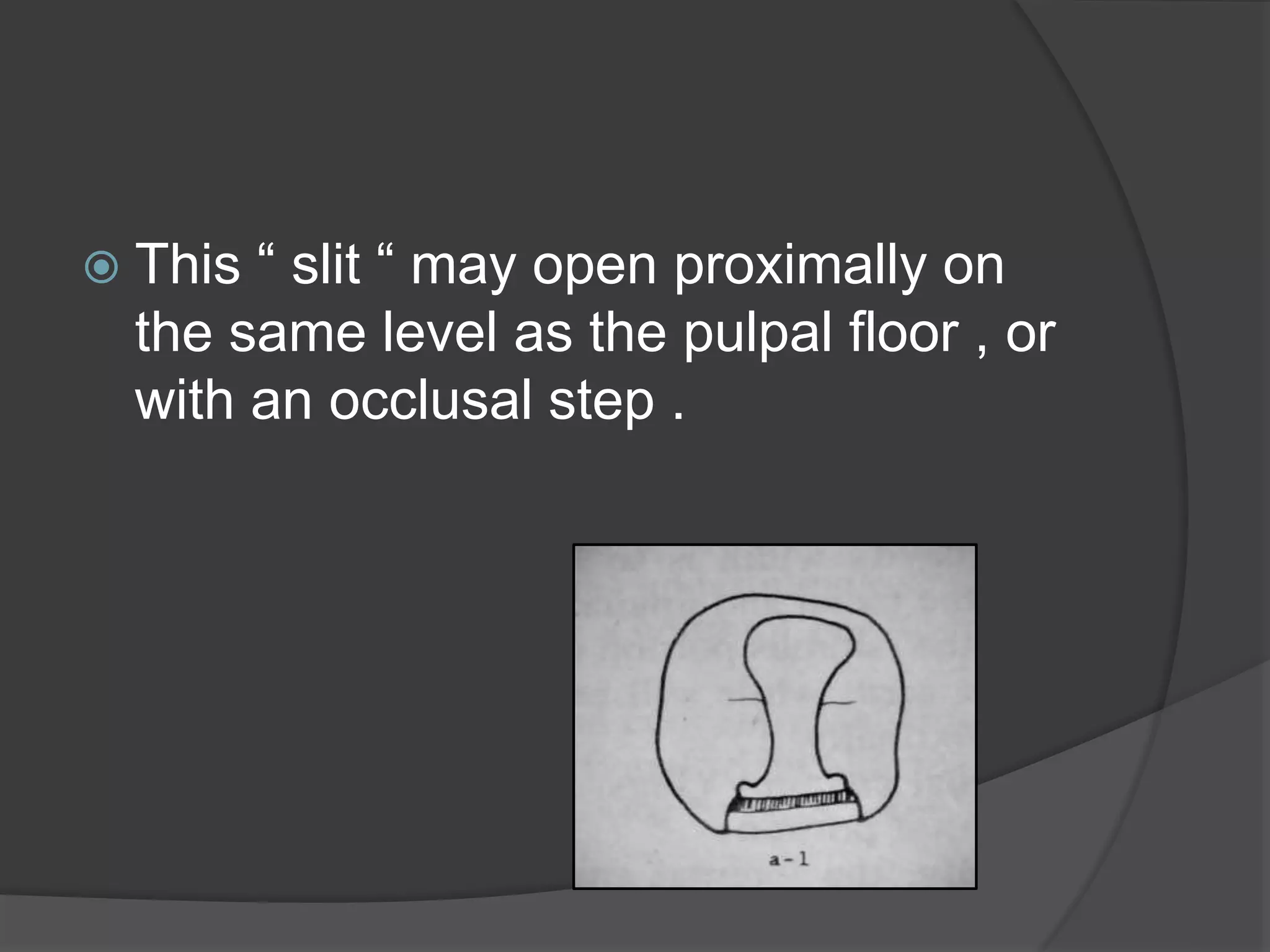
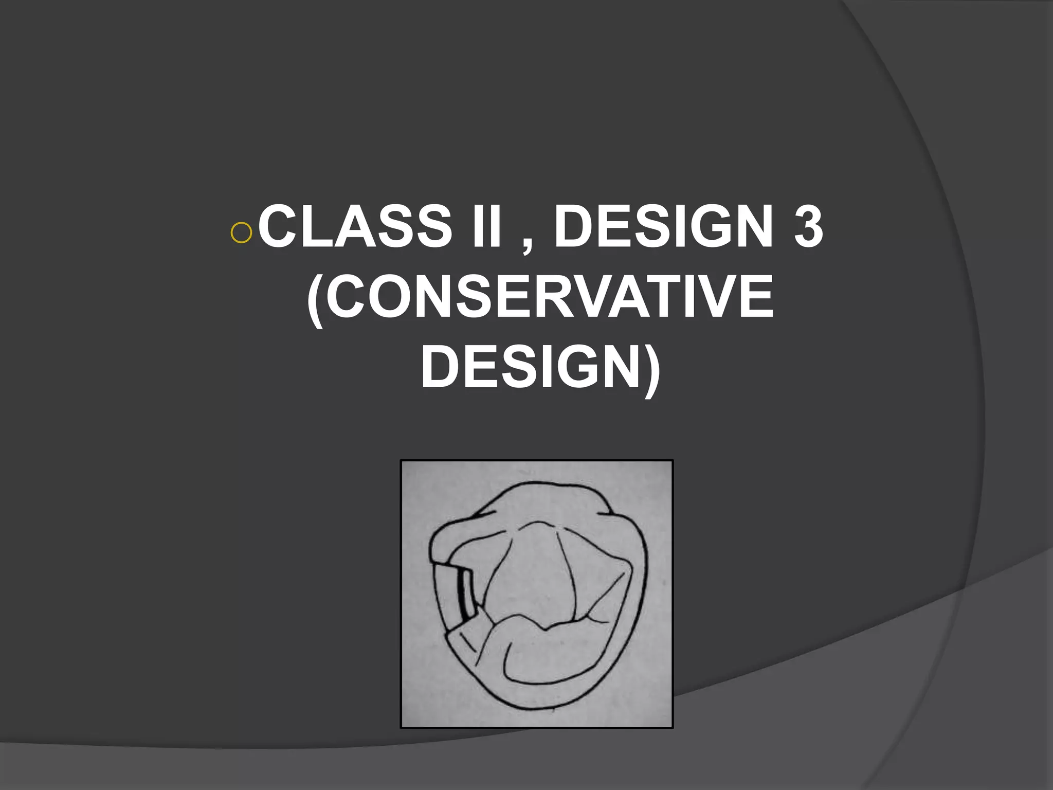
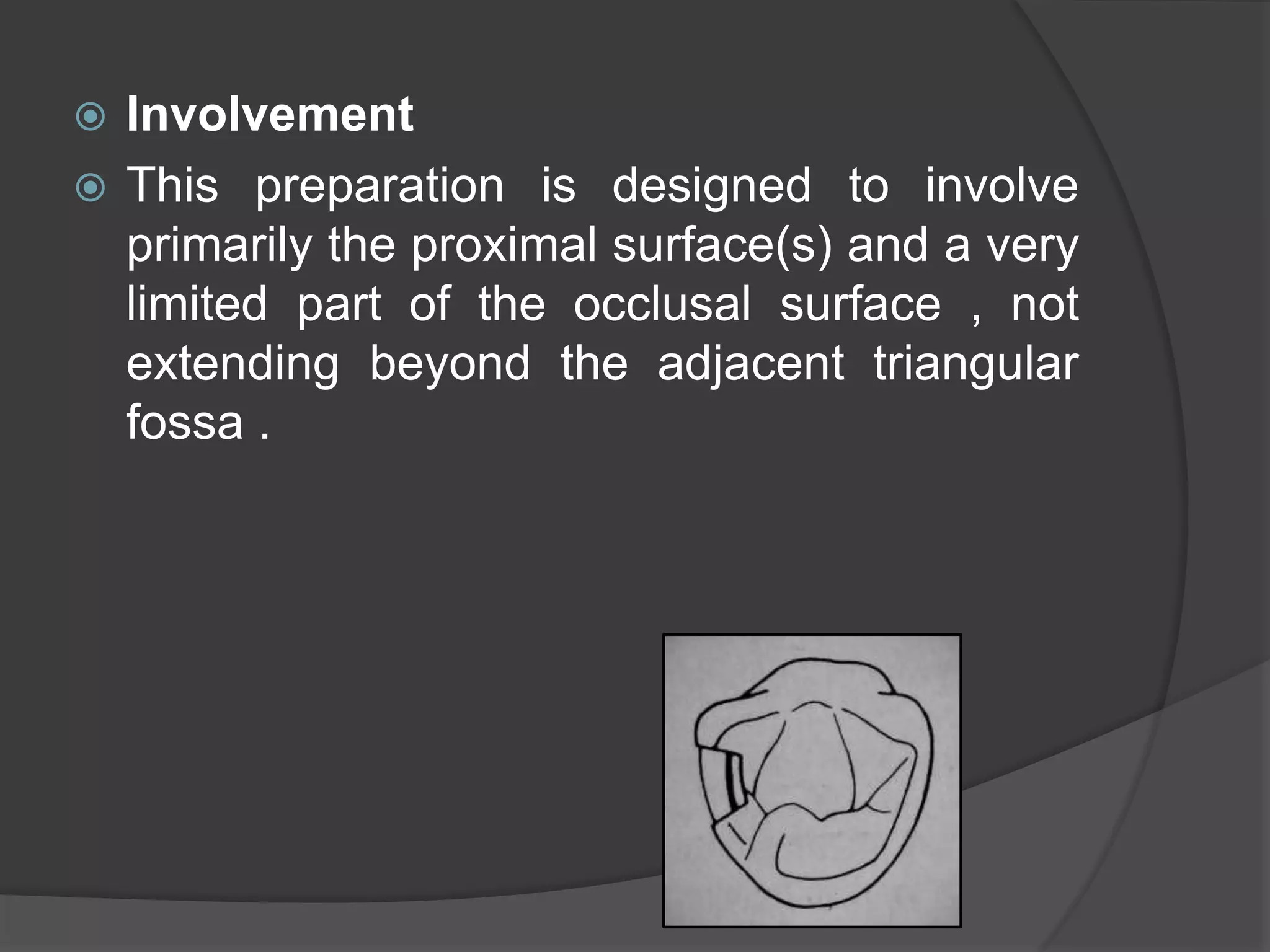
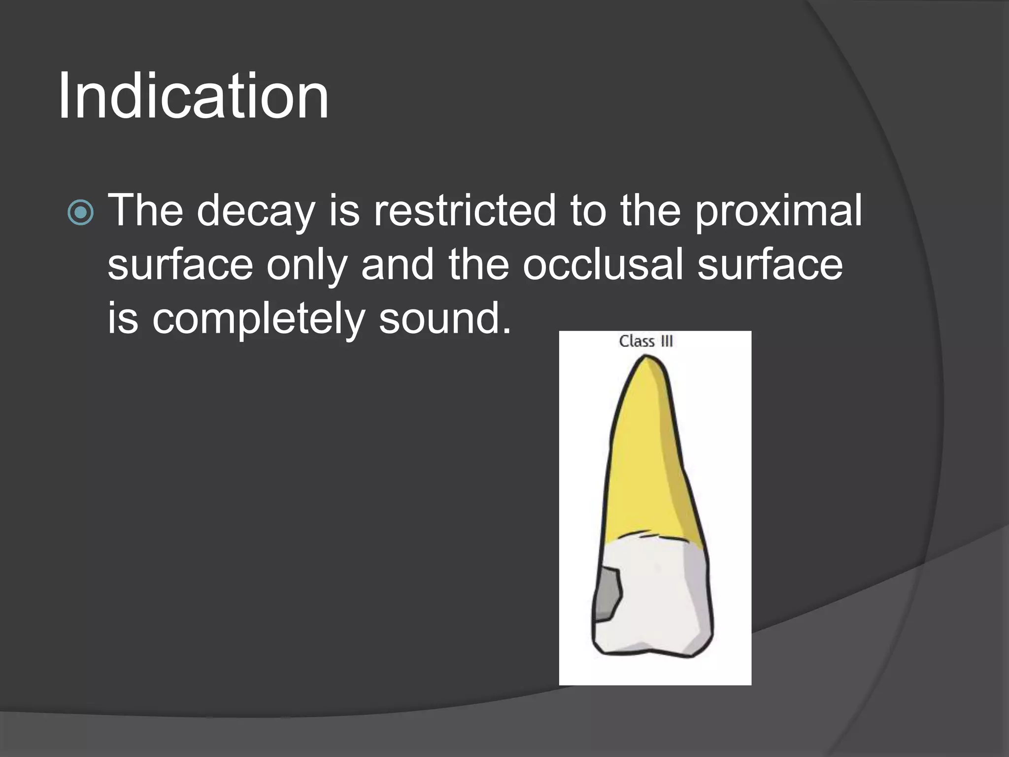
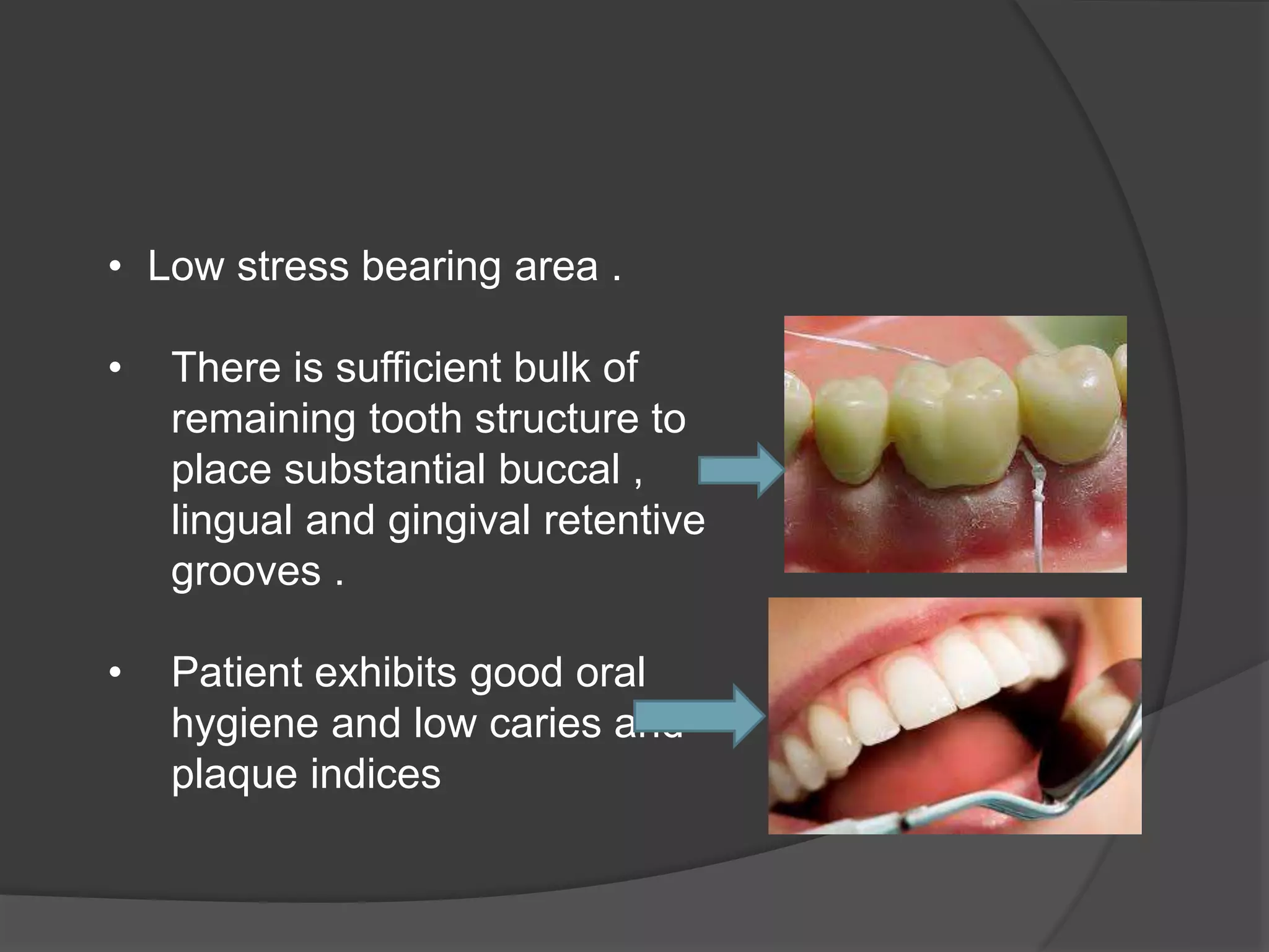
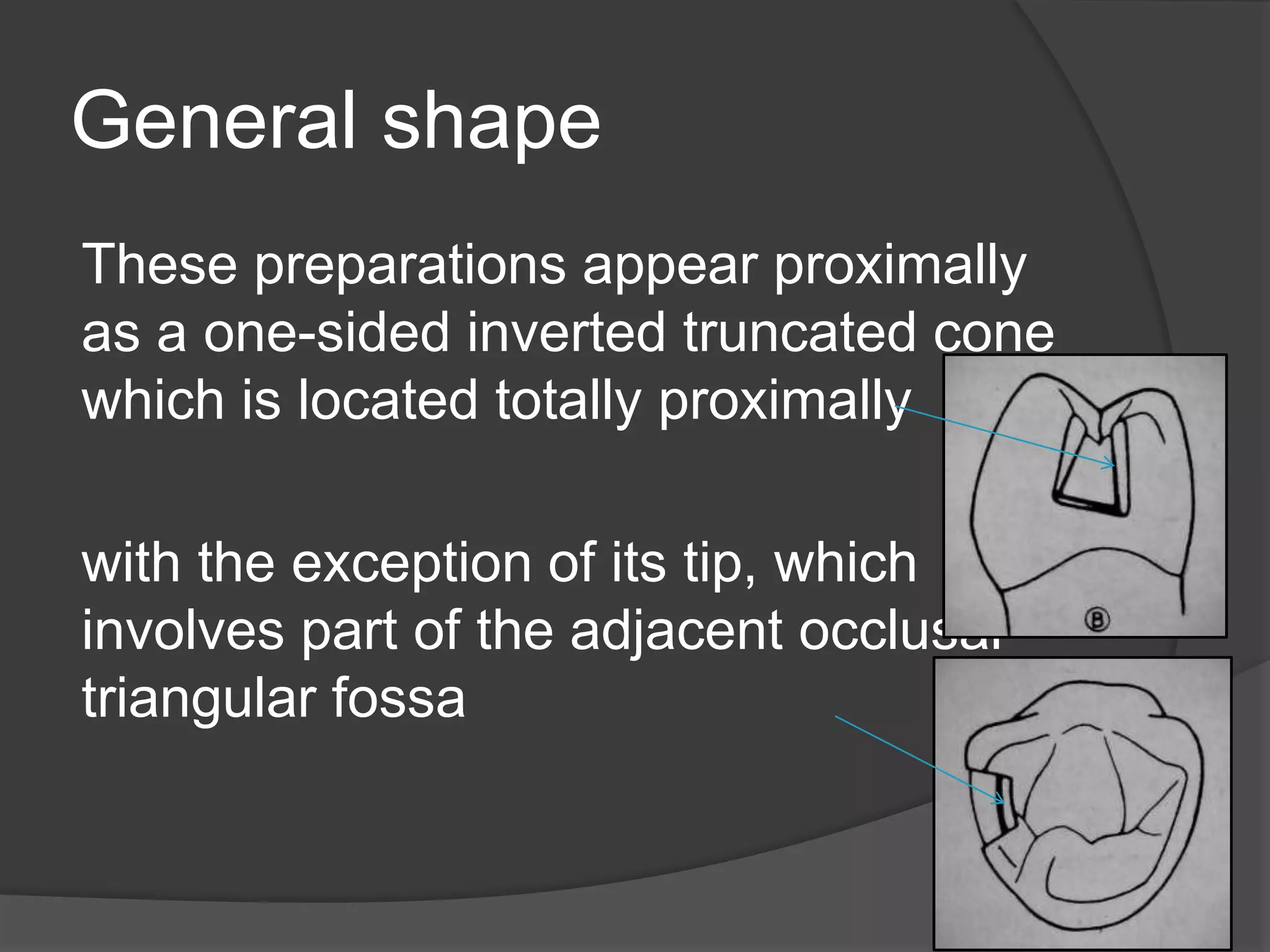
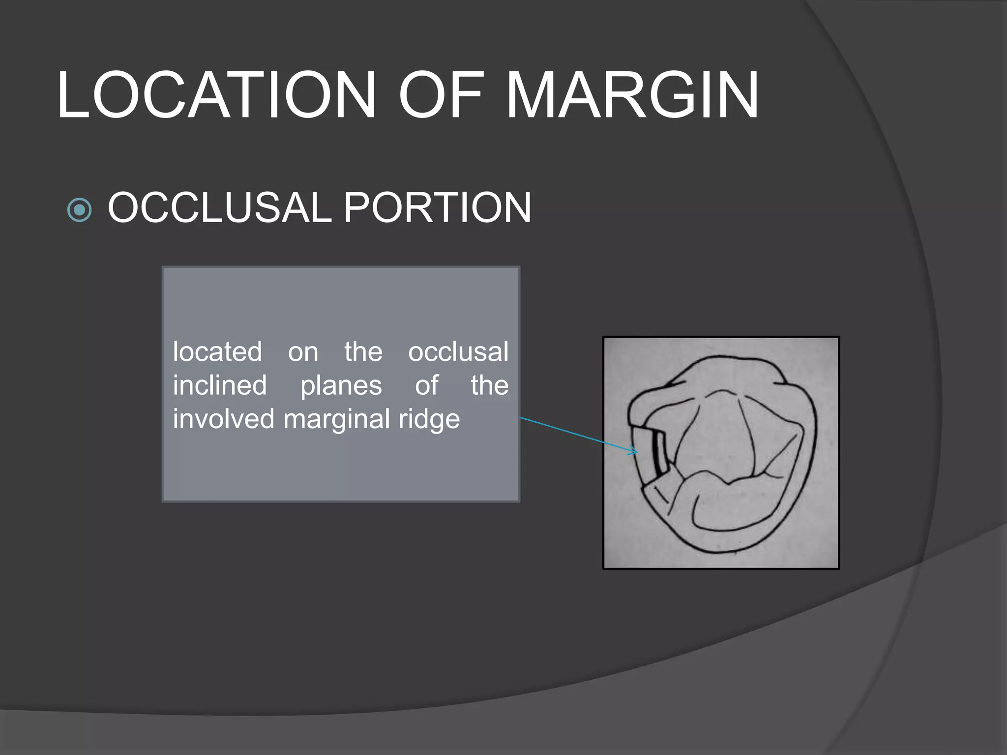
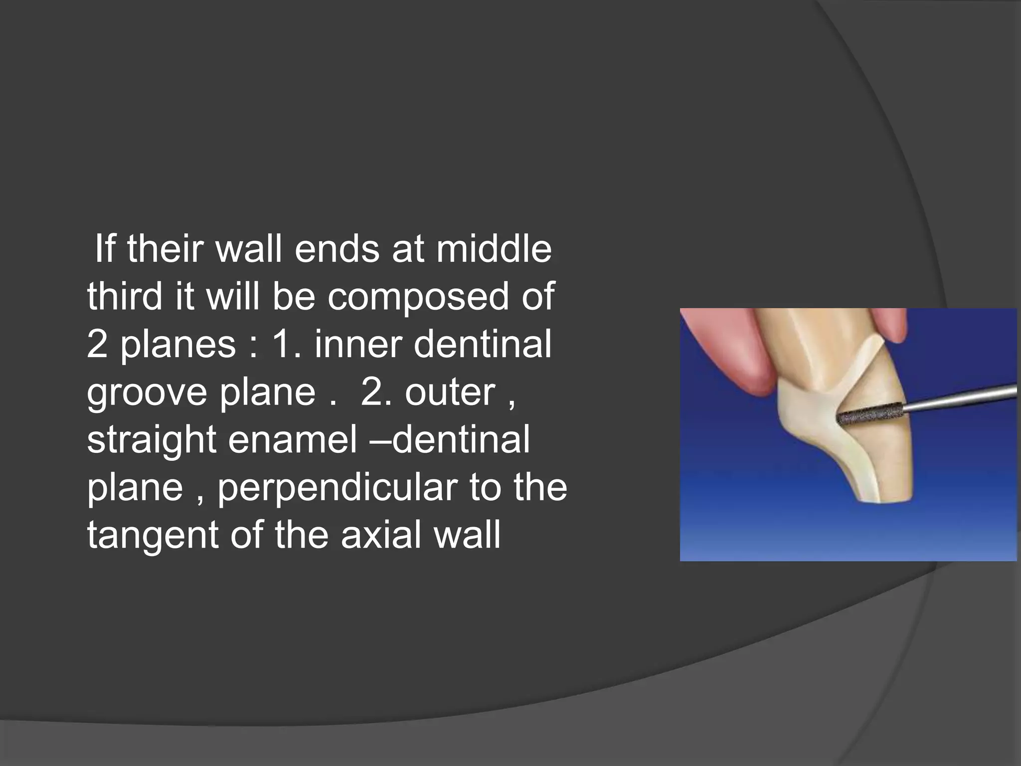
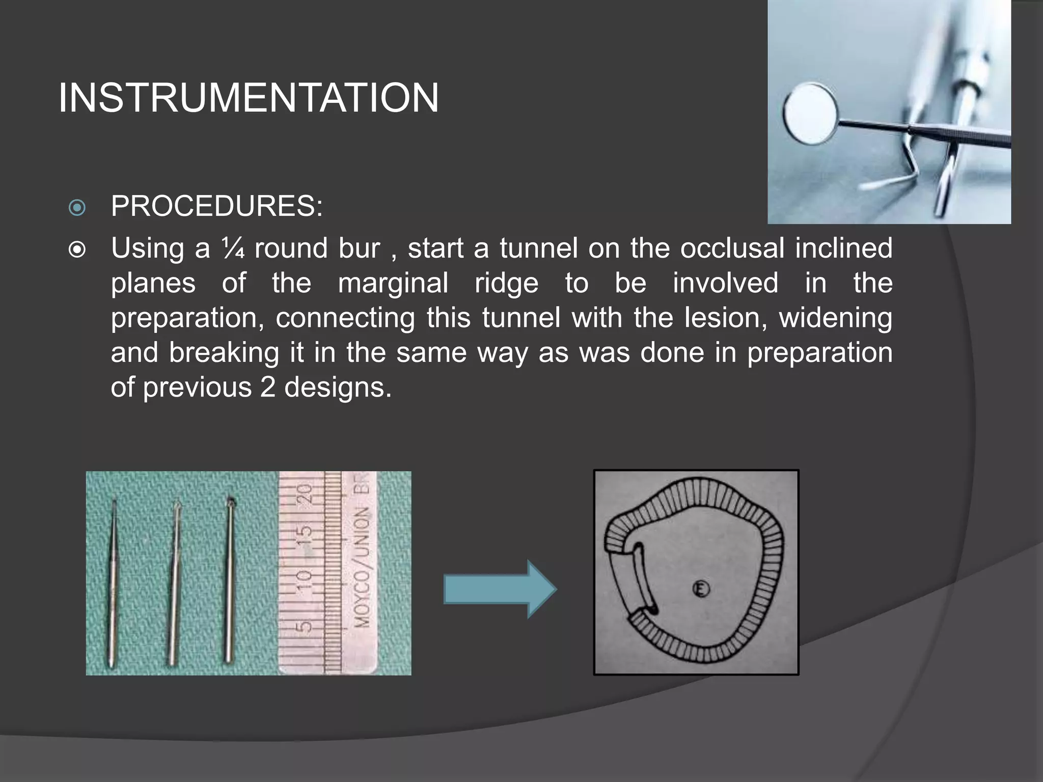
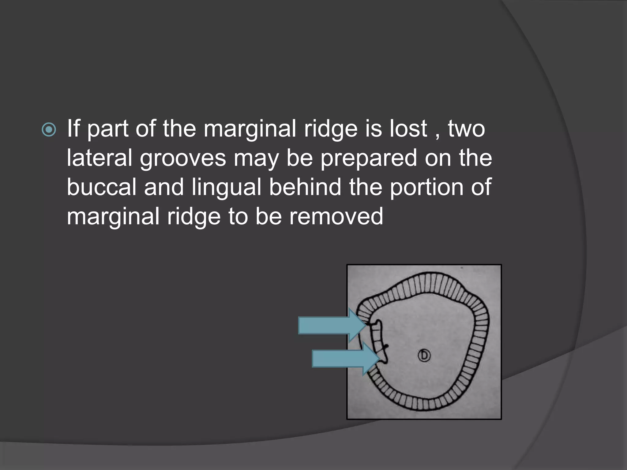
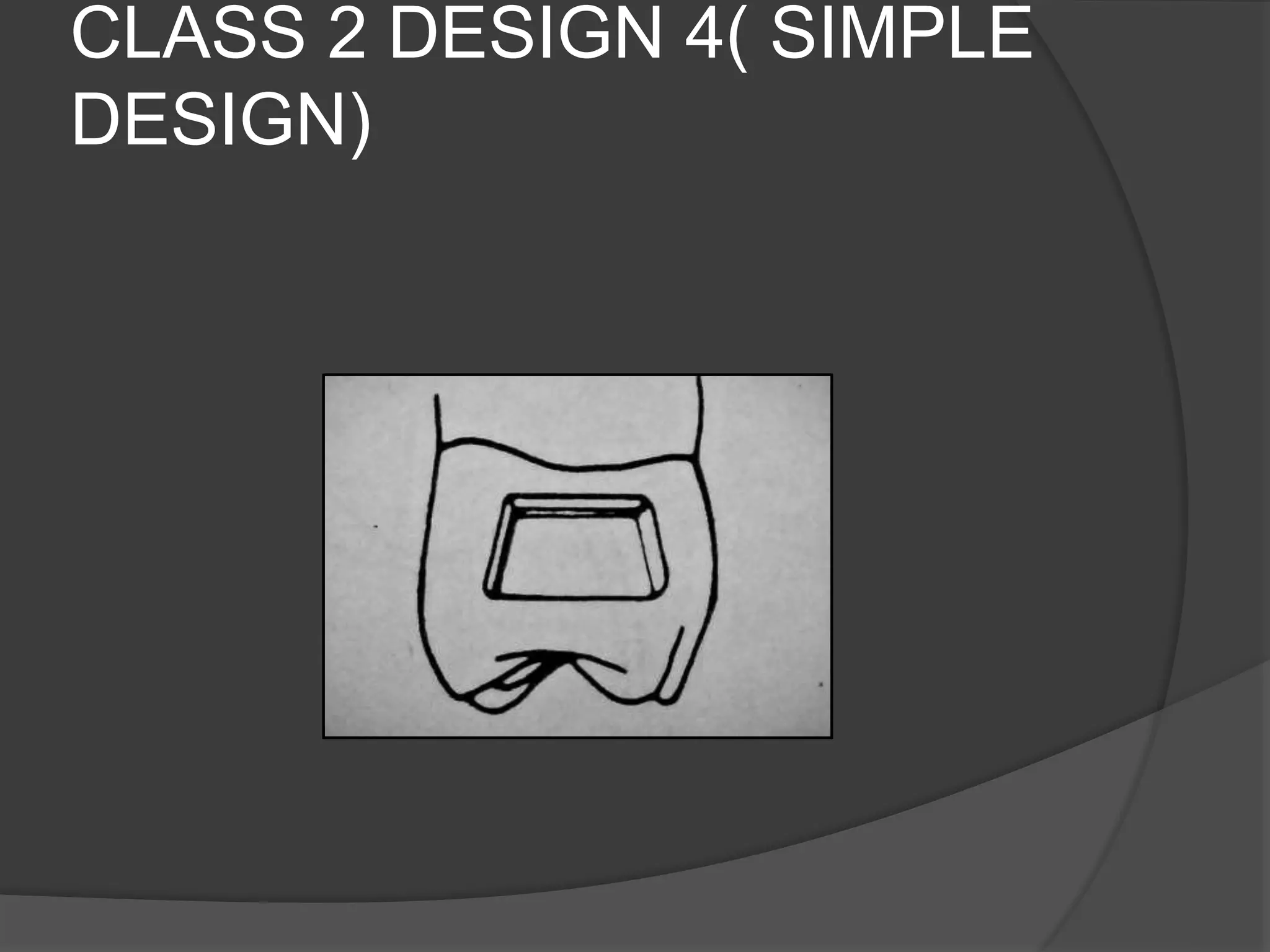
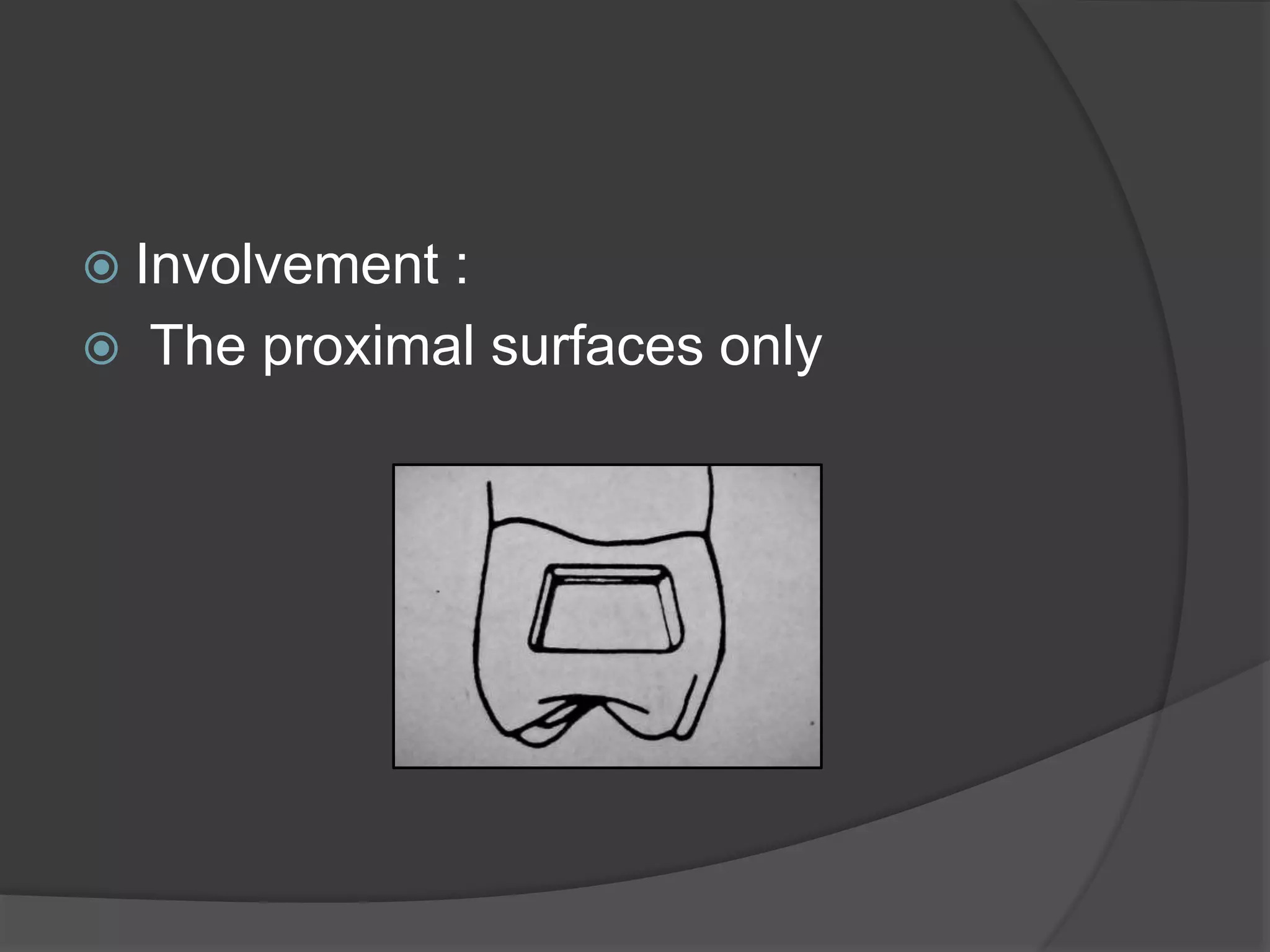
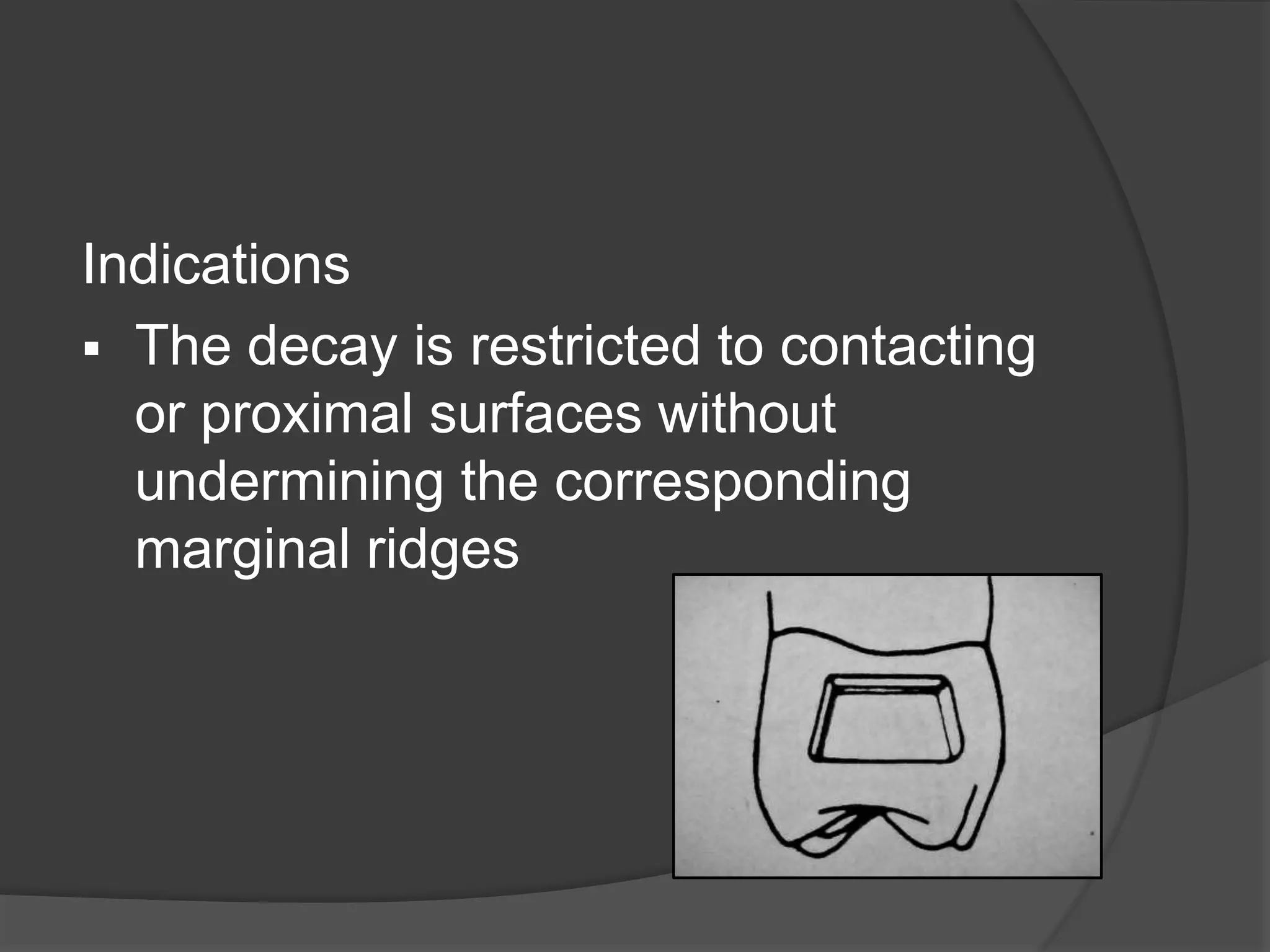
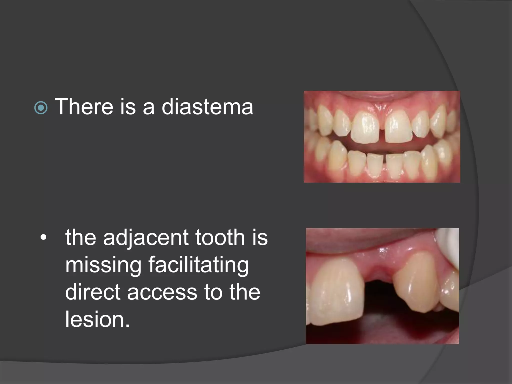
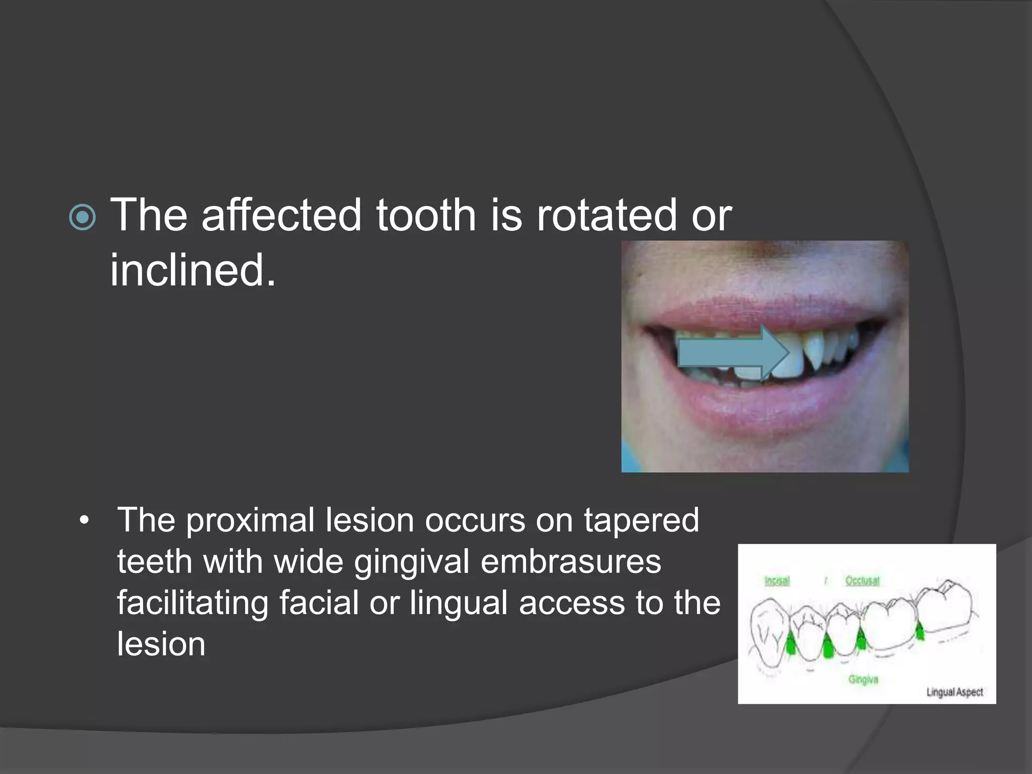
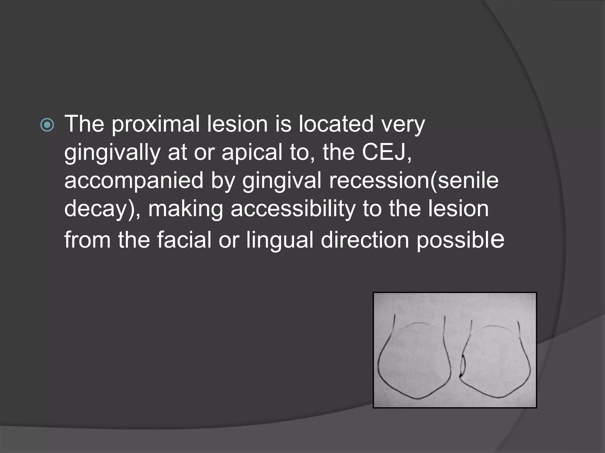
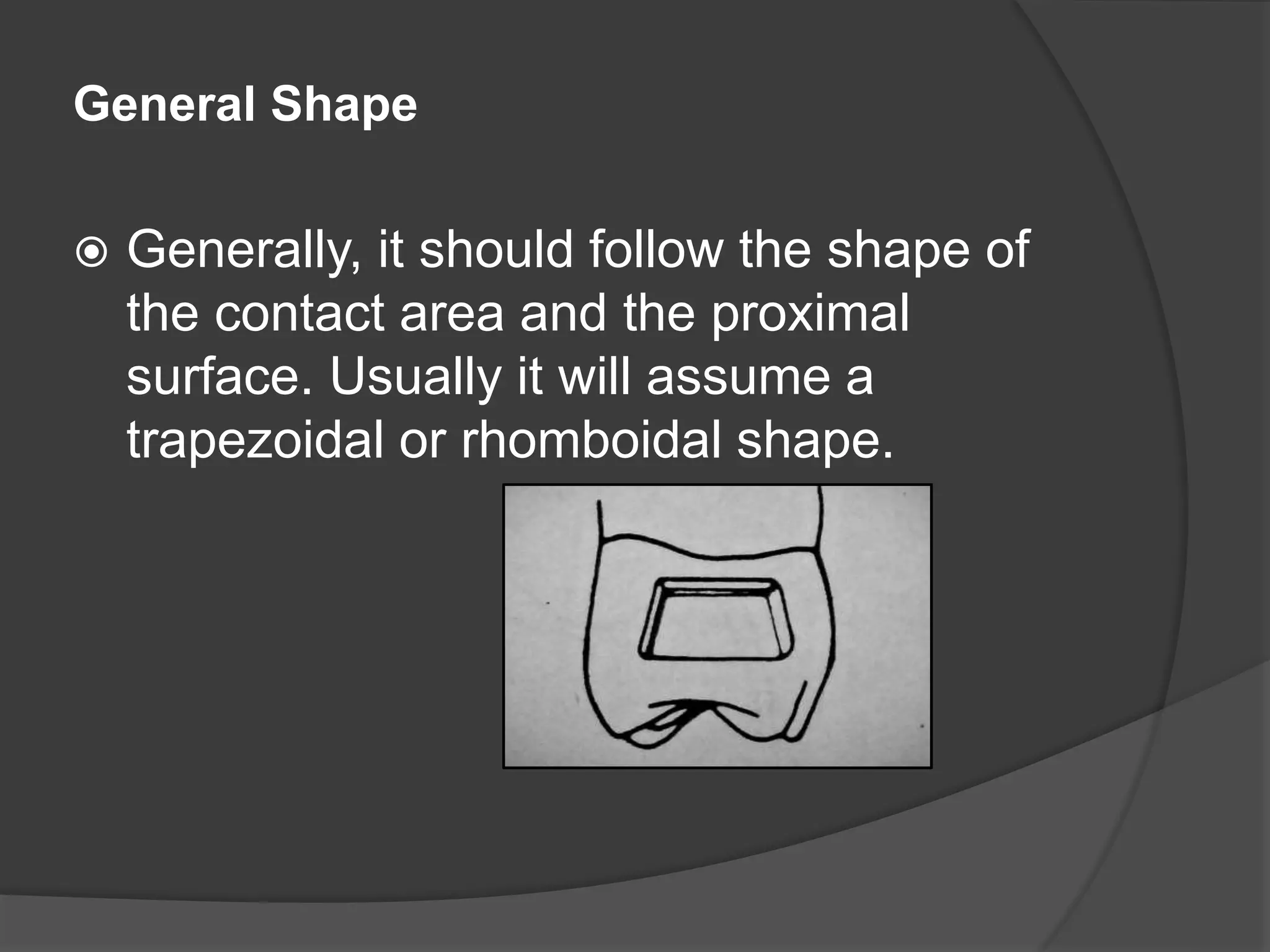
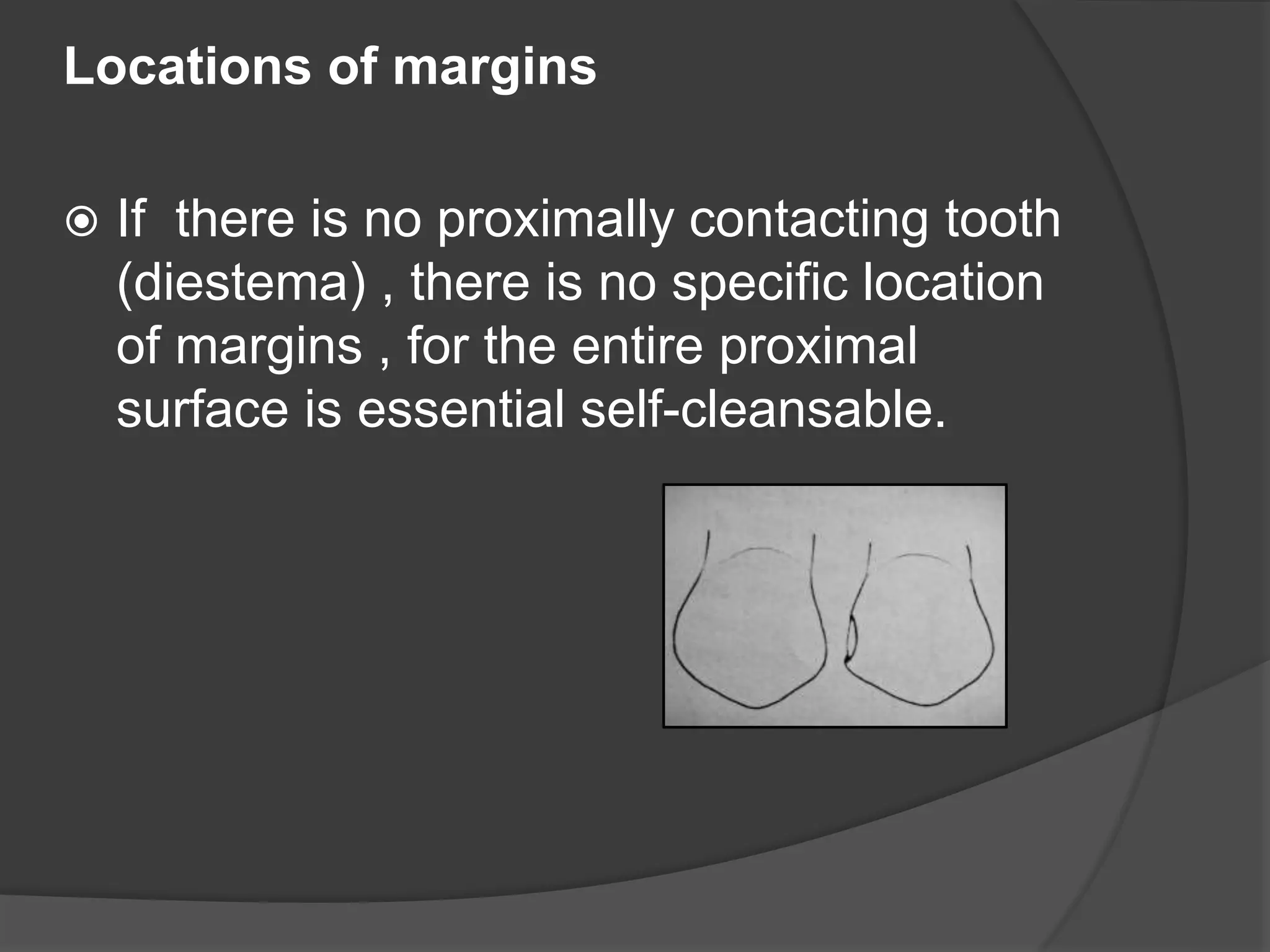
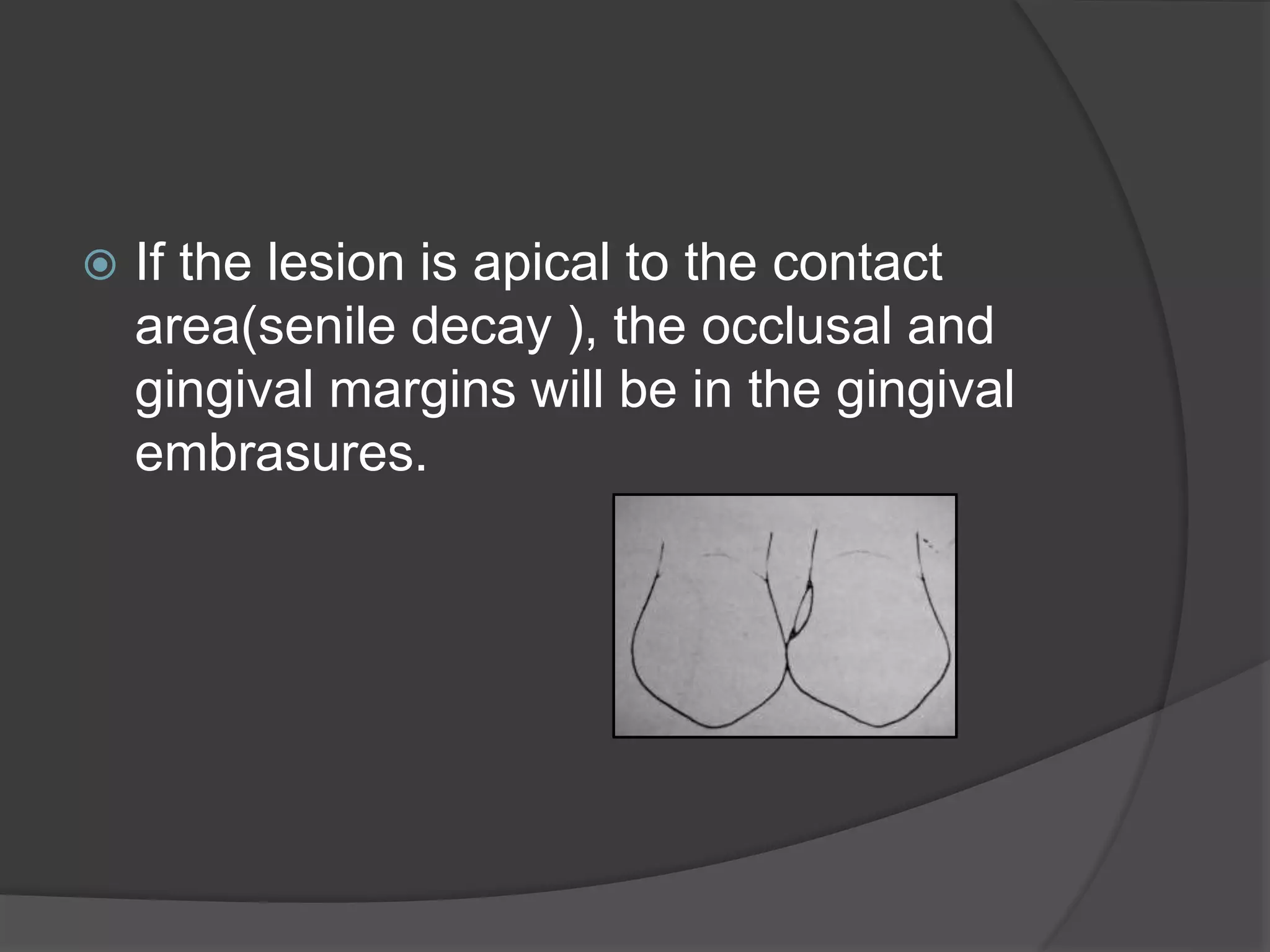
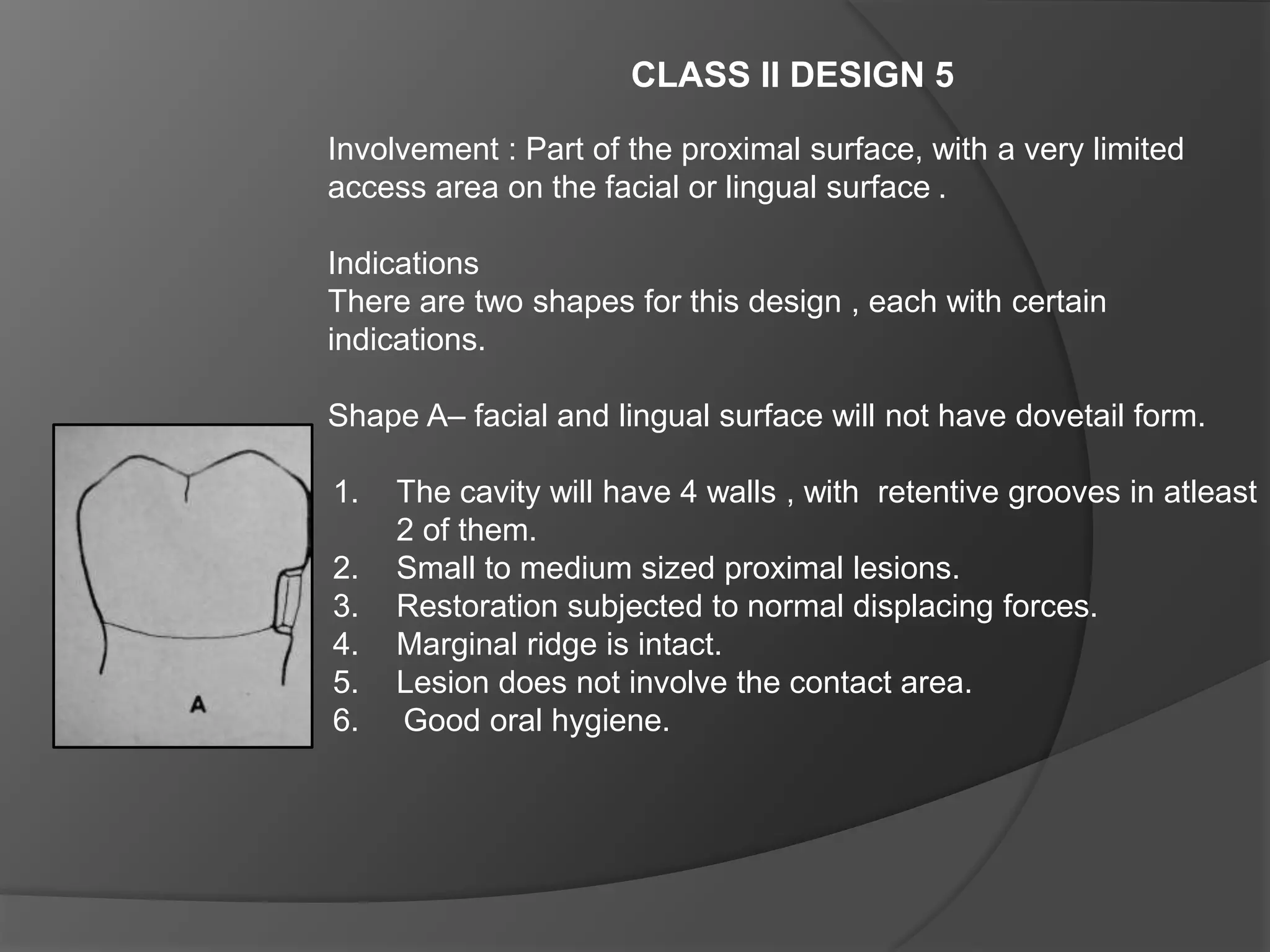
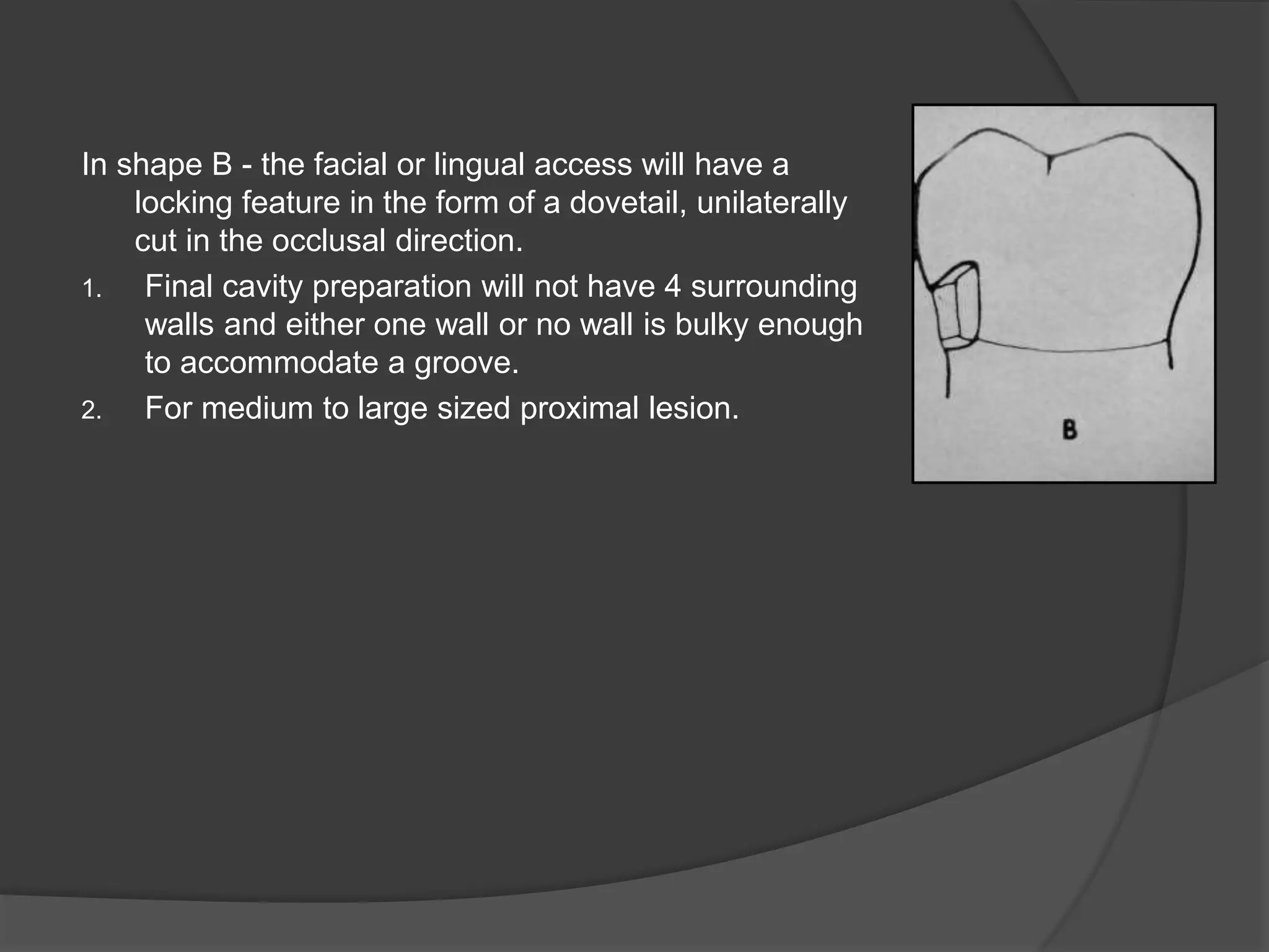
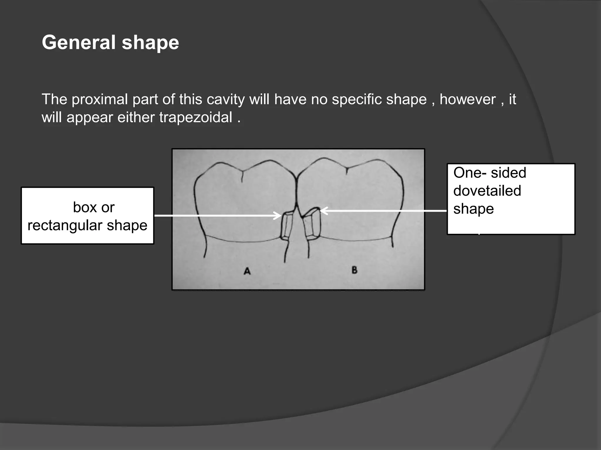
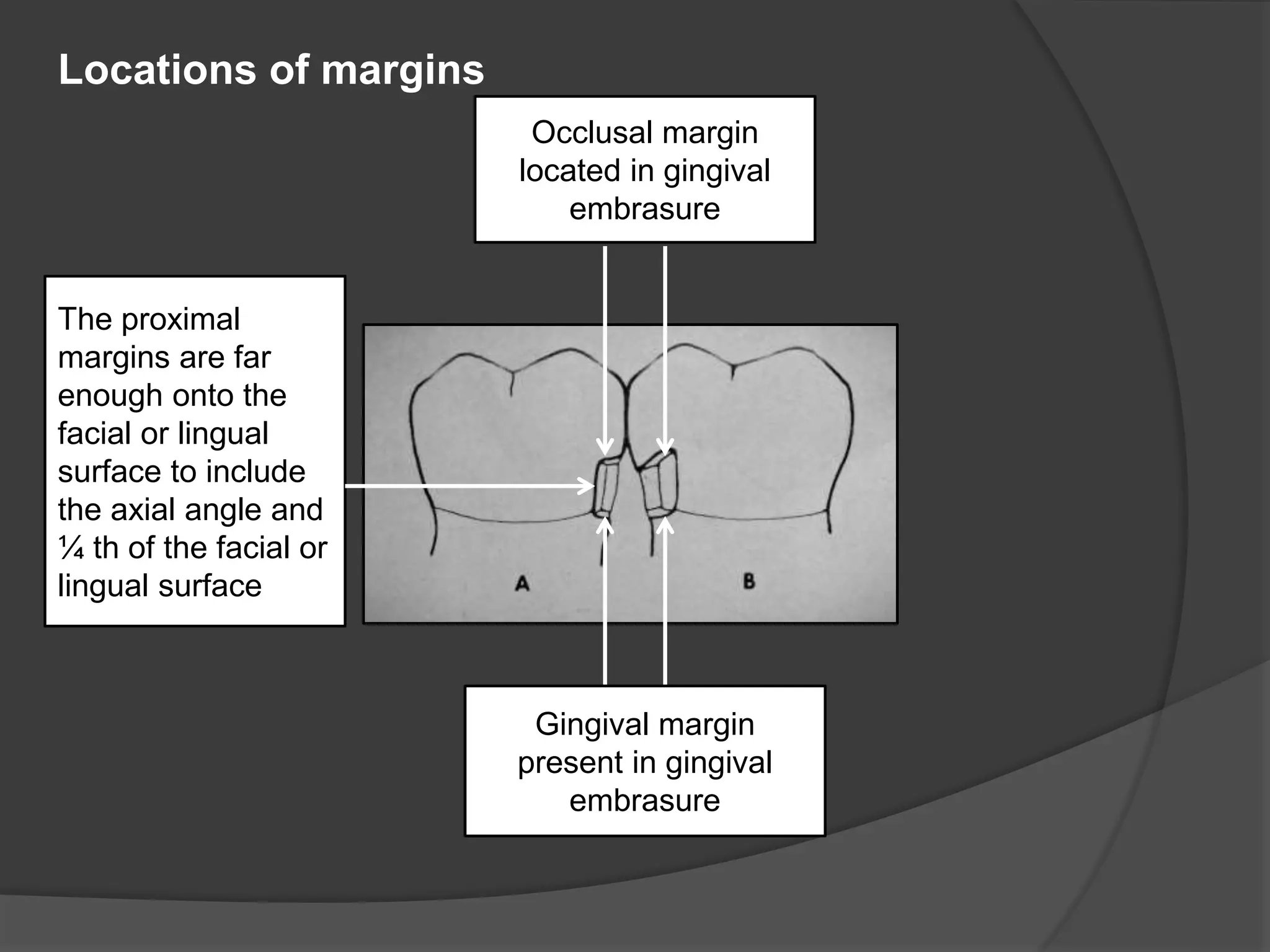
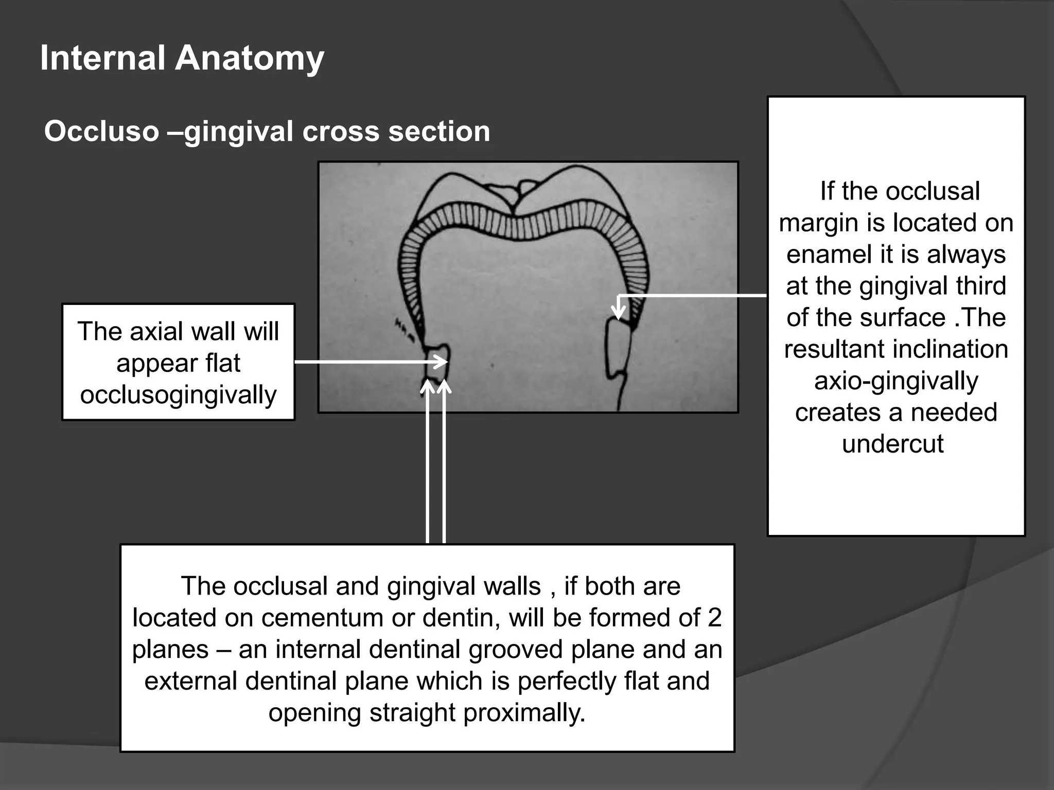
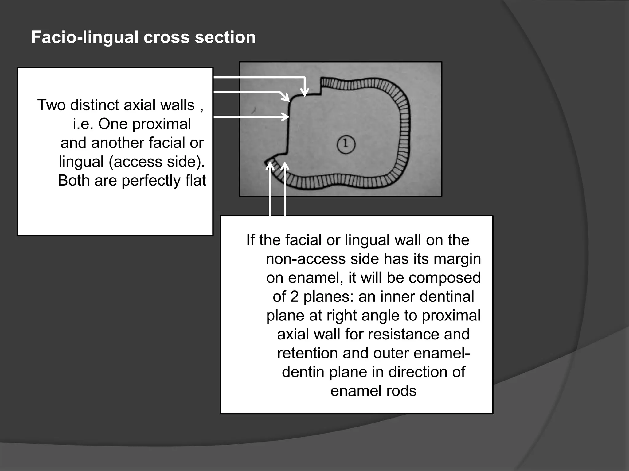
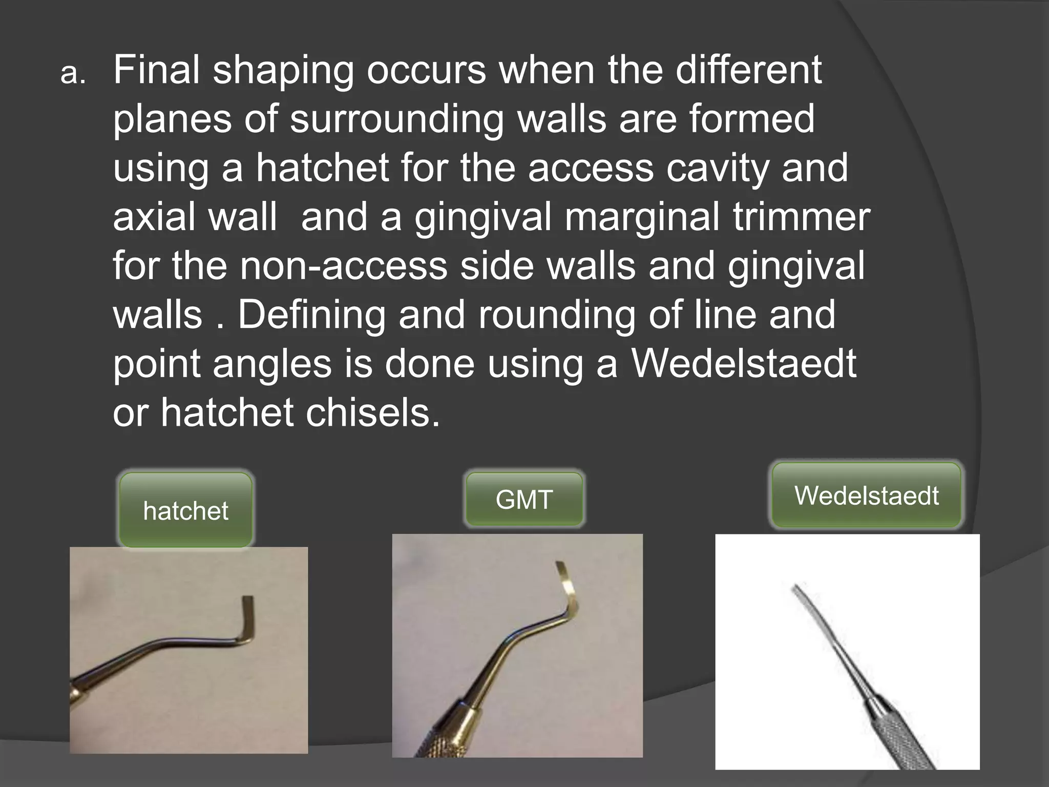
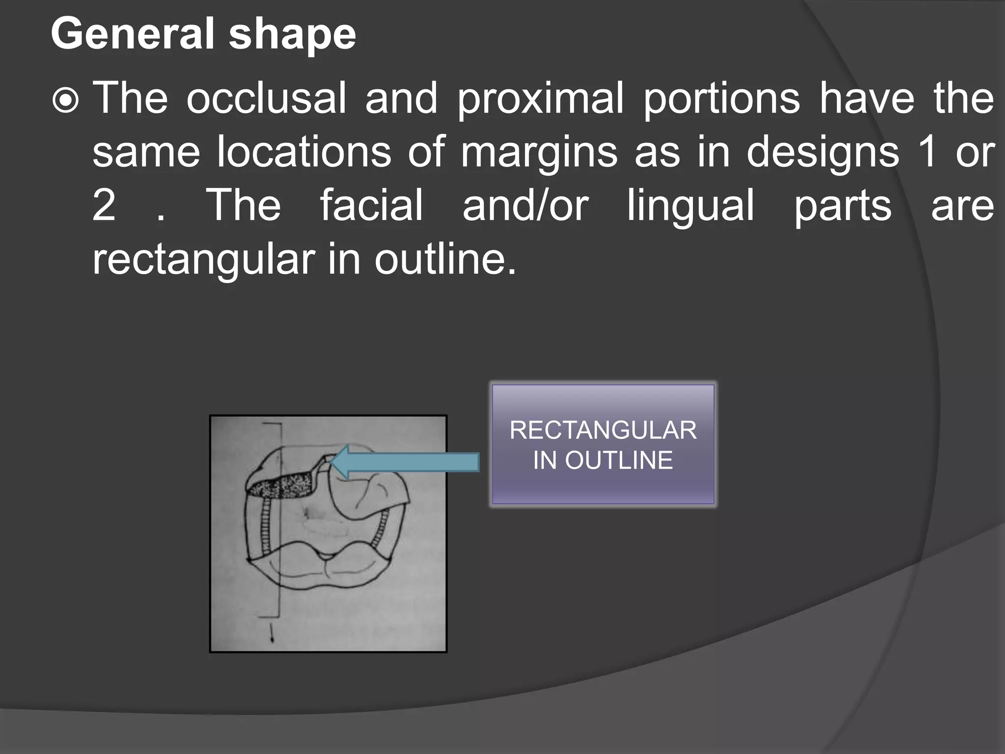
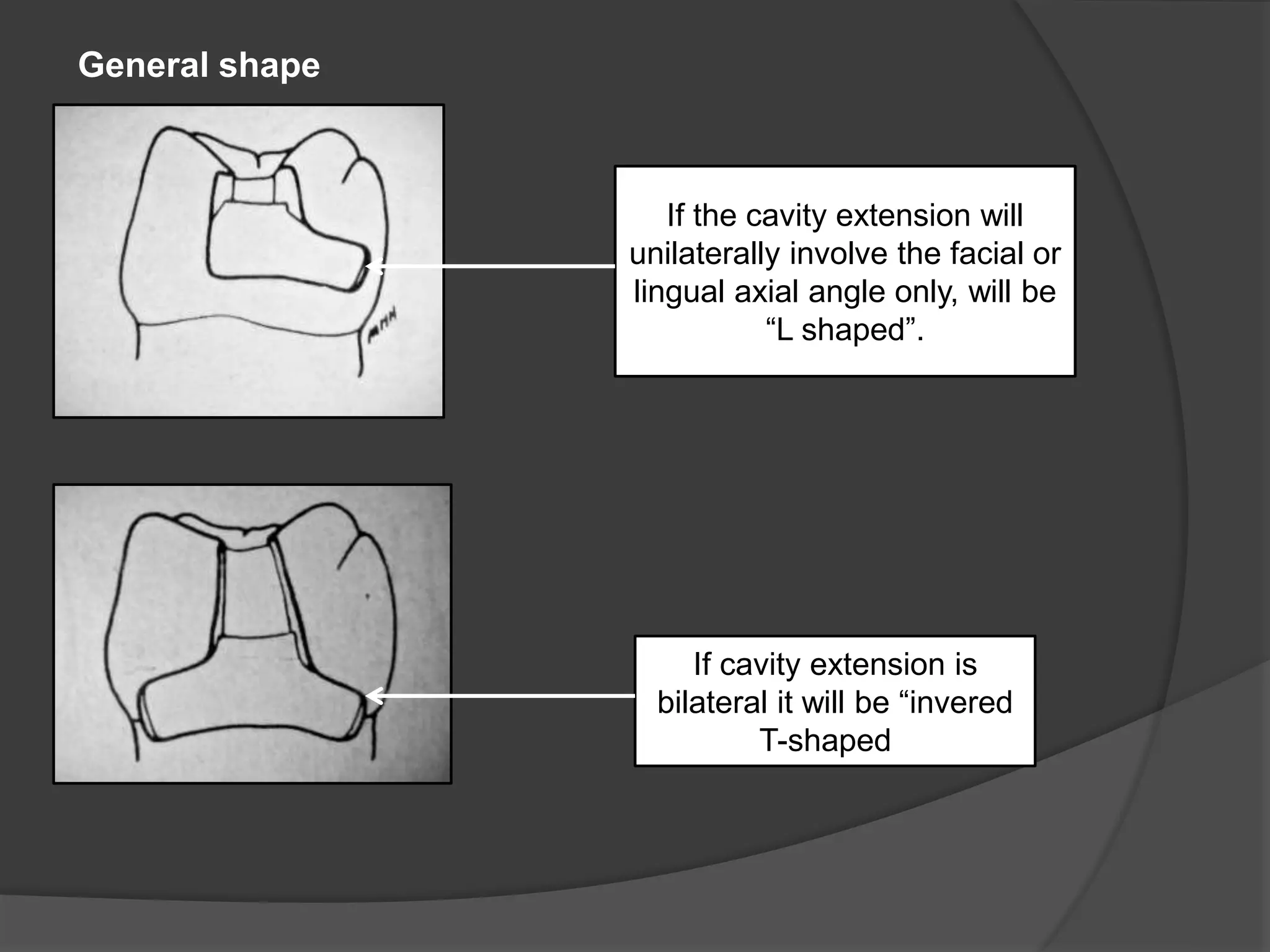
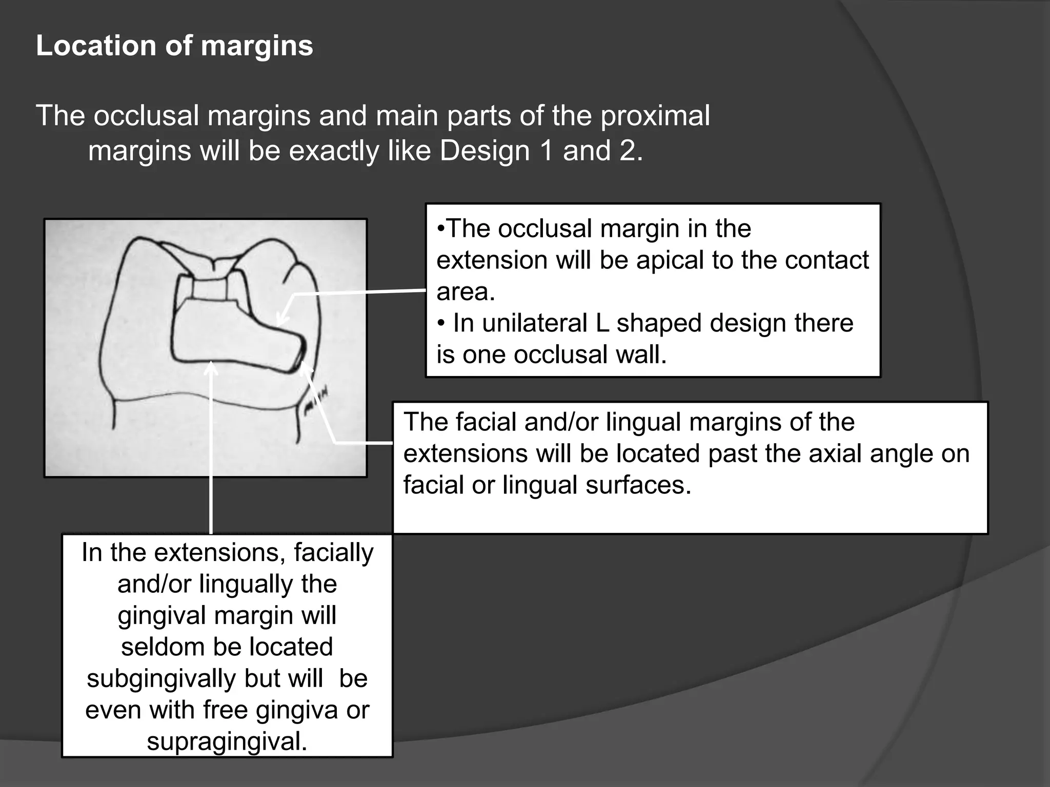
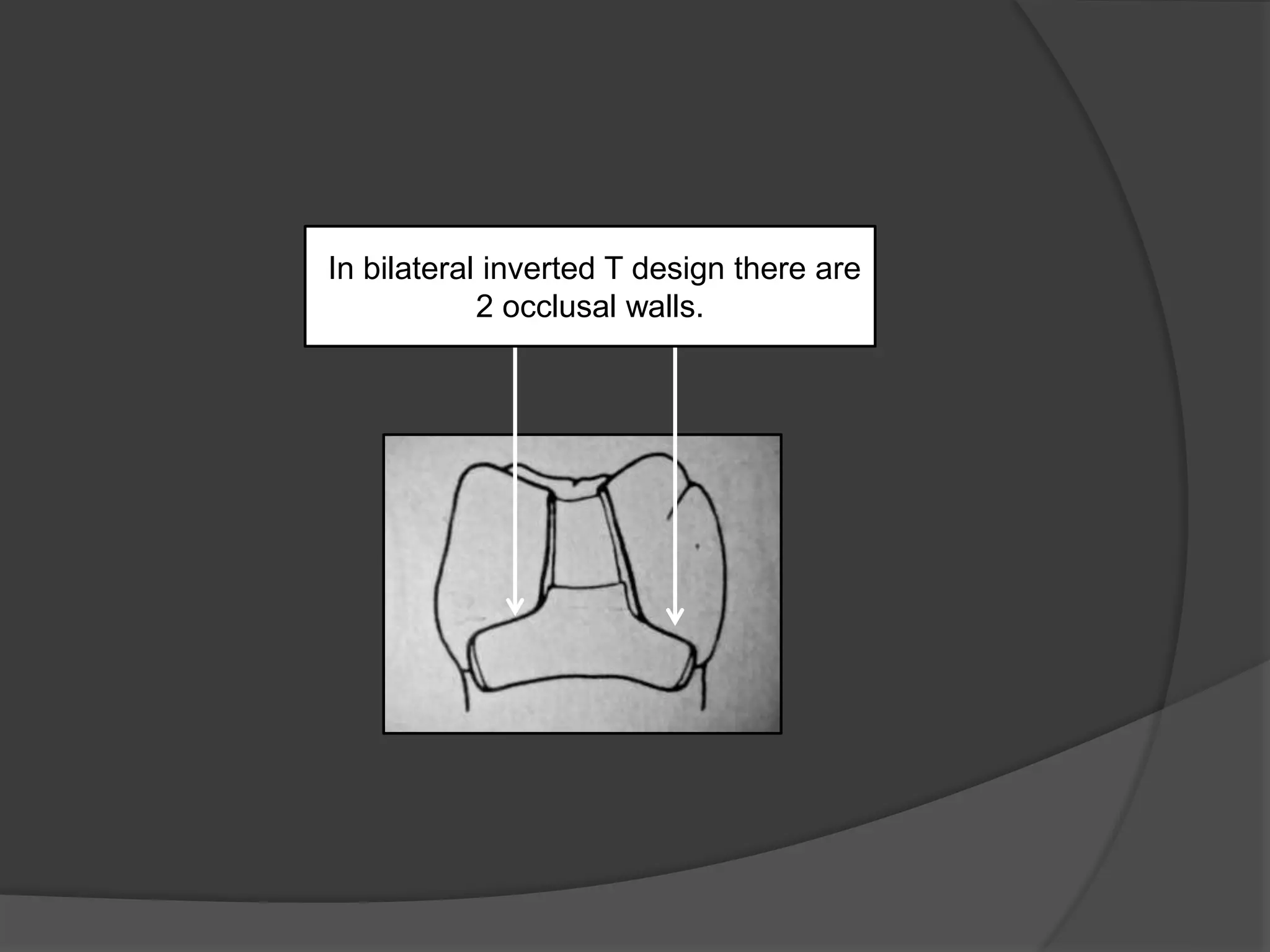
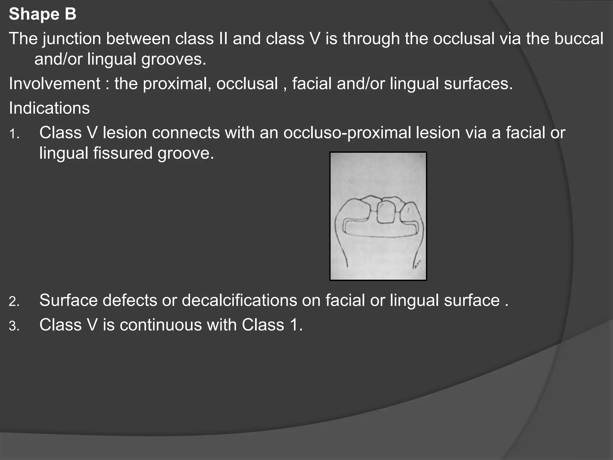
1) The document describes modifications to a standard Class 2 cavity preparation design based on factors like lesion size and location, tooth structure, and patient oral hygiene. 2) It presents 6 designs (labeled 1-6) that vary the cavity shape, locations of margins, and internal anatomy features to best suit different clinical situations. 3) Each design is indicated for specific caries patterns and locations, and involves different areas of the tooth structure, with variations in how the cavity preparation margins and walls are formed.
this contains the steps for the class 1 cavity preparation for amalgam in detail. also contains the difference between composite and amalgam cavity preparation.
A well pictured presentation on Endodontic Instrumentation for UG students. Best for getting a good grip on the topic as a whole. Meant to supplement not substitute standard texts.
Fundamentals in tooth preparation, Now many indications for treatment for teeth are not due to caries and, therefore, the preparation of the tooth is no longer referred to as cavity preparation but as tooth preparation, and the term cavity is used only as a historical reference.
NOMENCLATURE
This presentation specifically deals with the maxillary and mandibular Major connectors used in a cast partial denture. it also mentions the uses, advantages and disadvantages of each,
Bevels and flares are very important components of resin restoration procedure. This presentation focuses on bevels and flares in restorative procedure.
An inlay may cap none, or may cap all but one cusp.
Sturdevant’s 4th ed. page579
Inlays may be used as single-tooth restorations for proximo-occlusal or gingival lesions with minimal to moderate extensions
Shillingburg page 1
An inlay may be defined as a restoration which has been constructed out of mouth from gold, porcelain, or other material & then cemented into the prepared cavity of a tooth.
William McGehee pg410
a detailed account of the principles of tooth preparation with main reference from Shillingburg
The presentation is available on request. Mail me at apurvathampi@gmail.com
A concise and brief presentation on cleaning and shaping of root canals. Colorful and well pictured. Ideal for UG students and PG students to get a good understanding of BMP techniques.
Cavity design is an important step in tooth preparation.Slideshare also shows G.V black classification of cavity(class 1 to class 6). Steps in preparation of class 1 ad class 2 cavity.
silver Amalgam cavity preparation for class 1 /certified fixed orthodontic co...Indian dental academy
The Indian Dental Academy is the Leader in continuing dental education , training dentists in all aspects of dentistry and offering a wide range of dental certified courses in different formats.
Smoking is a major public health problem. Cigarette smoking acts as a nicotine delivery in humans, has found to produce profound changes in physiological architecture. Smoking’s as well as chronic pain are one of the major challenging health concerns faced in day to day life. During smoking nicotine is quickly absorbed into the blood stream within a time gap of 30 seconds it reaches the brain. It stimulates the brain to release various chemicals namely epinephrine which will give a pleasurable euphoric effect. It is a proven fact that smoking of tobacco will cause the production of Rheumatoid factors or anti-cyclic citrullinated peptide autoantibodies which is a risk factor for the development of Rheumatoid arthritis. There is a positive relation between smoking and depression and it has been seen smokers use more number of cigarettes when depressed and smoking also caused the individual who is depressed more prone to pain than a normal smoker. Quitting of smoking is quite difficult because of unpleasant withdrawal syndrome that consists of frustration, depression, anxiety, reduced heart rate, increased weight, depressed mood, difficulty in concentration. Because of all these withdrawal symptoms individuals who try to quit start up again very soon. Smoking is a health hazard, this is a well-known fact and the noxious effects are multiple so in management of pain in theseindividual’s, necessary steps has to be put forward in order to quit the habit. Cognitive behavioural therapy or antidepressant therapy in the management of pain of depressed patients who are smokers has shown good results in a rehabilitation centre on the course of the management of pain.
Taurodontism, a dental anomaly is defined as a change in tooth shape caused by the failure of
Hertwig’s epithelial root sheath to invaginate at the proper horizontal level. Enlarged and elongated
pulp chamber, apically shifted pulpal floor, and lack of constriction at the level of the cementoenamel
junction are the characteristic features. In performing root canal t
appreciate the complexity of the root canal system, canal obliteration, configuration, and the potential
for additional root canal systems. Careful exploration of the all orifices with the help of magnification,
ultrasonics and a modified filling technique are useful for its better management.
Smear layer is a controversial topic in the field of operative dentistry and endodontics. This presentation includes composition, concepts, structure, advantages, disadvantages, and removal methods of smear layer.
This presentation is all about restoration of endodontically treated teeth, prefabricated post and core, cast post and core, direct and indirect technique.
This presentation describes about evolution of nitinol (NiTi), its properties, manufacturing, metallurgy and various rotary systems in the field of endodontics.
About failures of root canal treatment and retreatment. This presentation describes about various techniques for gutta percha removal, posts removal, pastes removal, and removal of separated instrument
Ozempic: Preoperative Management of Patients on GLP-1 Receptor Agonists Saeid Safari
Preoperative Management of Patients on GLP-1 Receptor Agonists like Ozempic and Semiglutide
ASA GUIDELINE
NYSORA Guideline
2 Case Reports of Gastric Ultrasound
Report Back from SGO 2024: What’s the Latest in Cervical Cancer?bkling
Are you curious about what’s new in cervical cancer research or unsure what the findings mean? Join Dr. Emily Ko, a gynecologic oncologist at Penn Medicine, to learn about the latest updates from the Society of Gynecologic Oncology (SGO) 2024 Annual Meeting on Women’s Cancer. Dr. Ko will discuss what the research presented at the conference means for you and answer your questions about the new developments.
Recomendações da OMS sobre cuidados maternos e neonatais para uma experiência pós-natal positiva.
Em consonância com os ODS – Objetivos do Desenvolvimento Sustentável e a Estratégia Global para a Saúde das Mulheres, Crianças e Adolescentes, e aplicando uma abordagem baseada nos direitos humanos, os esforços de cuidados pós-natais devem expandir-se para além da cobertura e da simples sobrevivência, de modo a incluir cuidados de qualidade.
Estas diretrizes visam melhorar a qualidade dos cuidados pós-natais essenciais e de rotina prestados às mulheres e aos recém-nascidos, com o objetivo final de melhorar a saúde e o bem-estar materno e neonatal.
Uma “experiência pós-natal positiva” é um resultado importante para todas as mulheres que dão à luz e para os seus recém-nascidos, estabelecendo as bases para a melhoria da saúde e do bem-estar a curto e longo prazo. Uma experiência pós-natal positiva é definida como aquela em que as mulheres, pessoas que gestam, os recém-nascidos, os casais, os pais, os cuidadores e as famílias recebem informação consistente, garantia e apoio de profissionais de saúde motivados; e onde um sistema de saúde flexível e com recursos reconheça as necessidades das mulheres e dos bebês e respeite o seu contexto cultural.
Estas diretrizes consolidadas apresentam algumas recomendações novas e já bem fundamentadas sobre cuidados pós-natais de rotina para mulheres e neonatos que recebem cuidados no pós-parto em unidades de saúde ou na comunidade, independentemente dos recursos disponíveis.
É fornecido um conjunto abrangente de recomendações para cuidados durante o período puerperal, com ênfase nos cuidados essenciais que todas as mulheres e recém-nascidos devem receber, e com a devida atenção à qualidade dos cuidados; isto é, a entrega e a experiência do cuidado recebido. Estas diretrizes atualizam e ampliam as recomendações da OMS de 2014 sobre cuidados pós-natais da mãe e do recém-nascido e complementam as atuais diretrizes da OMS sobre a gestão de complicações pós-natais.
O estabelecimento da amamentação e o manejo das principais intercorrências é contemplada.
Recomendamos muito.
Vamos discutir essas recomendações no nosso curso de pós-graduação em Aleitamento no Instituto Ciclos.
Esta publicação só está disponível em inglês até o momento.
Prof. Marcus Renato de Carvalho
www.agostodourado.com
New Directions in Targeted Therapeutic Approaches for Older Adults With Mantl...i3 Health
i3 Health is pleased to make the speaker slides from this activity available for use as a non-accredited self-study or teaching resource.
This slide deck presented by Dr. Kami Maddocks, Professor-Clinical in the Division of Hematology and
Associate Division Director for Ambulatory Operations
The Ohio State University Comprehensive Cancer Center, will provide insight into new directions in targeted therapeutic approaches for older adults with mantle cell lymphoma.
STATEMENT OF NEED
Mantle cell lymphoma (MCL) is a rare, aggressive B-cell non-Hodgkin lymphoma (NHL) accounting for 5% to 7% of all lymphomas. Its prognosis ranges from indolent disease that does not require treatment for years to very aggressive disease, which is associated with poor survival (Silkenstedt et al, 2021). Typically, MCL is diagnosed at advanced stage and in older patients who cannot tolerate intensive therapy (NCCN, 2022). Although recent advances have slightly increased remission rates, recurrence and relapse remain very common, leading to a median overall survival between 3 and 6 years (LLS, 2021). Though there are several effective options, progress is still needed towards establishing an accepted frontline approach for MCL (Castellino et al, 2022). Treatment selection and management of MCL are complicated by the heterogeneity of prognosis, advanced age and comorbidities of patients, and lack of an established standard approach for treatment, making it vital that clinicians be familiar with the latest research and advances in this area. In this activity chaired by Michael Wang, MD, Professor in the Department of Lymphoma & Myeloma at MD Anderson Cancer Center, expert faculty will discuss prognostic factors informing treatment, the promising results of recent trials in new therapeutic approaches, and the implications of treatment resistance in therapeutic selection for MCL.
Target Audience
Hematology/oncology fellows, attending faculty, and other health care professionals involved in the treatment of patients with mantle cell lymphoma (MCL).
Learning Objectives
1.) Identify clinical and biological prognostic factors that can guide treatment decision making for older adults with MCL
2.) Evaluate emerging data on targeted therapeutic approaches for treatment-naive and relapsed/refractory MCL and their applicability to older adults
3.) Assess mechanisms of resistance to targeted therapies for MCL and their implications for treatment selection
Tom Selleck Health: A Comprehensive Look at the Iconic Actor’s Wellness Journeygreendigital
Tom Selleck, an enduring figure in Hollywood. has captivated audiences for decades with his rugged charm, iconic moustache. and memorable roles in television and film. From his breakout role as Thomas Magnum in Magnum P.I. to his current portrayal of Frank Reagan in Blue Bloods. Selleck's career has spanned over 50 years. But beyond his professional achievements. fans have often been curious about Tom Selleck Health. especially as he has aged in the public eye.
Follow us on: Pinterest
Introduction
Many have been interested in Tom Selleck health. not only because of his enduring presence on screen but also because of the challenges. and lifestyle choices he has faced and made over the years. This article delves into the various aspects of Tom Selleck health. exploring his fitness regimen, diet, mental health. and the challenges he has encountered as he ages. We'll look at how he maintains his well-being. the health issues he has faced, and his approach to ageing .
Early Life and Career
Childhood and Athletic Beginnings
Tom Selleck was born on January 29, 1945, in Detroit, Michigan, and grew up in Sherman Oaks, California. From an early age, he was involved in sports, particularly basketball. which played a significant role in his physical development. His athletic pursuits continued into college. where he attended the University of Southern California (USC) on a basketball scholarship. This early involvement in sports laid a strong foundation for his physical health and disciplined lifestyle.
Transition to Acting
Selleck's transition from an athlete to an actor came with its physical demands. His first significant role in "Magnum P.I." required him to perform various stunts and maintain a fit appearance. This role, which he played from 1980 to 1988. necessitated a rigorous fitness routine to meet the show's demands. setting the stage for his long-term commitment to health and wellness.
Fitness Regimen
Workout Routine
Tom Selleck health and fitness regimen has evolved. adapting to his changing roles and age. During his "Magnum, P.I." days. Selleck's workouts were intense and focused on building and maintaining muscle mass. His routine included weightlifting, cardiovascular exercises. and specific training for the stunts he performed on the show.
Selleck adjusted his fitness routine as he aged to suit his body's needs. Today, his workouts focus on maintaining flexibility, strength, and cardiovascular health. He incorporates low-impact exercises such as swimming, walking, and light weightlifting. This balanced approach helps him stay fit without putting undue strain on his joints and muscles.
Importance of Flexibility and Mobility
In recent years, Selleck has emphasized the importance of flexibility and mobility in his fitness regimen. Understanding the natural decline in muscle mass and joint flexibility with age. he includes stretching and yoga in his routine. These practices help prevent injuries, improve posture, and maintain mobilit
Acute scrotum is a general term referring to an emergency condition affecting the contents or the wall of the scrotum.
There are a number of conditions that present acutely, predominantly with pain and/or swelling
A careful and detailed history and examination, and in some cases, investigations allow differentiation between these diagnoses. A prompt diagnosis is essential as the patient may require urgent surgical intervention
Testicular torsion refers to twisting of the spermatic cord, causing ischaemia of the testicle.
Testicular torsion results from inadequate fixation of the testis to the tunica vaginalis producing ischemia from reduced arterial inflow and venous outflow obstruction.
The prevalence of testicular torsion in adult patients hospitalized with acute scrotal pain is approximately 25 to 50 percent
micro teaching on communication m.sc nursing.pdfAnurag Sharma
Microteaching is a unique model of practice teaching. It is a viable instrument for the. desired change in the teaching behavior or the behavior potential which, in specified types of real. classroom situations, tends to facilitate the achievement of specified types of objectives.
Knee anatomy and clinical tests 2024.pdfvimalpl1234
This includes all relevant anatomy and clinical tests compiled from standard textbooks, Campbell,netter etc..It is comprehensive and best suited for orthopaedicians and orthopaedic residents.
6. A patient with good oral hygiene, low
plaque and caries index.
7. A lesion where after removal of carious
dentin sufficient bulk in the buccal and
lingual wall present.
8. GENERAL SHAPE :
OCCLUSAL PORTION :
Outline resembles class 1 design 1 or 2
, except they should have some dovetail
formation which may be toward one side
only.
9. PROXIMAL PORTION:
These preparation assume an only
unilateral inverted truncated cone
shape
10. LOCATION OF MARGIN
OCCLUSAL PORTION :
Resemble those seen in class1 design 1
11. Proximal portion
Gingival margin may be located anywhere on the
proximal surface,provided
1. It is gingival to the contact area
13. ISTHMUS PORTION
facial and lingual margins at the isthmus are placed on
corresponding surfaces of inclined plane and the remaining
areas of marginal ridge. Because of the limited cavity width ,
the universal sweeping curves of facial and lingual margins
occlusally will always reverse in isthmus portion (in S-shaped
form) to include contact area.
15. Proximal portion
Mesio-distal cross-section :
Similar to the conventional design, except that rarely gingival
margin may be located on cementum. All line angles are
rounded, with exception of gingivo-axial line angle, which
should be kept sharp.
16. facio – lingual cross section
It is very similar to the conventional design
With following variations --
Buccal and lingual dentinal retention grooves will
be present
17. The axial depth of modern design
cavity is much less than that of the
conventional design .
18. Preparation modifications:
In tapered teeth (bell shaped teeth )
in line with the axio-pulpal line angle
facially and lingually a groove is prepared
on each of the facial and lingual walls
respectively.
19. . After preparing a Class II cavity preparation, if
there presents an intact marginal ridge, crossed by
a fissure, or carious groove, all that is needed is a
proximal extension in the form of slit .
21. This “ slit “ may open proximally on
the same level as the pulpal floor , or
with an occlusal step .
22. Instrumentation for Class II , Design 2
Procedure
Procedural steps are similar to those described for Design 1,
with exception that smaller instruments are used.
Eg. In gaining access and gross removal use a 168 bur, instead
of a 699 bur. In preliminary and final shaping use the
smallest sizes of chisels . The proximal retention grooves ,
which are mandatory here, are created with smaller gingival
marginal trimmers or angle former.
24. Involvement
This preparation is designed to involve
primarily the proximal surface(s) and a very
limited part of the occlusal surface , not
extending beyond the adjacent triangular
fossa .
25. Indication
The decay is restricted to the proximal
surface only and the occlusal surface
is completely sound.
26. • Low stress bearing area .
• There is sufficient bulk of
remaining tooth structure to
place substantial buccal ,
lingual and gingival retentive
grooves .
• Patient exhibits good oral
hygiene and low caries and
plaque indices
27. General shape
These preparations appear proximally
as a one-sided inverted truncated cone
which is located totally proximally
with the exception of its tip, which
involves part of the adjacent occlusal
triangular fossa
28. LOCATION OF MARGIN
OCCLUSAL PORTION
located on the occlusal
inclined planes of the
involved marginal ridge
30. Internal anatomy :
1. Mesio-distal cross section
The gingival floor may assume one of two forms
a. If the gingival margin is present at the
gingival third of the proximal surface , the
floor will be formed of 3 planes –
1. an inner dentinal plane in the form of a
groove.
2. middle transistional dentinal phase
3. an outer enamelo-dentinal plane
following the direction of the enamel rods
and inclining gingivo-proximally.
31. . If the gingival margin is located at the middle third
proximally the gingival floor will be formed of 2
planes –
1. an inner dentinal plane in the form of a groove.
2. straight plane formed of enamel and dentin
32. Facio-lingual cross section
This view shows the axial wall to be
perfectly convex.
The facial and lingual walls , If their margins
are at the facial or lingual thirds of the
proximal surface ,
It will be formed of 3 planes : 1. an inner
dentinal plane in form of groove . 2. a
transitional dentinal plane. 3. an outer enamel
–dentinal plane following the direction of
enamel rods proximo-buccally and lingually.
33. If their wall ends at middle
third it will be composed of
2 planes : 1. inner dentinal
groove plane . 2. outer ,
straight enamel –dentinal
plane , perpendicular to the
tangent of the axial wall
34. INSTRUMENTATION
PROCEDURES:
Using a ¼ round bur , start a tunnel on the occlusal inclined
planes of the marginal ridge to be involved in the
preparation, connecting this tunnel with the lesion, widening
and breaking it in the same way as was done in preparation
of previous 2 designs.
35. If part of the marginal ridge is lost , two
lateral grooves may be prepared on the
buccal and lingual behind the portion of
marginal ridge to be removed
38. Indications
The decay is restricted to contacting
or proximal surfaces without
undermining the corresponding
marginal ridges
39. There is a diastema
• the adjacent tooth is
missing facilitating
direct access to the
lesion.
40. The affected tooth is rotated or
inclined.
• The proximal lesion occurs on tapered
teeth with wide gingival embrasures
facilitating facial or lingual access to the
lesion
41. The proximal lesion is located very
gingivally at or apical to, the CEJ,
accompanied by gingival recession(senile
decay), making accessibility to the lesion
from the facial or lingual direction possible
42. General Shape
Generally, it should follow the shape of
the contact area and the proximal
surface. Usually it will assume a
trapezoidal or rhomboidal shape.
43. Locations of margins
If there is no proximally contacting tooth
(diestema) , there is no specific location
of margins , for the entire proximal
surface is essential self-cleansable.
44. If the lesion is apical to the contact
area(senile decay ), the occlusal and
gingival margins will be in the gingival
embrasures.
45. Internal anatomy
a. Facio –lingual cross section
The axial wall is seen to be flat to
slightly convex facio-lingually .
(interrupted line)
The buccal or lingual wall , on the
access side will be a one-planed
enamel dentinal wall
following the directions of the
enamel rods.
46. Occlusogingival cross section
The gingival floor may be seen to occur in one of
the two ways
If the gingival margin is located on cementum ,
cementum should be removed
gingival floor should be completely formed of
dentin and in 2 planes : an inner one formed of a
groove and an outer one perfectly straight and
flat
47. If the gingival margin is present on
enamel, it will be formed of 3 planes :
inner dentinal groove , a dentinal
transistional plane and an outer plane
of enamel and dentin
48. Instrumentation for class II design 4
Prerequisites :
These are similar to previous designs , with the exception that
wedges are used if they will interfere with access.
Procedures :
a. Gaining access and gross removal
From the access side, using a round bur ¼ the size of the proximal
preparation with axial pressure and lateral dragging. Remove decayed and
sound tooth structure within the outline.
49. b Preliminary shaping
This is done using the base of an inverted cone
bur . With buccal access ,at the future location of
the lingual wall ,form the lingual wall in gingivo-occlusal
movements .
50. • At the gingival floor location ,use the
same part of the bur moving it in a bucco-lingual
direction to form the gingival floor .
51. • Then using the side of the bur , in an occluso-apical
direction , form the buccal wall .
52. The occlusal wall could be formed
with a tapered fissure bur , using
bucco-lingual strokes.
53. If access is from the lingual, the exact reverse of the previous
steps are followed.
Final shaping is accompanished in several steps.
The different planes for the buccal and lingual walls can be
formed using a hatchet and wedelstaedt chisel .
54. Similarly different planes for the gingival and
occlusal walls could be formed using a
Wedelstaedt chisel from buccal or hatchet
from lingual
55. CLASS II DESIGN 5
Involvement : Part of the proximal surface, with a very limited
access area on the facial or lingual surface .
Indications
There are two shapes for this design , each with certain
indications.
Shape A– facial and lingual surface will not have dovetail form.
1. The cavity will have 4 walls , with retentive grooves in atleast
2 of them.
2. Small to medium sized proximal lesions.
3. Restoration subjected to normal displacing forces.
4. Marginal ridge is intact.
5. Lesion does not involve the contact area.
6. Good oral hygiene.
56. In shape B - the facial or lingual access will have a
locking feature in the form of a dovetail, unilaterally
cut in the occlusal direction.
1. Final cavity preparation will not have 4 surrounding
walls and either one wall or no wall is bulky enough
to accommodate a groove.
2. For medium to large sized proximal lesion.
57. General shape
The proximal part of this cavity will have no specific shape , however , it
will appear either trapezoidal .
One- sided
dovetailed
shapedovetailed
shape
a box or
rectangular shape
58. Occlusal margin
located in gingival
embrasure
Gingival margin
present in gingival
embrasure
Locations of margins
The proximal
margins are far
enough onto the
facial or lingual
surface to include
the axial angle and
¼ th of the facial or
lingual surface.
59. Internal Anatomy
Occluso –gingival cross section
The axial wall will
appear flat
occlusogingivally
The occlusal and gingival walls , if both are
located on cementum or dentin, will be formed of 2
planes – an internal dentinal grooved plane and an
external dentinal plane which is perfectly flat and
opening straight proximally.
If the occlusal
margin is located on
enamel it is always
at the gingival third
of the surface .The
resultant inclination
axio-gingivally
creates a needed
undercut.
60. Two distinct axial walls ,
i.e. One proximal
and another facial or
lingual (access side).
Both are perfectly flat
.
If the facial or lingual wall on the
non-access side has its margin
on enamel, it will be composed
of 2 planes: an inner dentinal
plane at right angle to proximal
axial wall for resistance and
retention and outer enamel-dentin
plane in direction of
enamel rods
Facio-lingual cross section
61. Facio-lingual cross section
In a very apically located
lesion, part or all of this
facial or lingual walls
will be completely
formed of dentin,
always at a right angle
to the axial wall.
The mesial and distal
wall on the access
side is always one
planed. It is formed
of enamel and
dentin following the
direction of enamel
rods.
62. Instrumentation
a. Gaining access and gross removal :
with a tapered fissure bur using axial pressure and lateral
dragging , on the access side, cut the access window
which usually will be in sound tooth structures.
Access window
in the sound
tooth structure
63. From this access preparation
introduce the same tapered fissure
bur proximally using axial and lingual
pressure and occluso-gingival
dragging.
Axial and
lingual
pressure
Bucco
lingual
direction
64. a. Preliminary shaping : this is done using the tip and side of a
700 or a 169 bur which creates definite surrounding walls,
and to formulate the proximal axial wall. Retention grooves if
indicated , may be cut using a ¼ round bur, dragged along
the axio-gingival and sometimes axio-occlusal line angle
with pressure gingivally and occlusally.
.
65. a. Final shaping occurs when the different
planes of surrounding walls are formed
using a hatchet for the access cavity and
axial wall and a gingival marginal trimmer
for the non-access side walls and gingival
walls . Defining and rounding of line and
point angles is done using a Wedelstaedt
or hatchet chisels.
hatchet GMT Wedelstaedt
66. Cavity finish is accomplished using
the same instruments but with lighter
and more frequent applications
67. CLASS II , DESIGN 6
Involvement : The occlusal, proximal(s) and
part of the facial and/or lingual surfaces.
68. Indications:
1. The cusp length is double or more its width ,
either throughout or at certain portions of
cusp.
2. A cusp is completely missing .
3. A foundation for cast restoration is required.
4. Teeth have a doubtful prognosis
endodontically and periodontically.
5. A badly broken down tooth needs to be
prepared prior to endodontic or orthodontic
treatment
69. General shape
The occlusal and proximal portions have the
same locations of margins as in designs 1 or
2 . The facial and/or lingual parts are
rectangular in outline.
RECTANGULAR
IN OUTLINE
70. Locations of margins
The occlusal and proximal portions have
same location of margins as design 1or 2.
With only partial mesio-distal replacement
of cusp, the margin should not end at the
tip of cusp rather it should be located
mesial or distal to it.
• Similarly if margins come near groove , the groove
is involved in cavity preparation
71. Cuspal elements , or parts
of them , which will
accomodate amalgam
must be cut flat, i.e. In the
form of a table with right
angled cavosurface
margins.
Reduce cusps or parts of a
cusp until there is minimum
length:width ratio of 1:1. If
length:width ratio is
different at different levels
tables can be prepared at
these different levels.
The junction between tables
should be rounded.
Cusps or parts of it to be replaced or covered with amalgam should be
reduced at least 1.5-2 mm from the opposing cuspal elements in both
static and dynamic contacts. This will accomodate sufficient amalgam
bulk to resist loading.
Internal Anatomy
72. It is always advisable to
have a retention form ,
for e.g. External box or
groove, adjacent to the
tabled cusp
• Undermined or
thinned enamel
should be tabled .
• Never place pins on
tables which will
accommodate
amalgam cusps.
73. Class II Design 7 (Combination of Class II with Class V)
Shape A
The junction between the Class II and Class V via the proximal,
crossing the axial angles.
Involvement
The occlusal, proximal and part or all of the gingival third of the
facial and/or lingual surfaces with the intervening part of the
axial angles
74. Indications
a. When at a location apical to the contact area, an
occluso-proximal lesion joins a senile decay lesion via
decalcification, or a defect that has spread laterally
beyond the regular cavity preparation.
b. A class V lesion undermines enamel or directly
involves tooth structure of the adjacent axial angles in
a tooth having a proximo-occlusal lesion.
c. Surface defects or decalcifications at the axial angles
of the tooth are continous with a proximo-occlusal
cavity preparation apical to contact area.
75. If the cavity extension will
unilaterally involve the facial or
lingual axial angle only, will be
“L shaped”.
If cavity extension is
bilateral it will be “invered
T-shaped”.
General shape
76. Location of margins
The occlusal margins and main parts of the proximal
margins will be exactly like Design 1 and 2.
In the extensions, facially
and/or lingually the
gingival margin will
seldom be located
subgingivally but will be
even with free gingiva or
supragingival.
•The occlusal margin in the
extension will be apical to the contact
area.
• In unilateral L shaped design there
is one occlusal wall.
The facial and/or lingual margins of the
extensions will be located past the axial angle on
facial or lingual surfaces.
78. Shape B
The junction between class II and class V is through the occlusal via the buccal
and/or lingual grooves.
Involvement : the proximal, occlusal , facial and/or lingual surfaces.
Indications
1. Class V lesion connects with an occluso-proximal lesion via a facial or
lingual fissured groove.
2. Surface defects or decalcifications on facial or lingual surface .
3. Class V is continuous with Class 1.
79. General shape
Occlusal and proximal
portions are exactly as design
1 and 2. The facial and / or
lingual parts are inverted T-shaped.
Internal Anatomy
The connection between the occlusal
and facial or lingual portion is in the form
of long arm of inverted T.
80. Class II Design 8
Involvement
Two or more surfaces of an endodontically treated that
does not require post retention .
Indications
1. The remaining tooth structure after endodontics can
support and retain an amalgam restoration.
2. The tooth has a sufficient pulp chamber to accomodate
retaining self – resisting amalgam bulk i.e. A minimum 2
mm thickness in three dimensions.
3. The post-endodontics pulp chamber has at least two
opposing intact walls.
4. The tooth contains sufficiently large root canals to
accomodate retaining resisting amalgam bulk at its
occlusal 1/3rd (i.e. Minimum 1.5 mm thickness )
5. A foundations is needed for a reinforcing restorations
(cast restoration)
81. 6. There is sufficient remaining tooth structure to permit the
preparation of flat planes at right angles to occluding forces.
7. There has been successful root canal therapy leaving an intact
subpulpal floor.
8. The tooth does not show any signs of cracking or crazing.
82. Excavate from the entire pulp
chamber any residual root canal
filling materials or debris. Bare
dentin should be exposed on
the surrounding walls and
subpulpal floor
Large root canals that
can accomodate an
amalgam thickness of
1.5 mm should have
the root canal filling
removed to a 3-4 mm
depth .
General shape
The outline will appear exactly as
described for Design 6.
Internal anatomy
83. Each flat portions of the tooth
preparation eg. Tables or ledges should
be opposed by a similar flat component
for proper reciprocation, to immobilize
the restoration and evenly distribute
stresses.
•Any external boxes for retention should not perforate to the pulp chamber or
cause thinning in the intervening walls.
• In preparing tables and ledges allow sufficient reduction depths to provide
enough thickness of amalgam to serve as a foundation for a reinforcing cast
restoration.
84. In the bulky portion of the
surrounding walls of the
pulp chamber, cut flat
ledges to receive most of
the occlusal loading ,
thereby minimizing
stresses on the subpulpal
floor during such loading .
85. If possible “square up” surrounding walls provided this
action will not perforate to the surface , furcation , or
thin tooth structure to the extent of making these
areas non-resistant .
Retain any residual
pulpal floor , placing
ledges in it and
making it as flat as
possible.
86. Instrumentation for Class II, Design 8
The occluso-proximal cavity preparation and capping of cusps , short of pulp
chamber and root canal preparation, is done in same way as described for Designs
1 and 6 .
For intrapulpal and intraradicular prepartion the following procedure may be done :
Ledges and shelves may be prepared using 555 and 556 burs in apical pressure
and lateral dragging .
APICAL
PRESURE
87. Intraradicular preparation is started by removing
the specified lengths of the indicated root canal
material using hot gutta-percha pluggers,peeso
reamers or bibevelled-sided reamers.
88. Then the specified length of the canal is
widened and side-paralleled using large
tapered, then cylindrical fissure burs
(704,558).
89. Following this , the junction between pulp
chamber and root canal preparations
should be rounded using round burs.
Finally , all junctional parts of the cavity
preparations are rounded using a round
bur or a very sharp gingival marginal
trimmer
90. References
Operative Dentistry – Modern theory and Practice –
Marzouk.
Art and Science of Operative Dentistry – Sturdevant.
Fundamental of operative dentistry – j.summit
Textbook of endodontics- Nisha garg