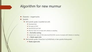
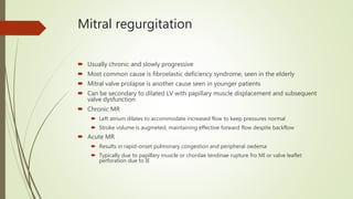
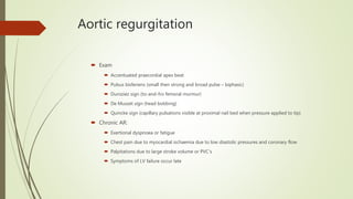
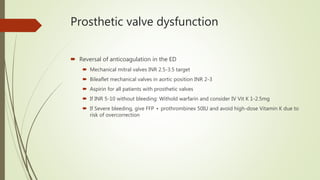
Valvular emergencies
Dr Andrew Crofton
Disclaimer: https://criticalcarecollaborative.com/disclaimer/
Introduction
 2.5-fold risk of death and 3-fold risk of stroke compared to general population
 The New Murmur
 Benign or physiological
 Do not cause symptoms or findings of cardiovascular disease
 Soft, systolic ejection murmurs that begin after S1, end before S2 and no abnormal heart sounds
 Systolic murmurs
 May be associated with anaemia, sepsis, volume overload or any other cause of increased CO
 Look for underlying cause vs. management of murmur itself
 Patients without chest pain, dyspnoea, fever or other signs attributable to valvular disease do not
need emergent echo but need referral
 Any diastolic murmur or new systolic murmur with symptoms at rest is pathological and needs
inpatient echo
 Always consider endocarditis, especially if valvular insufficiency
Algorithm for new murmur
 Diastolic – Urgent echo
 Systolic
 Mid-systolic grade 2 (audible but soft)
 Asymptomatic
 No signs of CVD
 Normal ECG and CXR
 Murmur does not increase with valsalva or standing
 = No further workup
 Symptomatic, signs of CVD, abnormal ECG/CXR, murmur increases with Valsalva or standing
 = Needs urgent echo
 Mid-systolic grade 3 (loud but no thrill)/Early or late systolic/Holosystolic
 Needs urgent echo
Grading system for murmurs
Grade Description
1 Faint, may not be heard in all positions
2 Quiet but heard immediately
3 Moderately loud
4 Loud
5 Heard with stethoscope partially off chest
wall
6 Heard when stethoscope entirely off chest
wall
Valve disorder Murmur Sounds and signs
Mitral stenosis Mid-diastolic rumble, crescendos
to S2
Loud S1, small apical pulse
Mitral regurgitation Acute: Harsh apical systolic
murmur starting with S1 and may
end before S2.
Chronic: High-pitched apical
holosystolic murmur radiating into
S2
S3 +/- S4
Mitral valve prolapse Click with late systolic murmur
crescendos into S2
Mid-systolic click; S2 may be
diminished by late-systolic
Aortic stenosis Harsh ejection systolic murmur Paradoxical split S2 +- S3/4
Small amplitude pulse, slow
rise/sustained peak
Aortic regurgitation High-pitched blowing diastolic
murmur immediately after S2
S3 and wide pulse pressure
Mitral stenosis
 Rheumatic heart disease is most common
 Usually presents with gradual onset pulmonary congestion and AF
 Mitral annular calcification is a non-rheumatic cause
 Most common in women, elderly and those with HTN/Chronic renal failure
 Rarely causes severe symptoms
 Clinical features
 Exertional dyspnoea, often precipitated by anaemia/sepsis (increased CO)
 Systemic emboli are a risk (particularly if in AF)
 Haemoptysis is rare but can be massive
 Recurrent bronchitis, fatigue, paroxysmal AF
 Signs:
 Mitral facies (peripheral cyanosis of cheeks)
 Mid-diastolic rumbling murmur with crescendo towards S2 (presystolic accentuation disappears if in AF)
 S1 usually loud with loud opening snap heard to right of apex
 Apical pulse is small and tapping due to underfilled LV
Mitral stenosis
 Diagnosis and treatment
 ECG: Notched or biphasic P waves (P mitrale) and right axis deviation
 CXR: Straightening of left heart border (LA enlargement) and later pulmonary congestion
 Echo: Severe <1cm2
 Medical management
 Symptom control
 Beta-blockers to reduce HR, increase diastolic filling time
 AF with RVR or dyspnoea on exertion may benefit from rate control
 Anticoagulation if LA diameter >55mm OR in AF, a left atrial thrombus or history of systemic emboli
 Surgical for symptomatic disease
 Balloon valvotomy, valve repair or valve replacement before onset of severe pulmonary hypertension
Mitral regurgitation
 Usually chronic and slowly progressive
 Most common cause is fibroelastic deficiency syndrome, seen in the elderly
 Mitral valve prolapse is another cause seen in younger patients
 Can be secondary to dilated LV with papillary muscle displacement and subsequent
valve dysfunction
 Chronic MR
 Left atrium dilates to accommodate increased flow to keep pressures normal
 Stroke volume is augmeted, maintaining effective forward flow despite backflow
 Acute MR
 Results in rapid-onset pulmonary congestion and peripheral oedema
 Typically due to papillary muscle or chordae tendinae rupture fro MI or valve leaflet
perforation due to IE
Mitral regurgitation
 Clinical features
 Acute MR: Severe dyspnoea, peripheral oedema, tachycardia, cardiogenic shock. S4
gallop and harsh apical systolic murmur loudest in early-mid systole diminishing before
S2
 Chronic MR: Eventual exertional dyspnoea, sometimes with AF
 Late systolic left parasternal lift and lateral displacement of apex beat
 High-pitched holosystolic murmur heard at apex
 S1 soft and obscured by murmur
 S3 often heard followed by short diastolic rumble, indicating increased flow into the ventricle
 Signs of systemic thromboembolism may be first suggestion of MR with AF
Mitral regurgitation
 Diagnosis
 Thinks of acute MR in any patient new-onset and marked pulmonary oedema
 Especially if normal heart size on CXR or do not respond to therapy
 Look at ECG for anteroinferior ischaemia
 Chronic MR
 ECG signs of LV hypertrophy and LA enlargement
 CXR shows LA and ventricular enlargement proportional to regurgitant volume
Mitral regurgitation
 Treatment
 Acute MR
 Oxygen, PPV, nitrates (reduce afteroad, to increase forward flow and restore mitral valve competence as
LV size diminishes
 Inotropic support as bridge to surgery (if structural disruption)
 Aortic balloon pump may be effective bridge
 Medical therapy is aimed at improving forward flow with emergent cardiology and surgical consultation
during optimisation
 Chronic MR
 Treat acute symptoms
 Control AF with RVR using beta-blockers or CCB’s
 Start anticoagulation to prevent systemic embolisation
 Cardiology consultation
Mitral valve prolapse
 Systolic billowing of one or both leaflets into the LA with/without mitral regurgitation
 Characterised by myxomatous degeneration of the valve due to inherited connective tissue defects
 Most common valvular disease in developed countries (2.4% of population)
 Presence of concomitant mitral regurgitation determines prognosis
 Clnical features
 Mostly asymptomatic
 May present with atypical chest pain, palpitations, fatigue, anxiety or dyspnoea unrelated to exertion
 Signs such as scoliosis, pectus excavatum and low body weight may be seen
 If exercise induces symptoms, morbidity increases
 Auscultation
 Mid-systolic click
 Maneuvers that decrease preload (Valsalva/standing) will cause the click to occur earlier in diastole
 Increased preload by squatting or hand gripping causes systolic click to move later into systole
 A late systolic murmur that crescendos into S2 is heard in some
Mitral valve prolapse
 Diagnosis and therapy
 Emergency evaluation focuses on identification of long-term complications such as AF or
heart failure
 Refer to cardiologist if suspected
 Palpitations attributable to mitral valve prolapse may respond to beta-blockers but
should be left to cardiologist
 Antithrombotic therapy only indicated if TIA/stroke or AF
 If prolapse and MR, require endocarditis prophylaxis
Aortic stenosis
 Most common cause in developed countries is degenerative calcification,
associated with increased age, smoking, dyslipidaemia and diabetes
 Rheumatic heart disease is the most common cause worldwide
 Bicuspid aortic valves and congenital heart disease are also seen
 3% prevalence if >74 years old
 Typically long asymptomatic period with LV hypertrophy to preserve EF
 Ultimately LV hypertrophy impairs filling and increases myocardial oxygen demand with
slow reduction in cardiac output, coronary and systemic blood flow
Aortic stenosis
 Clinical features
 Classic triad: Dyspnoea, chest pain and syncope
 Many patients with severe stenosis (<1cm2) are asymptomatic
 Often stepwise dyspnoea, followed by chest pain, then syncope and finally signs of heart failure
 Once symptoms start, mortality increases
 Physical exam
 Late peaking systolic murmur at right 2nd ICS, radiating to carotids, with single or paradoxically split S2, S4 gallop and
reduced carotid pulse with delayed upstroke and long plateau (pulsus parvus et tardus)
 Brachioradial delay is also seen
 Narrowed pulse pressure
 AS with AF = Dire consequences
 AS typically have diastolic dysfunction due to LV hypertrophy and are reliant on atrial kick for filling
 If then given GTN for chest pain or dyspnoea, can collapse
Aortic stenosis
 Diagnosis
 ECG: LV hypertrophy + LBBB or RBBB in 10%
 Late CXR findings are LV hypertrophy (not dilation) and pulmonary congestion
 Treatment
 APO: Oxygen, PPV. Use nitrates, beta-blockers, CCB’s and diuretics with caution
 Reducing preload or afterload can cause significant hypotension
 New-onset AF may require cardioversion urgently
 If newly symptomatic, admit
 Without surgery 40-50% mortality within 1 year
 If discharged, need to avoid vigorous activity and need prompt review by Cardiologist.
 Prophylactic antibiotics for endocarditis are not required
 Need valve replacement as soon as symptomatic, asymptomatic with LV dysfunction or if jet peak
velocity >4m/s
Aortic regurgitation
 Slowly progressive over years
 Ultimately leads to LV dilatation and hypertrophy
 Get wide pulse pressures
 Tachycardia shortens diastole, which decreases regurgitant volume and mutes symptoms early on
 In contrast, increased afterload e.g. exercise, exacerbates regurgitant flow and may precipitate
symptoms
 Over time, increased LV dilatation and hypertrophy leads to impaired systolic function and reduced
CO with failure symptoms
 50% of cases due to leaflet disorders secondary to bicuspid aortic valves, infective endocarditis or
rheumatic heart disease
 Non-valvular causes include aortic dissection, aort root dilatation (Marfan’s) or aortitis
 Also frequently associated with aortic stenosis and can be severe in this case
Aortic regurgitation
 Clinical features
 Acute:
 Rapid dyspnoea, pulmonary oedema, tachycardia and cardiogenic shock
 Sudden onset ripping or tearing pain suggests acute aortic dissection
 Fever or IVDU suggests endocarditis
 Exam
 High-pitched blowing diastolic murmur immediately after S2 at left sternal border 2/3rd ICS
 May get systolic ejection murmur due to increased SV and S3 due to ventricular dilatation also
 Austin Flint murmur (mid-diastolic rumble) may be heard in left lateral position at apex using Bell of
stethoscope
 Widened pulse pressure
 Corrigan (water-hammer) pulse
Aortic regurgitation
 Exam
 Accentuated praecordial apex beat
 Pulsus bisferiens (small then strong and broad pulse – biphasic)
 Duroziez sign (to-and-fro femoral murmur)
 De Musset sign (head bobbing)
 Quincke sign (capillary pulsations visible at proximal nail bed when pressure applied to tip)
 Chronic AR:
 Exertional dyspnoea or fatigue
 Chest pain due to myocardial ischaemia due to low diastolic pressures and coronary flow
 Palpitations due to large stroke volume or PVC’s
 Symptoms of LV failure occur late
Aortic regurgitation
 Diagnosis
 Acute AR
 CXR may show acute pulmonary oedema without cardiac enlargement (if acute)
 May shows signs of dissection
 ECG
 Sinus tachycardia
 Ischaemic changes or ST elevation due to dissection involving coronary arteries
 Chronic AR:
 CXR: Cardiomegaly, aortic dilatation, evidence of heart falure
 ECG: LV hypertrophy
Aortic regurgitation
 Treatment
 Acute: Immediate surgical intervention
 Medical: Oxygen, intubation for respiratory failure, nitroprusside + inotropes can augment forward flow
and reduce LV end-diastolic pressure
 Diuretics and nitrates are usually ineffective
 Beta-blockers are CI in acute aortic regurgitation (commonly used in aortic dissection but if acute
aortic regurgitation co-exists, do better with tachycardia to reduce time/volume of regurgitation flow
 Aortic balloon pumps are also contraindicated as worsen regurgitant flow
 If mild AR due to endocarditis, antibiotics may be sufficient without operative intervention
 Chronic AR:
 Vasodilators e.g. ACEi, dihydropyridines
 Those who are symptomatic, have low EF or who have significant LV dilatation – consider aortic valve
replacement
Right-sided valve disease
 Trivial TR and PR are common
 Pathological TR usually in setting of:
 Elevated right sided pressure or volume overload
 E.g. Pulmonary hypertension, chronic lung disease, PE or atrial septal defects
 Tricuspid stenosis is rare and usually accompanied by regurgitation
 Pulmonic valve is least likely to be affected by acquired disease
 Acute onset tricuspid disease is usually due to endocarditis, and typically
aggressive organisms (e.g. S. aureus)
Right-sided valve disease
 Clinical
 Right heart failure signs and symptoms
 Exertional dyspnoea is often the first symptoms if associated with pulmonary
hypertension
 Tricuspid regurgitation
 Murmur is soft, blowing and holosystolic along left lower sternal edge and increases with
respiration
 Systolic waveform may be seen in JVP in severe TR
 Tricuspid stenosis
 Rumbling crescendo-decrescendo diastolic murmur before S1
 Along left lower sternal edge, increases with inspiration and often preceded by opening snap
Right–sided valvular disease
 Exam
 Pulmonic stenosis
 Exertional dyspnoea, syncope, chest pain and signs of right heart failure
 Harsh systolic murmur, best head in left 2nd ICS, increased with inspiration
 Pulmonic regurgitation
 High-pitched and blowing diastolic murmur (Graham Steell murmur) increased with inspiration
 Best heard over left 2nd/3rd ICS
 Typically have right ventricular thrill and heave
Right-sided valvular disease
 Treatment
 Treat underlying cause
 Diuretics treated effects of elevated venous pressures but need to use with caution to
avoid volume depletion (as reliant on preload) and electrolyte abnormalities
 If symptomatic pulmonic or tricuspid stenosis, may be candidates for balloon valvotomy
 Severe TR due to structural valve abnormality may require valve replacement
Prosthetic valve disease
 Mechanical
 Valve thrombosis or thromboembolism rate 8% per year (1-2% with anticoagulation)
 Embolic risk highest in first 3 post-operative years
 Emboli more common from mitral than aortic valves
 Major bleeding complications on warfarin 1.4% per year
 Antiplatelet therapy is recommended for all patients with any prosthetic valve
 Complications
 Thrombosis
 Dehiscence of sutures
 Gradual degeneration
 Sudden fracture
 Symptoms are usually slowly progressive but acute failure and death can occur
Prosthetic valve disease
 Prosthetic valve endocarditis
 Occurs in 6% of patients within 5 years of surgery
 Early causes (first year) include S. epidermidis and S. aureus
 Late cases usually the same as native valves (Strep viridans, Serratia, Pseudomonas
Prosthetic valve disease
 Clinical features
 Cardiac remodelling persists despite valve replacement
 Patients are likely to have concomitant CAD, systemic hypertension, LV failure or AF
 Acute onset of respiratory distress, APO or cardiogenic shock
 Think mechanical valve failure, tearing of a bioprosthesis or large clot obstructing the valve and preventing closure
 Paravalvular leak can also present with AHF
 Slowly progressive HF can occur with gradual thrombus formation
 Mechanical valves often have loud metallic sounds
 Aortic valves often have systolic murmurs but diastolic murmurs should always be considered pathological
 A ‘quiet’ mechanical valve is concerning
 Aortic bioprostheses usually cause a short mid-systolic murmur
 Mitral bioprostheses usually cause a short diastolic rumble
 Loud holosystolic murmur indicates dysfunction
Prosthetic valve dysfunction
 Diagnosis of dysfunction and complications
 Consider this in any patient with a valve replacement and new or progressive dyspnoea, CCF,
decreased exercise tolerance or pain
 Suspect thromboembolism, septic embolism or ICH in any patient with prosthetic valve and
new neurological deficit (like AF)
 Consider endocarditis in patient with prosthetic valve and persistent fever/fever without source
 Treatment and disposition
 Cardiology and cardiothoracic consult
 Emergent surgery and thrombolytic therapy are options for thrombotic complications
 Lesser degrees of obstruction may simple require optimisation of anticoagulation
 Obtain consultation on all if suspicious for complication
Prosthetic valve dysfunction
 Reversal of anticoagulation in the ED
 Mechanical mitral valves INR 2.5-3.5 target
 Bileaflet mechanical valves in aortic position INR 2-3
 Aspirin for all patients with prosthetic valves
 If INR 5-10 without bleeding: Withold warfarin and consider IV Vit K 1-2.5mg
 If Severe bleeding, give FFP + prothrombinex 50IU and avoid high-dose Vitamin K due to
risk of overcorrection
Pregnant women with valvular disease
 Increased CO and blood volume
 Accentuate murmurs in mitral and aortic stenosis
 Reduced SVR
 May attenuate aortic or mitral regurgitant murmurs
 Asymptomatic mild lesions are well tolerated
 Symptomatic disease, pulmonary HTN or LV dysfunction requires high-risk obstetric follow-up
 Neonatal complications associated:
 Prematurity
 IUGR
 RDS
 Intraventricular haemorrhage
 Death
 Mild disease: medical Rx
 Moderate or severe stenotic valvular disease: Balloon valvotomy or surgery
 Aortic or mitral regurgitation who have severe symptoms may also require surgery
 Endocarditis prophylaxis is not required for delivery
Pregnant women with valvular disease
 Presentations during pregnancy
 Extremis, dyspnoea, pulmonary oedema, angina, syncope
 Hypercoagulable state makes native dysfunctional or prosthetic valve thrombosis more
likely and anticoagulation is recommended (LMWH or UFH)
 Low-dose aspirin is recommended in 2nd and 3rd trimesters for pregnant patients with
mechanical or bioprosthetic valves

Valvular disease

  • 1.
    Valvular emergencies Dr AndrewCrofton Disclaimer: https://criticalcarecollaborative.com/disclaimer/
  • 2.
    Introduction  2.5-fold riskof death and 3-fold risk of stroke compared to general population  The New Murmur  Benign or physiological  Do not cause symptoms or findings of cardiovascular disease  Soft, systolic ejection murmurs that begin after S1, end before S2 and no abnormal heart sounds  Systolic murmurs  May be associated with anaemia, sepsis, volume overload or any other cause of increased CO  Look for underlying cause vs. management of murmur itself  Patients without chest pain, dyspnoea, fever or other signs attributable to valvular disease do not need emergent echo but need referral  Any diastolic murmur or new systolic murmur with symptoms at rest is pathological and needs inpatient echo  Always consider endocarditis, especially if valvular insufficiency
  • 3.
    Algorithm for newmurmur  Diastolic – Urgent echo  Systolic  Mid-systolic grade 2 (audible but soft)  Asymptomatic  No signs of CVD  Normal ECG and CXR  Murmur does not increase with valsalva or standing  = No further workup  Symptomatic, signs of CVD, abnormal ECG/CXR, murmur increases with Valsalva or standing  = Needs urgent echo  Mid-systolic grade 3 (loud but no thrill)/Early or late systolic/Holosystolic  Needs urgent echo
  • 4.
    Grading system formurmurs Grade Description 1 Faint, may not be heard in all positions 2 Quiet but heard immediately 3 Moderately loud 4 Loud 5 Heard with stethoscope partially off chest wall 6 Heard when stethoscope entirely off chest wall
  • 5.
    Valve disorder MurmurSounds and signs Mitral stenosis Mid-diastolic rumble, crescendos to S2 Loud S1, small apical pulse Mitral regurgitation Acute: Harsh apical systolic murmur starting with S1 and may end before S2. Chronic: High-pitched apical holosystolic murmur radiating into S2 S3 +/- S4 Mitral valve prolapse Click with late systolic murmur crescendos into S2 Mid-systolic click; S2 may be diminished by late-systolic Aortic stenosis Harsh ejection systolic murmur Paradoxical split S2 +- S3/4 Small amplitude pulse, slow rise/sustained peak Aortic regurgitation High-pitched blowing diastolic murmur immediately after S2 S3 and wide pulse pressure
  • 6.
    Mitral stenosis  Rheumaticheart disease is most common  Usually presents with gradual onset pulmonary congestion and AF  Mitral annular calcification is a non-rheumatic cause  Most common in women, elderly and those with HTN/Chronic renal failure  Rarely causes severe symptoms  Clinical features  Exertional dyspnoea, often precipitated by anaemia/sepsis (increased CO)  Systemic emboli are a risk (particularly if in AF)  Haemoptysis is rare but can be massive  Recurrent bronchitis, fatigue, paroxysmal AF  Signs:  Mitral facies (peripheral cyanosis of cheeks)  Mid-diastolic rumbling murmur with crescendo towards S2 (presystolic accentuation disappears if in AF)  S1 usually loud with loud opening snap heard to right of apex  Apical pulse is small and tapping due to underfilled LV
  • 7.
    Mitral stenosis  Diagnosisand treatment  ECG: Notched or biphasic P waves (P mitrale) and right axis deviation  CXR: Straightening of left heart border (LA enlargement) and later pulmonary congestion  Echo: Severe <1cm2  Medical management  Symptom control  Beta-blockers to reduce HR, increase diastolic filling time  AF with RVR or dyspnoea on exertion may benefit from rate control  Anticoagulation if LA diameter >55mm OR in AF, a left atrial thrombus or history of systemic emboli  Surgical for symptomatic disease  Balloon valvotomy, valve repair or valve replacement before onset of severe pulmonary hypertension
  • 8.
    Mitral regurgitation  Usuallychronic and slowly progressive  Most common cause is fibroelastic deficiency syndrome, seen in the elderly  Mitral valve prolapse is another cause seen in younger patients  Can be secondary to dilated LV with papillary muscle displacement and subsequent valve dysfunction  Chronic MR  Left atrium dilates to accommodate increased flow to keep pressures normal  Stroke volume is augmeted, maintaining effective forward flow despite backflow  Acute MR  Results in rapid-onset pulmonary congestion and peripheral oedema  Typically due to papillary muscle or chordae tendinae rupture fro MI or valve leaflet perforation due to IE
  • 9.
    Mitral regurgitation  Clinicalfeatures  Acute MR: Severe dyspnoea, peripheral oedema, tachycardia, cardiogenic shock. S4 gallop and harsh apical systolic murmur loudest in early-mid systole diminishing before S2  Chronic MR: Eventual exertional dyspnoea, sometimes with AF  Late systolic left parasternal lift and lateral displacement of apex beat  High-pitched holosystolic murmur heard at apex  S1 soft and obscured by murmur  S3 often heard followed by short diastolic rumble, indicating increased flow into the ventricle  Signs of systemic thromboembolism may be first suggestion of MR with AF
  • 10.
    Mitral regurgitation  Diagnosis Thinks of acute MR in any patient new-onset and marked pulmonary oedema  Especially if normal heart size on CXR or do not respond to therapy  Look at ECG for anteroinferior ischaemia  Chronic MR  ECG signs of LV hypertrophy and LA enlargement  CXR shows LA and ventricular enlargement proportional to regurgitant volume
  • 11.
    Mitral regurgitation  Treatment Acute MR  Oxygen, PPV, nitrates (reduce afteroad, to increase forward flow and restore mitral valve competence as LV size diminishes  Inotropic support as bridge to surgery (if structural disruption)  Aortic balloon pump may be effective bridge  Medical therapy is aimed at improving forward flow with emergent cardiology and surgical consultation during optimisation  Chronic MR  Treat acute symptoms  Control AF with RVR using beta-blockers or CCB’s  Start anticoagulation to prevent systemic embolisation  Cardiology consultation
  • 12.
    Mitral valve prolapse Systolic billowing of one or both leaflets into the LA with/without mitral regurgitation  Characterised by myxomatous degeneration of the valve due to inherited connective tissue defects  Most common valvular disease in developed countries (2.4% of population)  Presence of concomitant mitral regurgitation determines prognosis  Clnical features  Mostly asymptomatic  May present with atypical chest pain, palpitations, fatigue, anxiety or dyspnoea unrelated to exertion  Signs such as scoliosis, pectus excavatum and low body weight may be seen  If exercise induces symptoms, morbidity increases  Auscultation  Mid-systolic click  Maneuvers that decrease preload (Valsalva/standing) will cause the click to occur earlier in diastole  Increased preload by squatting or hand gripping causes systolic click to move later into systole  A late systolic murmur that crescendos into S2 is heard in some
  • 13.
    Mitral valve prolapse Diagnosis and therapy  Emergency evaluation focuses on identification of long-term complications such as AF or heart failure  Refer to cardiologist if suspected  Palpitations attributable to mitral valve prolapse may respond to beta-blockers but should be left to cardiologist  Antithrombotic therapy only indicated if TIA/stroke or AF  If prolapse and MR, require endocarditis prophylaxis
  • 14.
    Aortic stenosis  Mostcommon cause in developed countries is degenerative calcification, associated with increased age, smoking, dyslipidaemia and diabetes  Rheumatic heart disease is the most common cause worldwide  Bicuspid aortic valves and congenital heart disease are also seen  3% prevalence if >74 years old  Typically long asymptomatic period with LV hypertrophy to preserve EF  Ultimately LV hypertrophy impairs filling and increases myocardial oxygen demand with slow reduction in cardiac output, coronary and systemic blood flow
  • 15.
    Aortic stenosis  Clinicalfeatures  Classic triad: Dyspnoea, chest pain and syncope  Many patients with severe stenosis (<1cm2) are asymptomatic  Often stepwise dyspnoea, followed by chest pain, then syncope and finally signs of heart failure  Once symptoms start, mortality increases  Physical exam  Late peaking systolic murmur at right 2nd ICS, radiating to carotids, with single or paradoxically split S2, S4 gallop and reduced carotid pulse with delayed upstroke and long plateau (pulsus parvus et tardus)  Brachioradial delay is also seen  Narrowed pulse pressure  AS with AF = Dire consequences  AS typically have diastolic dysfunction due to LV hypertrophy and are reliant on atrial kick for filling  If then given GTN for chest pain or dyspnoea, can collapse
  • 16.
    Aortic stenosis  Diagnosis ECG: LV hypertrophy + LBBB or RBBB in 10%  Late CXR findings are LV hypertrophy (not dilation) and pulmonary congestion  Treatment  APO: Oxygen, PPV. Use nitrates, beta-blockers, CCB’s and diuretics with caution  Reducing preload or afterload can cause significant hypotension  New-onset AF may require cardioversion urgently  If newly symptomatic, admit  Without surgery 40-50% mortality within 1 year  If discharged, need to avoid vigorous activity and need prompt review by Cardiologist.  Prophylactic antibiotics for endocarditis are not required  Need valve replacement as soon as symptomatic, asymptomatic with LV dysfunction or if jet peak velocity >4m/s
  • 17.
    Aortic regurgitation  Slowlyprogressive over years  Ultimately leads to LV dilatation and hypertrophy  Get wide pulse pressures  Tachycardia shortens diastole, which decreases regurgitant volume and mutes symptoms early on  In contrast, increased afterload e.g. exercise, exacerbates regurgitant flow and may precipitate symptoms  Over time, increased LV dilatation and hypertrophy leads to impaired systolic function and reduced CO with failure symptoms  50% of cases due to leaflet disorders secondary to bicuspid aortic valves, infective endocarditis or rheumatic heart disease  Non-valvular causes include aortic dissection, aort root dilatation (Marfan’s) or aortitis  Also frequently associated with aortic stenosis and can be severe in this case
  • 18.
    Aortic regurgitation  Clinicalfeatures  Acute:  Rapid dyspnoea, pulmonary oedema, tachycardia and cardiogenic shock  Sudden onset ripping or tearing pain suggests acute aortic dissection  Fever or IVDU suggests endocarditis  Exam  High-pitched blowing diastolic murmur immediately after S2 at left sternal border 2/3rd ICS  May get systolic ejection murmur due to increased SV and S3 due to ventricular dilatation also  Austin Flint murmur (mid-diastolic rumble) may be heard in left lateral position at apex using Bell of stethoscope  Widened pulse pressure  Corrigan (water-hammer) pulse
  • 19.
    Aortic regurgitation  Exam Accentuated praecordial apex beat  Pulsus bisferiens (small then strong and broad pulse – biphasic)  Duroziez sign (to-and-fro femoral murmur)  De Musset sign (head bobbing)  Quincke sign (capillary pulsations visible at proximal nail bed when pressure applied to tip)  Chronic AR:  Exertional dyspnoea or fatigue  Chest pain due to myocardial ischaemia due to low diastolic pressures and coronary flow  Palpitations due to large stroke volume or PVC’s  Symptoms of LV failure occur late
  • 20.
    Aortic regurgitation  Diagnosis Acute AR  CXR may show acute pulmonary oedema without cardiac enlargement (if acute)  May shows signs of dissection  ECG  Sinus tachycardia  Ischaemic changes or ST elevation due to dissection involving coronary arteries  Chronic AR:  CXR: Cardiomegaly, aortic dilatation, evidence of heart falure  ECG: LV hypertrophy
  • 21.
    Aortic regurgitation  Treatment Acute: Immediate surgical intervention  Medical: Oxygen, intubation for respiratory failure, nitroprusside + inotropes can augment forward flow and reduce LV end-diastolic pressure  Diuretics and nitrates are usually ineffective  Beta-blockers are CI in acute aortic regurgitation (commonly used in aortic dissection but if acute aortic regurgitation co-exists, do better with tachycardia to reduce time/volume of regurgitation flow  Aortic balloon pumps are also contraindicated as worsen regurgitant flow  If mild AR due to endocarditis, antibiotics may be sufficient without operative intervention  Chronic AR:  Vasodilators e.g. ACEi, dihydropyridines  Those who are symptomatic, have low EF or who have significant LV dilatation – consider aortic valve replacement
  • 22.
    Right-sided valve disease Trivial TR and PR are common  Pathological TR usually in setting of:  Elevated right sided pressure or volume overload  E.g. Pulmonary hypertension, chronic lung disease, PE or atrial septal defects  Tricuspid stenosis is rare and usually accompanied by regurgitation  Pulmonic valve is least likely to be affected by acquired disease  Acute onset tricuspid disease is usually due to endocarditis, and typically aggressive organisms (e.g. S. aureus)
  • 23.
    Right-sided valve disease Clinical  Right heart failure signs and symptoms  Exertional dyspnoea is often the first symptoms if associated with pulmonary hypertension  Tricuspid regurgitation  Murmur is soft, blowing and holosystolic along left lower sternal edge and increases with respiration  Systolic waveform may be seen in JVP in severe TR  Tricuspid stenosis  Rumbling crescendo-decrescendo diastolic murmur before S1  Along left lower sternal edge, increases with inspiration and often preceded by opening snap
  • 24.
    Right–sided valvular disease Exam  Pulmonic stenosis  Exertional dyspnoea, syncope, chest pain and signs of right heart failure  Harsh systolic murmur, best head in left 2nd ICS, increased with inspiration  Pulmonic regurgitation  High-pitched and blowing diastolic murmur (Graham Steell murmur) increased with inspiration  Best heard over left 2nd/3rd ICS  Typically have right ventricular thrill and heave
  • 25.
    Right-sided valvular disease Treatment  Treat underlying cause  Diuretics treated effects of elevated venous pressures but need to use with caution to avoid volume depletion (as reliant on preload) and electrolyte abnormalities  If symptomatic pulmonic or tricuspid stenosis, may be candidates for balloon valvotomy  Severe TR due to structural valve abnormality may require valve replacement
  • 26.
    Prosthetic valve disease Mechanical  Valve thrombosis or thromboembolism rate 8% per year (1-2% with anticoagulation)  Embolic risk highest in first 3 post-operative years  Emboli more common from mitral than aortic valves  Major bleeding complications on warfarin 1.4% per year  Antiplatelet therapy is recommended for all patients with any prosthetic valve  Complications  Thrombosis  Dehiscence of sutures  Gradual degeneration  Sudden fracture  Symptoms are usually slowly progressive but acute failure and death can occur
  • 27.
    Prosthetic valve disease Prosthetic valve endocarditis  Occurs in 6% of patients within 5 years of surgery  Early causes (first year) include S. epidermidis and S. aureus  Late cases usually the same as native valves (Strep viridans, Serratia, Pseudomonas
  • 28.
    Prosthetic valve disease Clinical features  Cardiac remodelling persists despite valve replacement  Patients are likely to have concomitant CAD, systemic hypertension, LV failure or AF  Acute onset of respiratory distress, APO or cardiogenic shock  Think mechanical valve failure, tearing of a bioprosthesis or large clot obstructing the valve and preventing closure  Paravalvular leak can also present with AHF  Slowly progressive HF can occur with gradual thrombus formation  Mechanical valves often have loud metallic sounds  Aortic valves often have systolic murmurs but diastolic murmurs should always be considered pathological  A ‘quiet’ mechanical valve is concerning  Aortic bioprostheses usually cause a short mid-systolic murmur  Mitral bioprostheses usually cause a short diastolic rumble  Loud holosystolic murmur indicates dysfunction
  • 29.
    Prosthetic valve dysfunction Diagnosis of dysfunction and complications  Consider this in any patient with a valve replacement and new or progressive dyspnoea, CCF, decreased exercise tolerance or pain  Suspect thromboembolism, septic embolism or ICH in any patient with prosthetic valve and new neurological deficit (like AF)  Consider endocarditis in patient with prosthetic valve and persistent fever/fever without source  Treatment and disposition  Cardiology and cardiothoracic consult  Emergent surgery and thrombolytic therapy are options for thrombotic complications  Lesser degrees of obstruction may simple require optimisation of anticoagulation  Obtain consultation on all if suspicious for complication
  • 30.
    Prosthetic valve dysfunction Reversal of anticoagulation in the ED  Mechanical mitral valves INR 2.5-3.5 target  Bileaflet mechanical valves in aortic position INR 2-3  Aspirin for all patients with prosthetic valves  If INR 5-10 without bleeding: Withold warfarin and consider IV Vit K 1-2.5mg  If Severe bleeding, give FFP + prothrombinex 50IU and avoid high-dose Vitamin K due to risk of overcorrection
  • 31.
    Pregnant women withvalvular disease  Increased CO and blood volume  Accentuate murmurs in mitral and aortic stenosis  Reduced SVR  May attenuate aortic or mitral regurgitant murmurs  Asymptomatic mild lesions are well tolerated  Symptomatic disease, pulmonary HTN or LV dysfunction requires high-risk obstetric follow-up  Neonatal complications associated:  Prematurity  IUGR  RDS  Intraventricular haemorrhage  Death  Mild disease: medical Rx  Moderate or severe stenotic valvular disease: Balloon valvotomy or surgery  Aortic or mitral regurgitation who have severe symptoms may also require surgery  Endocarditis prophylaxis is not required for delivery
  • 32.
    Pregnant women withvalvular disease  Presentations during pregnancy  Extremis, dyspnoea, pulmonary oedema, angina, syncope  Hypercoagulable state makes native dysfunctional or prosthetic valve thrombosis more likely and anticoagulation is recommended (LMWH or UFH)  Low-dose aspirin is recommended in 2nd and 3rd trimesters for pregnant patients with mechanical or bioprosthetic valves