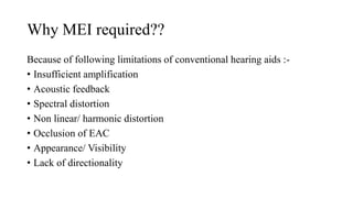
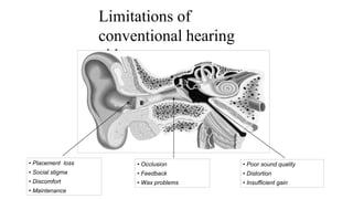
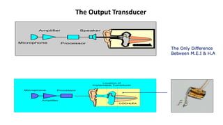
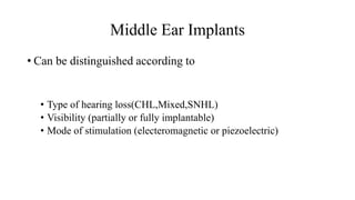
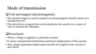
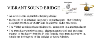
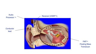
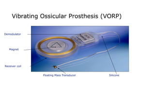
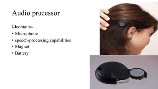
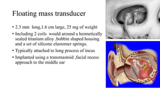
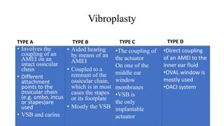
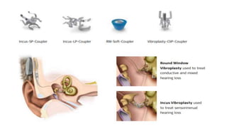
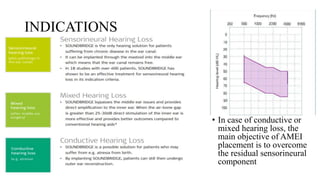
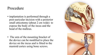
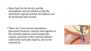
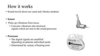
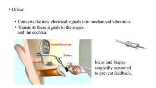
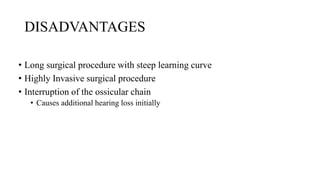
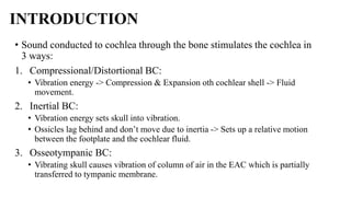
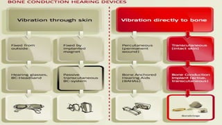
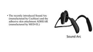
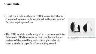
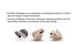
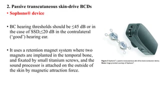
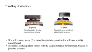
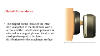
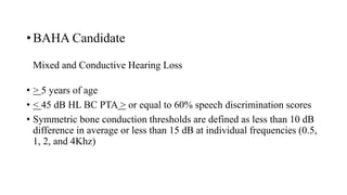
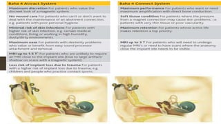
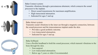
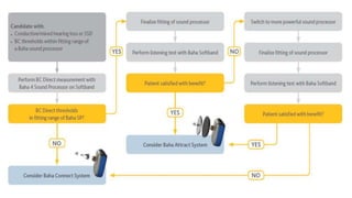
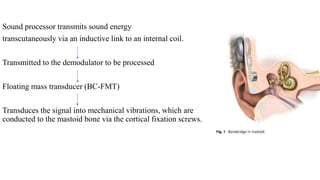
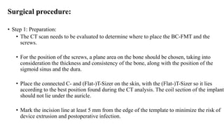
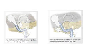
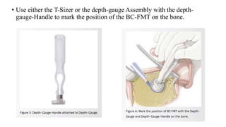
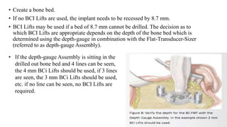
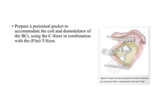
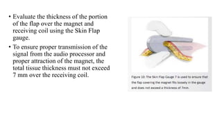
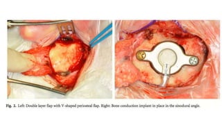
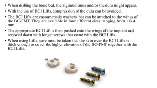
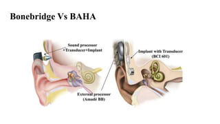
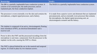
IMPLANTABLE MIDDLE
EAR & BONE CONDUCTION
HEARING DEVICES
Ashish K.Gupta
SMS Medical college jaipur
IMPLANTABLE
MIDDLE EAR DEVICES
• Bone Conduction Hearing Aids:
- Mild hearing loss
• Middle ear implants :
- Mild to profound
• Cochlear Implant :
- Profound hearing loss
Why MEI required??
Because of following limitations of conventional hearing aids :-
• Insufficient amplification
• Acoustic feedback
• Spectral distortion
• Non linear/ harmonic distortion
• Occlusion of EAC
• Appearance/ Visibility
• Lack of directionality
Insufficient Amplification
• The maximum gains for digital in-the-ear (ITE) , in-the-canal
(ITC), & completely-in-canal (CIC) aids currently are about
55-65 dB, 45-55 dB, 35-50 dB, respectively.
Acoustic Feedback
• Acoustic waves from the hearing aid speaker leak through the air
space between the hearing aid body & the EAC wall back to the
microphone, where ( for a subset of frequencies ) they add to existing
microphone input & are amplified further.
• The resulting positive feedback loop causes a low-frequency hum or
high-frequency squeal.
• Fitting aids tightly into the EAC can decrease feedback, but this
decrease comes at the cost of increased incidence of discomfort, otitis
externa, autophony, & blockage of natural sound input.
Limitations of
conventional hearing
aids
• Occlusion
• Feedback
• Wax problems
• Placement loss
• Social stigma
• Discomfort
• Maintenance
• Poor sound quality
• Distortion
• Insufficient gain
The Output Transducer
The Only Difference
Between M.E.I & H.A
Middle Ear Implants
• Can be distinguished according to
• Type of hearing loss(CHL,Mixed,SNHL)
• Visibility (partially or fully implantable)
• Mode of stimulation (electeromagnetic or piezoelectric)
Mode of transmission
 Coil and magnet (electromagnetic)
• The electrical signal is used to produce an electromagnetic field by means of a
transduction coil.
• This then drives a magnet that can be attached to the ossicles in a variety of
ways to transfer the vibrations.
Piezoelectric
• When a voltage is applied to a particular ceramic
• It causes a proportional deformation and hence displacement of that ceramic.
• This voltage-dependent displacement can then be coupled to the ossicles to
drive them
PARTIALLY IMPLANTABLE MED
VIBRANT SOUND BRIDGE
• An active semi-implantable hearing device
• It consists of an internal, surgically implanted part – the vibrating
ossicular prosthesis (VORP) and an external audio processor.
• The VORP consists of a receiving coil, conductor link and transducer
• The transducer employs a small electromagnetic coil and enclosed
magnet to produce vibrations in this floating mass transducer (FMT),
which can be coupled to the ossicles or round window
Audio
Processor TM
Receiver (VORPTM)
Conductor
lead
FMTTM
Floating Mass
Transducer
Vibrating Ossicular Prosthesis (VORP)
Demodulator
Receiver coil
Magnet
SiliconeFloating Mass Transducer
Audio processor
contains-
• Microphone
• speech-processing capabilities
• Magnet
• Battery
ADVANTAGES-
• No occlusion of the ear canal.
• Easily hidden under the hair.
• Held in place by magnetic attraction.
• No interference with glasses.
SAMBA AUDIO PROCESSOR
• SAMBA offers wireless connectivity via Bluetooth or telecoil and
plugs into external devices.
• Through this new feature users can connect to their mobile phones,
MP3 players, FM systems or other wireless devices without losing any
quality of sound
• SAMBA Remote Control-Users can simply control the volume and
change between programs with the easy-to operate remote control,
which is supplied with SAMBA.
• Adaptive Directional Microphones—Minimizes Background Noise
• Intelligent Sound Adapter—SAMBAAdapts to Users’s Listening
Habits
Floating mass transducer
• 2.3 mm long,1.6 cm large, 25 mg of weight
• Including 2 coils would around a hermetically
sealed titanium alloy ,bobbin shaped housing
and a set of silicone elastomer springs.
• Typically attached to long process of incus
• Implanted using a transmastoid ,facial recess
approach to the middle ear
Vibroplasty
TYPE A
• Involves the
coupling of an
AMEI on an
intact ossicular
chain
• Different
attachment
points to the
ossicular chain
(e.g. umbo, incus
or stapes)are
used
• VSB and carina
TYPE B
• Aided hearing
by means of an
AMEI
• Coupled to a
remnant of the
ossicular chain,
which is in most
cases the stapes
or its footplate
• Mostly the VSB
•The coupling of
the actuator
On one of the
middle ear
window
membranes
•VSB is
the only
implantable
actuator
•Direct coupling
of an AMEI to the
inner ear fluid
•OVAL window is
mostly used
•DACI system
TYPE C TYPE D
INDICATIONS
• In case of conductive or
mixed hearing loss, the
main objective of AMEI
placement is to overcome
the residual sensorineural
component
BENEFITS
• High frequency gain without feedback
• Elimination of the occlusion effect
• Improved comfort and ease of use
• Distortion-free signal
– No electronic receiver in the ear canal, which is a major source of distortion wi
hearing aids
• More natural sound quality
• Takes advantage of any low frequency residual hearing
– Low frequencies are still transmitted through the ear canal
• FMT is designed to be linear
– Mimics the natural vibratory pattern of the ossicular chain through 8000Hz
Direct acoustic cochlear implant (DACI)
• Initially called the direct acoustic cochlear stimulator this device
• Consists of an implantable electromagnetic transducer, which transfers
acoustic energy directly to the inner ear via a conventional stapes
prosthesis
• Indicated for profound mixed hearing loss.
• Beside the original use of the device, further applications involving the
stimulation of mobile middle ear structures or the round window are
imaginable
• The external behind-the-ear unit containing
the sound processor receives the acoustical
signal by two microphones and transmits it
transcutaneously to the implant by an
induction coil.
• The electro-magnetic Codacs actuator held by
a fixation system generates the vibration.
• The vibration is transmitted to the perilymph
fluid by a piston prosthesis crimped to the
angled Codacs actuator rod tip, the artificial
incus (AI), and inserted into the inner ear
through a stapes footplate fenestration
Codacs actuator
• An electromagnetic actuator based on the
“balanced armature principle”
• A disk-like part of the rod inside the actuator is
positioned between two permanent ring
magnets
• A titanium diaphragm encloses the rod and acts
as a spring
Middle ear transducer
• The semi-implantable MET uses an
external unit called the button external
audio processor, containing a
microphone,battery,signal processor and
transmitter.
• The transducer drives an electromagnetic
probe coupled to the body of the incus.
• The tip of the probe is made of aluminium
oxide, which forms a fibrous connection
with the incus body.
Fully implantable MED
CARINA
• The fully implantable Carina uses the same electromagnetic
transduction system, but consists of a subcutaneous microphone,
battery and an electronic receiver connected to a transducer.
• four main components: the implant,the programming system, the
charger, and the remote control.
• The microphone is housed under the skin behind the ear.
• It collects sound and sends it to the processor.
• The signal is then amplified, processed, and sent to the actuator At
this stage, the electrical signal is transduced in a mechanical
movement that will be transmitted to the ossicles or labyrinthine
windows.
• The transducer, which initially had a tip designed to couple it to the
body of the incus, was modified for application with different
extensions that allow coupling to the stapes, oval window , or round
window.
• This is used in patients with mixed hearing loss
Procedure
• implantation is performed through a
post-auricular incision with a posterior
small atticotomy (about 2 cm wide) to
expose the body of the incus and the
head of the malleus.
• The arm of the mounting bracket of
the device can be modified to place the
device on the incus and is fitted to the
mastoid cortex using bone screws.
• Bone beds for the device and the
microphone must be drilled so that the
electronics capsule and the microphone can
be positioned and secured
• There are 3 convenient microphone
placement locations: anterior and superior to
the external auditory canal (temporalis
region), posterior to the external auditory
canal (retro-auricular region), and on the
mastoid tip
ESTEEM
• A totally implantable hearing system that is implanted under the skin behind the
ear and within the middle ear space
• For moderate and severe sensorineural hearing loss
• Sensorineural hearing loss -- Loss of hearing resulting from problems in the
inner ear, the cochlear nerve, or in the brain.
• Has no microphone
• Uses the functioning eardrum to pick up vibrations
• Adjusts vibrations to individual hearing needs
• Pacemaker like battery
• Needs replacement after 4.5 to 9 years of continuous use.
• battery replaced in a minor outpatient surgical operation.
Parts
• Two piezoelectric transducers implanted in the middle ear
• Sensor – surgically attached to incus
• Driver – attached to stapes
• Sound Processor
• Implanted behind the ear
in a bony well
• Hold battery
• Personal Programmer
• Turns Esteem on/off
• Select volume levels
• Three program settings
PROCEDURE
• A post-auricular incision is made and a bone recess is fashioned
posterior to the mastoid to house the sound processor
• A tympanomastoidectomy is performed widely exposing the facial
recess to accommodate the driver.
• The incus and stapes are disarticulated and the distal 1 to 3 mm of the
long process of the incus is gently removed using either malleus
nipper or a cutting laser to prevent a mechanic feedback
• Transducers are contained in the mastoid cavity with hydroxyapatite
cement so their piezoelectric crystals are positioned.
• The sensor is interfaced with the incus using glass ionomeric cement
and the driver is cemented to the stapes
• Maximum vibrational motion of the middle ear is deliverable to the
piezoelectric transducer of esteem through the superior part of the
malleus head, on the lateral part of the incus body, and on the superior
part of the incus body near the incudomalleal joint
How it works
• Sound travels down ear canal and vibrates eardrum
• Sensor
• Picks up vibrations from incus.
• Converts vibrations into electrical
signals which are sent to the sound processor.
• Processor
• The digital signals are modified
depending on patients individual needs
• Determined by variety of hearing tests
• Driver
• Converts the new electrical signals into mechanical vibrations.
• Transmits these signals to the stapes
and the cochlea
Sound Processor
Sensor
Driver
Incus and Stapes
surgically separated
to prevent feedback.
• Indication
• Bilateral non progressive SNHL of moderate to severe degree ,
with at least 40%speech discrimination , age older than 18 years
and a normal middle ear function(evaluated by impedance
audiometry)
• Contraindication
• In case of allergy , abnormal tubal function or chronic naso-
paranasal pathology , tendency to keloid and staph infections
ADVANTAGES
• Completely invisible to self and others
• No acoustic feedback
• Maintenance free
DISADVANTAGES
• Long surgical procedure with steep learning curve
• Highly Invasive surgical procedure
• Interruption of the ossicular chain
• Causes additional hearing loss initially
Bone Conduction Hearing Devices
• The Bonebridge Bone Conduction Hearing Implant: indication
criteria, surgery and a systematic review of the literature
• Sprinzl, G.M. & Wolf-Magele, A.
Department of Otorhinolaryngology, Karl Landsteiner Private
University, St. Poelten, Austria
• Accepted for publication 8 June 2015 Clin. Otolaryngol. 2016, 41, 131–
143
INTRODUCTION
• Sound conducted to cochlea through the bone stimulates the cochlea in
3 ways:
1. Compressional/Distortional BC:
• Vibration energy -> Compression & Expansion oth cochlear shell -> Fluid
movement.
2. Inertial BC:
• Vibration energy sets skull into vibration.
• Ossicles lag behind and don’t move due to inertia -> Sets up a relative motion
between the footplate and the cochlear fluid.
3. Osseotympanic BC:
• Vibrating skull causes vibration of column of air in the EAC which is partially
transferred to tympanic membrane.
• Bone conduction hearing aid (BCHA) treatment has become the
standard of care for patients suffering with
Conductive and mixed losses
1. Congenital causes such as atresia or microtia
2. Acquired causes such as chronic otitis media or
3. Ossicular pathology
4. Chronic discharging ear (such as chronic suppurative
5. Otitis media (CSOM) or recurrent otitis externa) (
6. Inability to wear a hearing aid following radical Mastoid surgery
7. Unilateral mixed hearing loss.
 Single-sided deafness – sensorineural or conductive
1. Trauma resulting in hearing loss.
2. Unsuitable ear canal for a conventional hearing aid, e.g.
i. In a radical mastoid cavity
ii. An extremely narrow ear canal
iii. ‘Blind sac’ ear canal closure
iv. Lateral temporal bone resection
v. Extensive cranial base surgery.
BCHDs will not be commissioned for:
• patients with a bone disease that is unable to support an implant
• patients who have a sensitivity or allergy to the materials used
• patients with physical, emotional or psychological disorders that,
despite suitable treatment and support,would interfere with surgery or
the ability to allow suitable rehabilitation such that significant benefit
would be unlikely.
Transcutaneous vs Percutaneous
• BC sensitivity is fairly similar between percutaneous and
transcutaneous modes of transmission for frequencies below
1 kHz.
• There would be an expected 5–15 dB improvement in
efficiency with percutaneous systems when a BC transducer
is directly attached to the skull at higher frequencies.
Skin-drive BCDs
• Vibrations are transmitted through the skin, which is kept
intact.
• In conventional skin-drive BCDs, all components are kept
outside the skin, while the passive transcutaneous skin-drive
BCDs contain implanted magnet(s).
1. Conventional skin-drive BCD
• It is attached with a soft headband (softband), a steel spring headband, or with
spectacles for glasses.
• BAHAs are sometimes used with a softband/headband instead of a titanium
screw, thus behaving as a conventional skin-drive device.
• The use of BAHA on a softband (elastic fabric) or headband (diadem
type) is a valuable method of hearing rehabilitation in children who
are too young for implantation, and it is the gold standard for
preoperative assessment.
• The recently introduced Sound Arc
(manufactured by Cochlear) and the
adhesive skin attachment ADHEAR
(manufactured by MED-EL)
Sound Arc
In the mouth
• Ludwig van Beethoven, the well-known 18th
century composer who was completely deaf, was
the first person to use bone conduction.
• It has been theorized that Beethoven discovered
a way to hear the sound of his piano through his
jawbone by putting a rod between his teeth and
touching his piano with this rod.
• This was the first proof that sound can also reach
our brain by making use of the bones in our
skull.
• Soundbite
• It utilizes a behind-the-ear (BTE) transmitter that is
connected to a microphone placed in the ear canal of
the hearing-impaired ear.
• The BTE module sends a signal to a custom made in-
the-mouth (ITM) transducer that couples the buccal
surface of the maxillary molars to a piezoelectric
bone stimulator capable of conducting sound.
• It offers advantages over traditional osseointegrated devices in that it
does not require surgical placement.
• Acoustic feedback is the most commonly reported problem with the
Sound-Bite, and this is minimized with proper fitting.
2. Passive transcutaneous skin-drive BCDs
• Sophono® device
• BC hearing thresholds should be ≤45 dB or in
the case of SSD,≤20 dB in the contralateral
(‘good’) hearing ear.
• It uses a retention magnet system where two
magnets are implanted in the temporal bone,
and fixated by small titanium screws, and the
sound processor is attached on the outside of
the skin by magnetic attraction force.
• The vibrations of the transducer are transmitted through the soft
tissues, and the skin is most often thinned to 4–5 mm thickness (only
in adults).
• In order to overcome skin problems related to high skin pressure, the
Sophono® Alpha 1 uses a larger contact area than is used in
conventional BCDs.
Travelling of vibrations
• Skin will conduct sound (Force) and in certain frequencies skin will even amplify
sound (Force)
• The size of the baseplate in contact with the skin is important for maximum transfer of
power to the bone
• Baha® Attract device
• The magnet on the inside of the intact
skin is attached to the skull bone with a
screw, and the Baha® sound processor is
attached to a magnet plate on the skin via
a soft pad to equalize the force
distribution over the attachment surface.
Direct-drive BCDs
• The vibrations are transmitted directly to the bone via a screw or a flat
surface attachment.
• Divided into percutaneous and active transcutaneous devices.
OSSEOINTEGRATION
• It combines the concept of osseointegration and bone conduction
transmission to aid hearing.
• Osseointegration refers to the direct structural and functional
connection between ordered living bone and the surface of a load-
carrying implant.
• It was found in the 1950s by Professor Branemark that implanted
titanium would fuse with human bone in harmony and become part of
the bone, instead of giving rise to a foreign body reaction.
• Tjellstron was the pioneer in utilizing the concept of osseointegration
in hearing implants and established the use of BAHA in 1977.
1. Percutaneous direct-drive BCD
• BAHA
• BAHAs are mainly indicated for conductive and mixed hearing loss as well as
for single-sided deafness (SSD), and are used both on adults and on children
• surgery not be undertaken prior to age 2–3 years.
• This was to allow the skull to reach a minimum thickness and conditions to be
suitable to fit the fixture and allow osseointegration.
INDICATIONS
1. Patient using a conventional BC hearing aid
2. Conventional AC hearing aid user with
a. chronic otorrhea
b. chronic otitis media/externa
c. uncontrollable feedback due to a radical mastoidectomy
or a large meatoplasty.
3. Otosclerosis, tympanosclerosis, canal atresia with a
contraindication to repair, eg,
a. only hearing ear
b. combination with 2a-c.
4. SSD with better ear BC PTA better than 45db HL and
SDS >60%
5.Mixed hearing loss
Components of BAHA
71
2
1 Titanium implant
placed in the bone just
behind the ear
Abutment which
coupled with the fixture
and act as a connector
to the speech processor1
2
3
3
External sound
processor which
connects to the implant
• The titanium fixture is implanted in the skull bone of the patient and attached
to a percutaneous abutment and sound processor.
• The sound processor will convert sound energy to vibration, transmitted via
the abutment and the titanium fixture and then the skull, directly to the
functioning cochlea via bone conduction
• A 4 mm titanium fixture is secured to the bone.
• The abutment is then joined to the fixture after thinning the
periabutment subcutaneous tissue.
• The sound processor is fitted 3 months after the fixture has fully
osseointegrated with the skull bone.
• A skull thickness of 3 to 4 mm and good bone quality are essential for
successful BAHA surgery.
• How does the BAHA system work for Mixed and Conductive Hearing
Loss?
Sound conduction in the case of two normal cochleae (above) or with only a
functioning right cochlea and left SSD
• In paediatric patients with a thinner skull, it is recommended that
surgery is performed in two stages.
• The first stage is the insertion of the titanium fixture.
• In the second stage, which is approximately 3 months later, the
abutment is placed.
• but the newer tissue-preserving single-stage implants mean this is not
always necessary
• The sound processor can be fitted 3 weeks after that.
• A two-stage procedure is also indicated for patients with a history of
irradiation to the skull
• BAHA Candidate
Mixed and Conductive Hearing Loss
• > 5 years of age
• < 45 dB HL BC PTA > or equal to 60% speech discrimination scores
• Symmetric bone conduction thresholds are defined as less than 10 dB
difference in average or less than 15 dB at individual frequencies (0.5,
1, 2, and 4Khz)
Why BAHA for Single Sided Deafness
• Effective approach in patients with unilateral
deafness
• Alleviates the degree of hearing handicap
resulting from the head shadow effect
• Improves speech intelligibility in noise
• Improves the patient’s quality of life
• Provided a greater perceived benefit compared to
CROS system
POST OPERATIVE DIFFICULTIES
• The protrusion of the abutment through the skin.
• Cosmetic issues
• Skin requires daily hygienic care
• Can be skin overgrowth around the abutment
• Skin infection and low-grade inflammation.
BAHA attract vs BAHA
Baha Connect system:
Transmits vibrations through a percutaneous abutment, which connects the sound
processor to the implant.
• Direct sound transmission for maximum amplification.
• High transcranial attenuation.
• Indicated for ages 5 and up
Baha Attract system:
Transmits sound vibrations to the inner ear through a magnetic connection, between
the sound processor and the transcutaneous implant under the skin.
• Provides a good aesthetic outcome.
• Low transcranial attenuation.
• Indicated for ages 5 and up.
Baha Softband:
Uses a flexible headband to hold the sound processor, which transmit vibrations to the
bone through the skin.
• Non-surgical.
• Low transcranial attenuation.
• Below age 5 years or older if recommended.
2. Active transcutaneous direct-drive BCD
• In active transcutaneous direct-drive BCDs, the transducer is implanted under
intact skin. Hence, the vibrations are transmitted from the transducer directly
to the skull bone.
• The reason for calling it transcutaneous is that the electro- magnetic signal
from the sound processor is transmitted through the skin, not the vibrations.
• The sound processor is attached to the skin by retention magnets in the
implanted unit, and the sound signal is transmitted via an inductive link to the
implanted transducer.
Stimulation Implant generates vibrational
stimulation that is directly
applied to the bone (“direct
drive bone conduction
stimulation”). optimum BC
sound transmission.
Sound processor generates stimulation
that is applied from outside onto the skin
(like hearing glasses or BC-head-bands).
Skin attenuates sound before it reaches
the bone.
Sound processor Audio processor picks up sound and
generates signal that is transmitted to
the implant.
Transducer (“BC-FMT”) is part of
the implant and is directly attached to
the bone.
Sound processor picks up sound and
generates vibration that is applied
onto the skin. Sound processor needs
transducer and plate that adds to the
sound processor size and weight.
Implant Implant accepts signal and BC-FMT
generates vibration (“active implant”)
that is applied directly to bone.
Implant consists of a receiving coil, a
holding magnet, electronics and a
BC-FMT (transducer).
Implant’s main function is to hold
vibrating sound processor in place
and generate skin pressure (“passive
implant”).
Implant consists mainly of a magnet.
Transducer Transducer (“BC-FMT”) is part of
the implant and is directly attached to
the bone.
Transducer is part of the sound
processor and drives a plate that sits
on the skin. The plate is held by an
implanted magnet.
BONEBRIDGE
• The Bonebridge (BB) from MED-EL first active,
intact skin bone conduction implant system,
intended for individuals with conductive or mixed
hearing loss and Single-Sided Deafness (SSD).
• It is a semi-implantable hearing system consisting
of an implantable part, the BCI (Bone Conduction
Implant), and the external audio processor,
• The Bonebridge was first implanted in June
2011 as part of a clinical trial and was
launched in September 2012.
Sound processor transmits sound energy
transcutaneously via an inductive link to an internal coil.
Transmitted to the demodulator to be processed
Floating mass transducer (BC-FMT)
Transduces the signal into mechanical vibrations, which are
conducted to the mastoid bone via the cortical fixation screws.
• Indications for the Bonebridge
• Intended for adults and children aged 5 years and above.
• Patients who have a conductive or mixed mild-to- moderate hearing loss and still can
benefit from sound amplification. The pure tone average (PTA) BC threshold should be
better than or equal to 45 dB HL.
• Patients who have severe-to-profound sensorineural hearing loss in one ear and normal
hearing in the opposite ear (i.e. ‘SSD’). The PTA air conduction hearing thresholds of
the hearing ear should be better than or equal to 20 dB HL
• The Bonebridge for SSD is also indicated for any patient who is indicated for an air
conduction contralateral routing of signals (AC CROS) hearing aid.
• In addition to the above indications, individuals’ medical background has to
be evaluated to check for the following:
1.Revision tympanoplasty, ear canal stenosis or chronically draining ears
where conventional hearing aids are not suitable due to poor wearability.
2. Otosclerosis or tympanosclerosis that cannot be rectified to a
sufficient extent by surgery, or wearing conventional hearing aids.
3. Congenital malformations where ear canals are absent and cannot be
restored through conventional surgery.
4.Sudden deafness, acoustic neuroma or other reasons which cause severe
to profound sensorineural hearing loss on one side.
5. Anatomy which allows for appropriate placement of the bonebridge
implant as determined by CT scan.
6. The absence of retrocochlear and central auditory disorders.
7. Psychological and emotional stability with realistic expectations of the
benefits and limitations of the bonebridge.
Surgical procedure:
• Step 1: Preparation:
• The CT scan needs to be evaluated to determine where to place the BC-FMT and the
screws.
• For the position of the screws, a plane area on the bone should be chosen, taking into
consideration the thickness and consistency of the bone, along with the position of the
sigmoid sinus and the dura.
• Place the connected C- and (Flat-)T-Sizer on the skin, with the (Flat-)T-Sizer so it lies
according to the best position found during the CT analysis. The coil section of the implant
should not lie under the auricle.
• Mark the incision line at least 5 mm from the edge of the template to minimize the risk of
device extrusion and postoperative infection.
• Step 2: Incision
• Postauricular incision, incise the skin to thelevel of
the temporalis fascia. next, make an anteriorly-based
pericranial fascia incision.
• The portion of the pericranial flap overlying the
receiving coil and magnet may be excised, but the
anterior portion of the flap must be preserved to
provide a continuous tissue layer over the anterior
portion of the demodulator and the BC-FMT.
• In atresia cases, where a pinna reconstruction is
planned at a later stage, the incision should be made
further posteriorly.
• Note that only bipolar electrocautery should be used
once the BC is in the surgical field, or if the patient
already has an implant on the other side.
• Step 3: Creation of Bone Bed for BC-FMT And Periosteal Pocket For Coil
• The position of the bone bed for the BC-FMT as well as the position of the
fixation holes depends on the findings of the CT scan.
• If the BC-FMT is placed in the sinodural angle, the reference points are the
spine of Henle, the temporal line and the tip of the mastoid.
• It is usually made as close as possible to the EAC and with one of the BC-
FMT wings sitting on the temporal line.
• If the BC-FMT is placed behind the sinus, the attachment of the digastric
muscle and the mastoid tip are to be used as reference points.
• Use either the T-Sizer or the depth-gauge Assembly with the depth-
gauge-Handle to mark the position of the BC-FMT on the bone.
• Create a bone bed.
• If no BCI Lifts are used, the implant needs to be recessed by 8.7 mm.
• BCI Lifts may be used if a bed of 8.7 mm cannot be drilled. The decision as to
which BCI Lifts are appropriate depends on the depth of the bone bed which is
determined using the depth-gauge in combination with the Flat-Transducer-Sizer
(referred to as depth-gauge Assembly).
• If the depth-gauge Assembly is sitting in the
drilled out bone bed and 4 lines can be seen,
the 4 mm BCi Lifts should be used, if 3 lines
are seen, the 3 mm BCi Lifts should be used,
etc. if no line can be seen, no BCI Lifts are
required.
• Prepare a periosteal pocket to
accommodate the coil and demodulator of
the BCi, using the C-Sizer in combination
with the (Flat) T-Sizer.
• Evaluate the thickness of the portion
of the flap over the magnet and
receiving coil using the Skin Flap
gauge.
• To ensure proper transmission of the
signal from the audio processor and
proper attraction of the magnet, the
total tissue thickness must not exceed
7 mm over the receiving coil.
• Step 4: Preparing BCI Fixation
• Drill the fixation points.
• The diameter of the drill bit holes is 1.5 mm.
• Step 5: Fixation of the BCI
• In case BCI Lifts are used, attach the appropriate size and push them onto the wings of
the BC-FMT.
• Arrange the BCi over the surgical site so that the magnet protrusion is towards the skull,
with the triangle shape on the magnet facing towards the skin.
• Bend the transition of the implant according to the final position required.
• Place the implant coil and the demodulator under the periosteum so that it resides under
the desired external position of the audio processor (previously marked) and the BC-FMT
into the bed that has been prepared.
• Place one regular cortical screw in each anchor hole of the BC-FMT and secure tightly.
The regular screws have a diameter of 2 mm, and a golden surface finish.
• There is no clicking sound to indicate when enough force has been used.
• For tightening the screws of the BCI a force of about 10 ncm is sufficient.
• in case the screws are not adequate, alternative screw lengths can be used. The
demodulator of the implant does not need to be sutured down. The fixation of the
implant via the 2 screws is enough to hold the implant in place.
• Step 6: Closure
• Inspect the BC-FMT under the microscope.
• Palpate the main body of the BC-FMT to make
certain it is secure.
• The BC-FMT should be installed solidly and free
from any minor slackness when palpated. ensure
that the receiver coil is in the desired position.
• Close the scalp wound in layers, then suture the
skin flap with a double layer closure, taking care
not to make contact with the installed BCI during
the closure process.
• Clean the incision area and apply a pressure
dressing to the wound.
• To smoothen the transition of the wings to the
bone, add bone pate around the wings of the BCI.
• When drilling the bone bed, the sigmoid sinus and/or the dura might appear.
• With the use of BCI Lifts, compression of the dura can be avoided.
• The BCI Lifts are custom-made washers that can be attached to the wings of
the BC-FMT. They are available in four different sizes, ranging from 1 to 4
mm.
• The appropriate BCI Lift is then pushed onto the wings of the implant and
screwed down with longer screws that come with the BCI Lifts.
• When using Lifts, care must be taken that the skin over the BCI Lifts is
thick enough to cover the higher elevation of the BC-FMT together with the
BCI Lifts.
Bonebridge Vs BAHA
%
Bonebridge BAHA
The BB is a partially implantable bone conduction system. It
consists of an external part, the audio processor, and an
implanted part, the bone conduction implant.
The BAHA is a partially implantable bone conduction system.
The processor is worn externally over the skin and contains the
microphones, a digital signal processor, and a battery.
It is made from a passive screw and abutment that are implanted
percutaneously, and an externally worn processor that contains
the microphones, the digital signal processing unit, an
electromagnetic actuator and the battery.
The implant is composed of an active, electromagnetic floating
mass transducer (FMT), an electrical demodulator and a
receiver coil.
Power to drive the FMT and the processed recordings from the
microphone is sent trans- cutaneously from the processor to the
implant via the coils, causing the FMT to vibrate.
The FMT is placed behind the ear in the mastoid and temporal
regions. It is held in place by two titanium screws.
•The 3 Reasons for BONEBRIDGE’s Sound Quality
1. A Smaller, Lighter Processor
• Because the part of the BONEBRIDGE that vibrates the bone is entirely under
the skin, the external audio processor can be thin and light. It needs only a
battery and the computer chip which converts sound waves into the wireless
electrical signals.
• That means it’s not just nicer to wear, but you can wear it longer.
2. Under the Skin, or Through the Skin?
• A small and light audio processor isn’t the only reason to want an implant
that’s under the skin. It also makes for more comfortable hearing.
• The alternatives are a headband which can be uncomfortable and cause the
skull bones to deform, or a BAHA with its abutment and complications.
• With BONEBRIDGE the skin heals completely and the implant is totally
hidden.
• The audio processor is held onto the skin by magnetic attraction that can be
secure enough to keep it from falling off while not so tight as to cause
discomfort—it’s similar to how a cochlear implant uses its magnet.
3. Active Bone Conduction
• And since it has the implant under the skin, it sends sound vibrations directly into
the bone. This doesn’t just make hearing more comfortable, it makes for better
hearing quality.
• The skin above the temporal bone varies between 2–8 mm, which means that
when using a headband or BAHA you have to vibrate all of this skin even before
sound waves get to the bone. The skin reduces the loudness of higher-pitched
sounds and therefore distorts the sound.
• This means the BONEBRIDGE is both more efficient, because it vibrates the bone
directly, and sounds better.
Conclusion
• The Bonebridge BC implant system provides a valuable and stable audiological
benefit to patients suffering from conductive or mixed hearing loss and SSD.
• With its active transcutaneous design, it offers a lower complication rate to
percutaneous systems and higher and more reliable hearing gain compared to
other transcutaneous or percutaneous systems.
• Moreover, the fast activation of the implant system enables the recipient of the
system to benefit in a short time frame postoperatively from the intervention.
Summary
• The Bonebridge offers the following advantages over passive devices:
• It provides more output, making it more suitable for mixed hearing
loss and single-sided deafness.
• It provides stable output, independent of skin thickness and hair
growth.
• It does not require skin pressure for stimulation.
• It is comfortable to wear, leading to 50% longer wearing time per day
(extra 3.5 hrs/day).
• Its audio processor has a much lower profile and is therefore more
inconspicuous.
• Published and presented clinical experience shows excellent results
and high user satisfaction. Very low complication rates have been
confirmed.
THANK YOU

Implantable Middle ear and bone conduction hearing devices

  • 1.
    IMPLANTABLE MIDDLE EAR &BONE CONDUCTION HEARING DEVICES Ashish K.Gupta SMS Medical college jaipur
  • 2.
  • 3.
    • Bone ConductionHearing Aids: - Mild hearing loss • Middle ear implants : - Mild to profound • Cochlear Implant : - Profound hearing loss
  • 4.
    Why MEI required?? Becauseof following limitations of conventional hearing aids :- • Insufficient amplification • Acoustic feedback • Spectral distortion • Non linear/ harmonic distortion • Occlusion of EAC • Appearance/ Visibility • Lack of directionality
  • 5.
    Insufficient Amplification • Themaximum gains for digital in-the-ear (ITE) , in-the-canal (ITC), & completely-in-canal (CIC) aids currently are about 55-65 dB, 45-55 dB, 35-50 dB, respectively.
  • 6.
    Acoustic Feedback • Acousticwaves from the hearing aid speaker leak through the air space between the hearing aid body & the EAC wall back to the microphone, where ( for a subset of frequencies ) they add to existing microphone input & are amplified further. • The resulting positive feedback loop causes a low-frequency hum or high-frequency squeal. • Fitting aids tightly into the EAC can decrease feedback, but this decrease comes at the cost of increased incidence of discomfort, otitis externa, autophony, & blockage of natural sound input.
  • 7.
    Limitations of conventional hearing aids •Occlusion • Feedback • Wax problems • Placement loss • Social stigma • Discomfort • Maintenance • Poor sound quality • Distortion • Insufficient gain
  • 8.
    The Output Transducer TheOnly Difference Between M.E.I & H.A
  • 9.
    Middle Ear Implants •Can be distinguished according to • Type of hearing loss(CHL,Mixed,SNHL) • Visibility (partially or fully implantable) • Mode of stimulation (electeromagnetic or piezoelectric)
  • 10.
    Mode of transmission Coil and magnet (electromagnetic) • The electrical signal is used to produce an electromagnetic field by means of a transduction coil. • This then drives a magnet that can be attached to the ossicles in a variety of ways to transfer the vibrations. Piezoelectric • When a voltage is applied to a particular ceramic • It causes a proportional deformation and hence displacement of that ceramic. • This voltage-dependent displacement can then be coupled to the ossicles to drive them
  • 11.
  • 12.
    VIBRANT SOUND BRIDGE •An active semi-implantable hearing device • It consists of an internal, surgically implanted part – the vibrating ossicular prosthesis (VORP) and an external audio processor. • The VORP consists of a receiving coil, conductor link and transducer • The transducer employs a small electromagnetic coil and enclosed magnet to produce vibrations in this floating mass transducer (FMT), which can be coupled to the ossicles or round window
  • 13.
  • 14.
    Vibrating Ossicular Prosthesis(VORP) Demodulator Receiver coil Magnet SiliconeFloating Mass Transducer
  • 15.
    Audio processor contains- • Microphone •speech-processing capabilities • Magnet • Battery
  • 16.
    ADVANTAGES- • No occlusionof the ear canal. • Easily hidden under the hair. • Held in place by magnetic attraction. • No interference with glasses.
  • 17.
    SAMBA AUDIO PROCESSOR •SAMBA offers wireless connectivity via Bluetooth or telecoil and plugs into external devices. • Through this new feature users can connect to their mobile phones, MP3 players, FM systems or other wireless devices without losing any quality of sound • SAMBA Remote Control-Users can simply control the volume and change between programs with the easy-to operate remote control, which is supplied with SAMBA. • Adaptive Directional Microphones—Minimizes Background Noise • Intelligent Sound Adapter—SAMBAAdapts to Users’s Listening Habits
  • 19.
    Floating mass transducer •2.3 mm long,1.6 cm large, 25 mg of weight • Including 2 coils would around a hermetically sealed titanium alloy ,bobbin shaped housing and a set of silicone elastomer springs. • Typically attached to long process of incus • Implanted using a transmastoid ,facial recess approach to the middle ear
  • 21.
    Vibroplasty TYPE A • Involvesthe coupling of an AMEI on an intact ossicular chain • Different attachment points to the ossicular chain (e.g. umbo, incus or stapes)are used • VSB and carina TYPE B • Aided hearing by means of an AMEI • Coupled to a remnant of the ossicular chain, which is in most cases the stapes or its footplate • Mostly the VSB •The coupling of the actuator On one of the middle ear window membranes •VSB is the only implantable actuator •Direct coupling of an AMEI to the inner ear fluid •OVAL window is mostly used •DACI system TYPE C TYPE D
  • 24.
    INDICATIONS • In caseof conductive or mixed hearing loss, the main objective of AMEI placement is to overcome the residual sensorineural component
  • 25.
    BENEFITS • High frequencygain without feedback • Elimination of the occlusion effect • Improved comfort and ease of use • Distortion-free signal – No electronic receiver in the ear canal, which is a major source of distortion wi hearing aids • More natural sound quality • Takes advantage of any low frequency residual hearing – Low frequencies are still transmitted through the ear canal • FMT is designed to be linear – Mimics the natural vibratory pattern of the ossicular chain through 8000Hz
  • 26.
    Direct acoustic cochlearimplant (DACI) • Initially called the direct acoustic cochlear stimulator this device • Consists of an implantable electromagnetic transducer, which transfers acoustic energy directly to the inner ear via a conventional stapes prosthesis • Indicated for profound mixed hearing loss. • Beside the original use of the device, further applications involving the stimulation of mobile middle ear structures or the round window are imaginable
  • 27.
    • The externalbehind-the-ear unit containing the sound processor receives the acoustical signal by two microphones and transmits it transcutaneously to the implant by an induction coil. • The electro-magnetic Codacs actuator held by a fixation system generates the vibration. • The vibration is transmitted to the perilymph fluid by a piston prosthesis crimped to the angled Codacs actuator rod tip, the artificial incus (AI), and inserted into the inner ear through a stapes footplate fenestration
  • 28.
    Codacs actuator • Anelectromagnetic actuator based on the “balanced armature principle” • A disk-like part of the rod inside the actuator is positioned between two permanent ring magnets • A titanium diaphragm encloses the rod and acts as a spring
  • 29.
    Middle ear transducer •The semi-implantable MET uses an external unit called the button external audio processor, containing a microphone,battery,signal processor and transmitter. • The transducer drives an electromagnetic probe coupled to the body of the incus. • The tip of the probe is made of aluminium oxide, which forms a fibrous connection with the incus body.
  • 30.
  • 31.
    CARINA • The fullyimplantable Carina uses the same electromagnetic transduction system, but consists of a subcutaneous microphone, battery and an electronic receiver connected to a transducer. • four main components: the implant,the programming system, the charger, and the remote control.
  • 32.
    • The microphoneis housed under the skin behind the ear. • It collects sound and sends it to the processor. • The signal is then amplified, processed, and sent to the actuator At this stage, the electrical signal is transduced in a mechanical movement that will be transmitted to the ossicles or labyrinthine windows. • The transducer, which initially had a tip designed to couple it to the body of the incus, was modified for application with different extensions that allow coupling to the stapes, oval window , or round window. • This is used in patients with mixed hearing loss
  • 33.
    Procedure • implantation isperformed through a post-auricular incision with a posterior small atticotomy (about 2 cm wide) to expose the body of the incus and the head of the malleus. • The arm of the mounting bracket of the device can be modified to place the device on the incus and is fitted to the mastoid cortex using bone screws.
  • 34.
    • Bone bedsfor the device and the microphone must be drilled so that the electronics capsule and the microphone can be positioned and secured • There are 3 convenient microphone placement locations: anterior and superior to the external auditory canal (temporalis region), posterior to the external auditory canal (retro-auricular region), and on the mastoid tip
  • 35.
    ESTEEM • A totallyimplantable hearing system that is implanted under the skin behind the ear and within the middle ear space • For moderate and severe sensorineural hearing loss • Sensorineural hearing loss -- Loss of hearing resulting from problems in the inner ear, the cochlear nerve, or in the brain. • Has no microphone • Uses the functioning eardrum to pick up vibrations • Adjusts vibrations to individual hearing needs • Pacemaker like battery • Needs replacement after 4.5 to 9 years of continuous use. • battery replaced in a minor outpatient surgical operation.
  • 36.
    Parts • Two piezoelectrictransducers implanted in the middle ear • Sensor – surgically attached to incus • Driver – attached to stapes • Sound Processor • Implanted behind the ear in a bony well • Hold battery • Personal Programmer • Turns Esteem on/off • Select volume levels • Three program settings
  • 37.
    PROCEDURE • A post-auricularincision is made and a bone recess is fashioned posterior to the mastoid to house the sound processor • A tympanomastoidectomy is performed widely exposing the facial recess to accommodate the driver. • The incus and stapes are disarticulated and the distal 1 to 3 mm of the long process of the incus is gently removed using either malleus nipper or a cutting laser to prevent a mechanic feedback • Transducers are contained in the mastoid cavity with hydroxyapatite cement so their piezoelectric crystals are positioned.
  • 38.
    • The sensoris interfaced with the incus using glass ionomeric cement and the driver is cemented to the stapes • Maximum vibrational motion of the middle ear is deliverable to the piezoelectric transducer of esteem through the superior part of the malleus head, on the lateral part of the incus body, and on the superior part of the incus body near the incudomalleal joint
  • 39.
    How it works •Sound travels down ear canal and vibrates eardrum • Sensor • Picks up vibrations from incus. • Converts vibrations into electrical signals which are sent to the sound processor. • Processor • The digital signals are modified depending on patients individual needs • Determined by variety of hearing tests
  • 40.
    • Driver • Convertsthe new electrical signals into mechanical vibrations. • Transmits these signals to the stapes and the cochlea Sound Processor Sensor Driver Incus and Stapes surgically separated to prevent feedback.
  • 42.
    • Indication • Bilateralnon progressive SNHL of moderate to severe degree , with at least 40%speech discrimination , age older than 18 years and a normal middle ear function(evaluated by impedance audiometry) • Contraindication • In case of allergy , abnormal tubal function or chronic naso- paranasal pathology , tendency to keloid and staph infections
  • 43.
    ADVANTAGES • Completely invisibleto self and others • No acoustic feedback • Maintenance free
  • 44.
    DISADVANTAGES • Long surgicalprocedure with steep learning curve • Highly Invasive surgical procedure • Interruption of the ossicular chain • Causes additional hearing loss initially
  • 45.
    Bone Conduction HearingDevices • The Bonebridge Bone Conduction Hearing Implant: indication criteria, surgery and a systematic review of the literature • Sprinzl, G.M. & Wolf-Magele, A. Department of Otorhinolaryngology, Karl Landsteiner Private University, St. Poelten, Austria • Accepted for publication 8 June 2015 Clin. Otolaryngol. 2016, 41, 131– 143
  • 46.
    INTRODUCTION • Sound conductedto cochlea through the bone stimulates the cochlea in 3 ways: 1. Compressional/Distortional BC: • Vibration energy -> Compression & Expansion oth cochlear shell -> Fluid movement. 2. Inertial BC: • Vibration energy sets skull into vibration. • Ossicles lag behind and don’t move due to inertia -> Sets up a relative motion between the footplate and the cochlear fluid. 3. Osseotympanic BC: • Vibrating skull causes vibration of column of air in the EAC which is partially transferred to tympanic membrane.
  • 47.
    • Bone conductionhearing aid (BCHA) treatment has become the standard of care for patients suffering with Conductive and mixed losses 1. Congenital causes such as atresia or microtia 2. Acquired causes such as chronic otitis media or 3. Ossicular pathology 4. Chronic discharging ear (such as chronic suppurative 5. Otitis media (CSOM) or recurrent otitis externa) ( 6. Inability to wear a hearing aid following radical Mastoid surgery 7. Unilateral mixed hearing loss.
  • 48.
     Single-sided deafness– sensorineural or conductive 1. Trauma resulting in hearing loss. 2. Unsuitable ear canal for a conventional hearing aid, e.g. i. In a radical mastoid cavity ii. An extremely narrow ear canal iii. ‘Blind sac’ ear canal closure iv. Lateral temporal bone resection v. Extensive cranial base surgery.
  • 49.
    BCHDs will notbe commissioned for: • patients with a bone disease that is unable to support an implant • patients who have a sensitivity or allergy to the materials used • patients with physical, emotional or psychological disorders that, despite suitable treatment and support,would interfere with surgery or the ability to allow suitable rehabilitation such that significant benefit would be unlikely.
  • 51.
  • 52.
    • BC sensitivityis fairly similar between percutaneous and transcutaneous modes of transmission for frequencies below 1 kHz. • There would be an expected 5–15 dB improvement in efficiency with percutaneous systems when a BC transducer is directly attached to the skull at higher frequencies.
  • 53.
    Skin-drive BCDs • Vibrationsare transmitted through the skin, which is kept intact. • In conventional skin-drive BCDs, all components are kept outside the skin, while the passive transcutaneous skin-drive BCDs contain implanted magnet(s).
  • 54.
    1. Conventional skin-driveBCD • It is attached with a soft headband (softband), a steel spring headband, or with spectacles for glasses. • BAHAs are sometimes used with a softband/headband instead of a titanium screw, thus behaving as a conventional skin-drive device.
  • 55.
    • The useof BAHA on a softband (elastic fabric) or headband (diadem type) is a valuable method of hearing rehabilitation in children who are too young for implantation, and it is the gold standard for preoperative assessment.
  • 56.
    • The recentlyintroduced Sound Arc (manufactured by Cochlear) and the adhesive skin attachment ADHEAR (manufactured by MED-EL) Sound Arc
  • 58.
    In the mouth •Ludwig van Beethoven, the well-known 18th century composer who was completely deaf, was the first person to use bone conduction. • It has been theorized that Beethoven discovered a way to hear the sound of his piano through his jawbone by putting a rod between his teeth and touching his piano with this rod. • This was the first proof that sound can also reach our brain by making use of the bones in our skull.
  • 59.
    • Soundbite • Itutilizes a behind-the-ear (BTE) transmitter that is connected to a microphone placed in the ear canal of the hearing-impaired ear. • The BTE module sends a signal to a custom made in- the-mouth (ITM) transducer that couples the buccal surface of the maxillary molars to a piezoelectric bone stimulator capable of conducting sound.
  • 60.
    • It offersadvantages over traditional osseointegrated devices in that it does not require surgical placement. • Acoustic feedback is the most commonly reported problem with the Sound-Bite, and this is minimized with proper fitting.
  • 61.
    2. Passive transcutaneousskin-drive BCDs • Sophono® device • BC hearing thresholds should be ≤45 dB or in the case of SSD,≤20 dB in the contralateral (‘good’) hearing ear. • It uses a retention magnet system where two magnets are implanted in the temporal bone, and fixated by small titanium screws, and the sound processor is attached on the outside of the skin by magnetic attraction force.
  • 62.
    • The vibrationsof the transducer are transmitted through the soft tissues, and the skin is most often thinned to 4–5 mm thickness (only in adults). • In order to overcome skin problems related to high skin pressure, the Sophono® Alpha 1 uses a larger contact area than is used in conventional BCDs.
  • 63.
    Travelling of vibrations •Skin will conduct sound (Force) and in certain frequencies skin will even amplify sound (Force) • The size of the baseplate in contact with the skin is important for maximum transfer of power to the bone
  • 65.
    • Baha® Attractdevice • The magnet on the inside of the intact skin is attached to the skull bone with a screw, and the Baha® sound processor is attached to a magnet plate on the skin via a soft pad to equalize the force distribution over the attachment surface.
  • 67.
    Direct-drive BCDs • Thevibrations are transmitted directly to the bone via a screw or a flat surface attachment. • Divided into percutaneous and active transcutaneous devices.
  • 68.
    OSSEOINTEGRATION • It combinesthe concept of osseointegration and bone conduction transmission to aid hearing. • Osseointegration refers to the direct structural and functional connection between ordered living bone and the surface of a load- carrying implant. • It was found in the 1950s by Professor Branemark that implanted titanium would fuse with human bone in harmony and become part of the bone, instead of giving rise to a foreign body reaction. • Tjellstron was the pioneer in utilizing the concept of osseointegration in hearing implants and established the use of BAHA in 1977.
  • 69.
    1. Percutaneous direct-driveBCD • BAHA • BAHAs are mainly indicated for conductive and mixed hearing loss as well as for single-sided deafness (SSD), and are used both on adults and on children • surgery not be undertaken prior to age 2–3 years. • This was to allow the skull to reach a minimum thickness and conditions to be suitable to fit the fixture and allow osseointegration.
  • 70.
    INDICATIONS 1. Patient usinga conventional BC hearing aid 2. Conventional AC hearing aid user with a. chronic otorrhea b. chronic otitis media/externa c. uncontrollable feedback due to a radical mastoidectomy or a large meatoplasty. 3. Otosclerosis, tympanosclerosis, canal atresia with a contraindication to repair, eg, a. only hearing ear b. combination with 2a-c. 4. SSD with better ear BC PTA better than 45db HL and SDS >60% 5.Mixed hearing loss
  • 71.
    Components of BAHA 71 2 1Titanium implant placed in the bone just behind the ear Abutment which coupled with the fixture and act as a connector to the speech processor1 2 3 3 External sound processor which connects to the implant
  • 73.
    • The titaniumfixture is implanted in the skull bone of the patient and attached to a percutaneous abutment and sound processor. • The sound processor will convert sound energy to vibration, transmitted via the abutment and the titanium fixture and then the skull, directly to the functioning cochlea via bone conduction
  • 74.
    • A 4mm titanium fixture is secured to the bone. • The abutment is then joined to the fixture after thinning the periabutment subcutaneous tissue. • The sound processor is fitted 3 months after the fixture has fully osseointegrated with the skull bone. • A skull thickness of 3 to 4 mm and good bone quality are essential for successful BAHA surgery.
  • 76.
    • How doesthe BAHA system work for Mixed and Conductive Hearing Loss?
  • 77.
    Sound conduction inthe case of two normal cochleae (above) or with only a functioning right cochlea and left SSD
  • 78.
    • In paediatricpatients with a thinner skull, it is recommended that surgery is performed in two stages. • The first stage is the insertion of the titanium fixture. • In the second stage, which is approximately 3 months later, the abutment is placed. • but the newer tissue-preserving single-stage implants mean this is not always necessary • The sound processor can be fitted 3 weeks after that. • A two-stage procedure is also indicated for patients with a history of irradiation to the skull
  • 79.
    • BAHA Candidate Mixedand Conductive Hearing Loss • > 5 years of age • < 45 dB HL BC PTA > or equal to 60% speech discrimination scores • Symmetric bone conduction thresholds are defined as less than 10 dB difference in average or less than 15 dB at individual frequencies (0.5, 1, 2, and 4Khz)
  • 80.
    Why BAHA forSingle Sided Deafness • Effective approach in patients with unilateral deafness • Alleviates the degree of hearing handicap resulting from the head shadow effect • Improves speech intelligibility in noise • Improves the patient’s quality of life • Provided a greater perceived benefit compared to CROS system
  • 81.
    POST OPERATIVE DIFFICULTIES •The protrusion of the abutment through the skin. • Cosmetic issues • Skin requires daily hygienic care • Can be skin overgrowth around the abutment • Skin infection and low-grade inflammation.
  • 83.
  • 85.
    Baha Connect system: Transmitsvibrations through a percutaneous abutment, which connects the sound processor to the implant. • Direct sound transmission for maximum amplification. • High transcranial attenuation. • Indicated for ages 5 and up Baha Attract system: Transmits sound vibrations to the inner ear through a magnetic connection, between the sound processor and the transcutaneous implant under the skin. • Provides a good aesthetic outcome. • Low transcranial attenuation. • Indicated for ages 5 and up. Baha Softband: Uses a flexible headband to hold the sound processor, which transmit vibrations to the bone through the skin. • Non-surgical. • Low transcranial attenuation. • Below age 5 years or older if recommended.
  • 87.
    2. Active transcutaneousdirect-drive BCD • In active transcutaneous direct-drive BCDs, the transducer is implanted under intact skin. Hence, the vibrations are transmitted from the transducer directly to the skull bone. • The reason for calling it transcutaneous is that the electro- magnetic signal from the sound processor is transmitted through the skin, not the vibrations. • The sound processor is attached to the skin by retention magnets in the implanted unit, and the sound signal is transmitted via an inductive link to the implanted transducer.
  • 88.
    Stimulation Implant generatesvibrational stimulation that is directly applied to the bone (“direct drive bone conduction stimulation”). optimum BC sound transmission. Sound processor generates stimulation that is applied from outside onto the skin (like hearing glasses or BC-head-bands). Skin attenuates sound before it reaches the bone.
  • 89.
    Sound processor Audioprocessor picks up sound and generates signal that is transmitted to the implant. Transducer (“BC-FMT”) is part of the implant and is directly attached to the bone. Sound processor picks up sound and generates vibration that is applied onto the skin. Sound processor needs transducer and plate that adds to the sound processor size and weight. Implant Implant accepts signal and BC-FMT generates vibration (“active implant”) that is applied directly to bone. Implant consists of a receiving coil, a holding magnet, electronics and a BC-FMT (transducer). Implant’s main function is to hold vibrating sound processor in place and generate skin pressure (“passive implant”). Implant consists mainly of a magnet. Transducer Transducer (“BC-FMT”) is part of the implant and is directly attached to the bone. Transducer is part of the sound processor and drives a plate that sits on the skin. The plate is held by an implanted magnet.
  • 90.
    BONEBRIDGE • The Bonebridge(BB) from MED-EL first active, intact skin bone conduction implant system, intended for individuals with conductive or mixed hearing loss and Single-Sided Deafness (SSD). • It is a semi-implantable hearing system consisting of an implantable part, the BCI (Bone Conduction Implant), and the external audio processor, • The Bonebridge was first implanted in June 2011 as part of a clinical trial and was launched in September 2012.
  • 91.
    Sound processor transmitssound energy transcutaneously via an inductive link to an internal coil. Transmitted to the demodulator to be processed Floating mass transducer (BC-FMT) Transduces the signal into mechanical vibrations, which are conducted to the mastoid bone via the cortical fixation screws.
  • 92.
    • Indications forthe Bonebridge • Intended for adults and children aged 5 years and above. • Patients who have a conductive or mixed mild-to- moderate hearing loss and still can benefit from sound amplification. The pure tone average (PTA) BC threshold should be better than or equal to 45 dB HL. • Patients who have severe-to-profound sensorineural hearing loss in one ear and normal hearing in the opposite ear (i.e. ‘SSD’). The PTA air conduction hearing thresholds of the hearing ear should be better than or equal to 20 dB HL • The Bonebridge for SSD is also indicated for any patient who is indicated for an air conduction contralateral routing of signals (AC CROS) hearing aid.
  • 93.
    • In additionto the above indications, individuals’ medical background has to be evaluated to check for the following: 1.Revision tympanoplasty, ear canal stenosis or chronically draining ears where conventional hearing aids are not suitable due to poor wearability. 2. Otosclerosis or tympanosclerosis that cannot be rectified to a sufficient extent by surgery, or wearing conventional hearing aids. 3. Congenital malformations where ear canals are absent and cannot be restored through conventional surgery.
  • 94.
    4.Sudden deafness, acousticneuroma or other reasons which cause severe to profound sensorineural hearing loss on one side. 5. Anatomy which allows for appropriate placement of the bonebridge implant as determined by CT scan. 6. The absence of retrocochlear and central auditory disorders. 7. Psychological and emotional stability with realistic expectations of the benefits and limitations of the bonebridge.
  • 96.
    Surgical procedure: • Step1: Preparation: • The CT scan needs to be evaluated to determine where to place the BC-FMT and the screws. • For the position of the screws, a plane area on the bone should be chosen, taking into consideration the thickness and consistency of the bone, along with the position of the sigmoid sinus and the dura. • Place the connected C- and (Flat-)T-Sizer on the skin, with the (Flat-)T-Sizer so it lies according to the best position found during the CT analysis. The coil section of the implant should not lie under the auricle. • Mark the incision line at least 5 mm from the edge of the template to minimize the risk of device extrusion and postoperative infection.
  • 98.
    • Step 2:Incision • Postauricular incision, incise the skin to thelevel of the temporalis fascia. next, make an anteriorly-based pericranial fascia incision. • The portion of the pericranial flap overlying the receiving coil and magnet may be excised, but the anterior portion of the flap must be preserved to provide a continuous tissue layer over the anterior portion of the demodulator and the BC-FMT. • In atresia cases, where a pinna reconstruction is planned at a later stage, the incision should be made further posteriorly. • Note that only bipolar electrocautery should be used once the BC is in the surgical field, or if the patient already has an implant on the other side.
  • 99.
    • Step 3:Creation of Bone Bed for BC-FMT And Periosteal Pocket For Coil • The position of the bone bed for the BC-FMT as well as the position of the fixation holes depends on the findings of the CT scan. • If the BC-FMT is placed in the sinodural angle, the reference points are the spine of Henle, the temporal line and the tip of the mastoid. • It is usually made as close as possible to the EAC and with one of the BC- FMT wings sitting on the temporal line. • If the BC-FMT is placed behind the sinus, the attachment of the digastric muscle and the mastoid tip are to be used as reference points.
  • 100.
    • Use eitherthe T-Sizer or the depth-gauge Assembly with the depth- gauge-Handle to mark the position of the BC-FMT on the bone.
  • 101.
    • Create abone bed. • If no BCI Lifts are used, the implant needs to be recessed by 8.7 mm. • BCI Lifts may be used if a bed of 8.7 mm cannot be drilled. The decision as to which BCI Lifts are appropriate depends on the depth of the bone bed which is determined using the depth-gauge in combination with the Flat-Transducer-Sizer (referred to as depth-gauge Assembly). • If the depth-gauge Assembly is sitting in the drilled out bone bed and 4 lines can be seen, the 4 mm BCi Lifts should be used, if 3 lines are seen, the 3 mm BCi Lifts should be used, etc. if no line can be seen, no BCI Lifts are required.
  • 102.
    • Prepare aperiosteal pocket to accommodate the coil and demodulator of the BCi, using the C-Sizer in combination with the (Flat) T-Sizer.
  • 103.
    • Evaluate thethickness of the portion of the flap over the magnet and receiving coil using the Skin Flap gauge. • To ensure proper transmission of the signal from the audio processor and proper attraction of the magnet, the total tissue thickness must not exceed 7 mm over the receiving coil.
  • 104.
    • Step 4:Preparing BCI Fixation • Drill the fixation points. • The diameter of the drill bit holes is 1.5 mm.
  • 105.
    • Step 5:Fixation of the BCI • In case BCI Lifts are used, attach the appropriate size and push them onto the wings of the BC-FMT. • Arrange the BCi over the surgical site so that the magnet protrusion is towards the skull, with the triangle shape on the magnet facing towards the skin. • Bend the transition of the implant according to the final position required. • Place the implant coil and the demodulator under the periosteum so that it resides under the desired external position of the audio processor (previously marked) and the BC-FMT into the bed that has been prepared. • Place one regular cortical screw in each anchor hole of the BC-FMT and secure tightly. The regular screws have a diameter of 2 mm, and a golden surface finish.
  • 106.
    • There isno clicking sound to indicate when enough force has been used. • For tightening the screws of the BCI a force of about 10 ncm is sufficient. • in case the screws are not adequate, alternative screw lengths can be used. The demodulator of the implant does not need to be sutured down. The fixation of the implant via the 2 screws is enough to hold the implant in place.
  • 107.
    • Step 6:Closure • Inspect the BC-FMT under the microscope. • Palpate the main body of the BC-FMT to make certain it is secure. • The BC-FMT should be installed solidly and free from any minor slackness when palpated. ensure that the receiver coil is in the desired position. • Close the scalp wound in layers, then suture the skin flap with a double layer closure, taking care not to make contact with the installed BCI during the closure process. • Clean the incision area and apply a pressure dressing to the wound. • To smoothen the transition of the wings to the bone, add bone pate around the wings of the BCI.
  • 109.
    • When drillingthe bone bed, the sigmoid sinus and/or the dura might appear. • With the use of BCI Lifts, compression of the dura can be avoided. • The BCI Lifts are custom-made washers that can be attached to the wings of the BC-FMT. They are available in four different sizes, ranging from 1 to 4 mm. • The appropriate BCI Lift is then pushed onto the wings of the implant and screwed down with longer screws that come with the BCI Lifts. • When using Lifts, care must be taken that the skin over the BCI Lifts is thick enough to cover the higher elevation of the BC-FMT together with the BCI Lifts.
  • 111.
  • 112.
    % Bonebridge BAHA The BBis a partially implantable bone conduction system. It consists of an external part, the audio processor, and an implanted part, the bone conduction implant. The BAHA is a partially implantable bone conduction system. The processor is worn externally over the skin and contains the microphones, a digital signal processor, and a battery. It is made from a passive screw and abutment that are implanted percutaneously, and an externally worn processor that contains the microphones, the digital signal processing unit, an electromagnetic actuator and the battery. The implant is composed of an active, electromagnetic floating mass transducer (FMT), an electrical demodulator and a receiver coil. Power to drive the FMT and the processed recordings from the microphone is sent trans- cutaneously from the processor to the implant via the coils, causing the FMT to vibrate. The FMT is placed behind the ear in the mastoid and temporal regions. It is held in place by two titanium screws.
  • 113.
    •The 3 Reasonsfor BONEBRIDGE’s Sound Quality 1. A Smaller, Lighter Processor • Because the part of the BONEBRIDGE that vibrates the bone is entirely under the skin, the external audio processor can be thin and light. It needs only a battery and the computer chip which converts sound waves into the wireless electrical signals. • That means it’s not just nicer to wear, but you can wear it longer.
  • 114.
    2. Under theSkin, or Through the Skin? • A small and light audio processor isn’t the only reason to want an implant that’s under the skin. It also makes for more comfortable hearing. • The alternatives are a headband which can be uncomfortable and cause the skull bones to deform, or a BAHA with its abutment and complications. • With BONEBRIDGE the skin heals completely and the implant is totally hidden. • The audio processor is held onto the skin by magnetic attraction that can be secure enough to keep it from falling off while not so tight as to cause discomfort—it’s similar to how a cochlear implant uses its magnet.
  • 115.
    3. Active BoneConduction • And since it has the implant under the skin, it sends sound vibrations directly into the bone. This doesn’t just make hearing more comfortable, it makes for better hearing quality. • The skin above the temporal bone varies between 2–8 mm, which means that when using a headband or BAHA you have to vibrate all of this skin even before sound waves get to the bone. The skin reduces the loudness of higher-pitched sounds and therefore distorts the sound. • This means the BONEBRIDGE is both more efficient, because it vibrates the bone directly, and sounds better.
  • 116.
    Conclusion • The BonebridgeBC implant system provides a valuable and stable audiological benefit to patients suffering from conductive or mixed hearing loss and SSD. • With its active transcutaneous design, it offers a lower complication rate to percutaneous systems and higher and more reliable hearing gain compared to other transcutaneous or percutaneous systems. • Moreover, the fast activation of the implant system enables the recipient of the system to benefit in a short time frame postoperatively from the intervention.
  • 117.
    Summary • The Bonebridgeoffers the following advantages over passive devices: • It provides more output, making it more suitable for mixed hearing loss and single-sided deafness. • It provides stable output, independent of skin thickness and hair growth. • It does not require skin pressure for stimulation. • It is comfortable to wear, leading to 50% longer wearing time per day (extra 3.5 hrs/day). • Its audio processor has a much lower profile and is therefore more inconspicuous. • Published and presented clinical experience shows excellent results and high user satisfaction. Very low complication rates have been confirmed.
  • 118.

Editor's Notes

  • #25 bridge is indicated in patients with moderate to severe SNHL of up to 70 dB PTA hearing thresholds. T h e shaded area of the figure corresponds to the pure-tone audiometric implant criteria
  • #40 The Esteem device uses a piezoelectric sensor to monitor the eardrum and convert the signal into digital for processing. The processed information is then converted back into physical vibrations and applied to the stapes capitulum