SlideShare a Scribd company logo
Diseases of External Ear and its
Management
Presented by
Dr. Binod Chaudhary
Department of ENT
Chitwan Medical
College
Anatomy
Anatomy
Disease of external ear
Divided into
1. Disease of the pinna
a. Congenital disorders
b. Trauma to the auricle
c. Inflammatory disorders
d. Tumors
2. Disease of external auditory canal
a. Congenital disorders
b. Trauma
c. Inflammation
d. Tumors
e. Miscellaneous condition
Congenital disorders of pinna
• Due to developmental abnormalities
• May be minor variations or major
abnormalities
• Minor anomalies
– Accessory auricular tag
– Bat ear
– Lop ear
– Preauricular sinus
• Major anomalies
– Anotia
– Microtia
Anotia
Complete absence of pinna and lobule
Usually forms the part of first arch syndrome
Microtia
• A major developmental anomaly
• Frequently associated with anomalies of EAC,
middle ear and inner ear
• Hearing loss- frequent
• Degree of microtia may vary
– Grade I microtia: Small external ear and a small but
present external ear canal
– Grade II microtia: A partially developed ear (usu. top
portion underdeveloped) with a closed external ear
canal producing a CHL
– Grade III microtia: Most common form of microtia
with an absent external ear and small peanut-like
vestige structure and canal atresia
– Grade IV microtia: aka anotia, complete absence of
the external ear with canal atresia
Treatment options
• Do nothing
• Surgery
– Ear reconstruction using own body’s skin and
cartilage
– Ear reconstruction using MEDPOR implant
• Prosthesis
Macrotia
Lop/cup ear Preauricular tag
Bat ear
Congenital anomalies of pinna
Preauricular sinus
• A depression in front of the crus of helix or
above tragus
• Is an epithelial track and is due to incomplete
fusion of tubercles.
• Gets repeatedly infected causing purulent
discharge, may form abscess.
• Treatment of choice: surgical excision of the
track.
Trauma to the auricle
Hematoma of auricle
• Due to injury leading to collection of blood
and serum between the auricular cartilage
and its perichondrium.
• Extravasated blood may clot and then,
organize, resulting in a typical deformity called
cauliflower ear.
• Clinical features
– Commonly seen on the anterior surface
– Swelling, bluish and tender auricle
– If left untreated, necrosis of cartilage and scarring
of the auricle
– Superadded infection results perichondritis ad
abscess formation.
Treatment
• Aspiration of the hematoma under aseptic condition
and pressure dressing to prevent re-accumulation
• Incision and drainage if aspiration fails.
• All cases should receive antibiotic prophylaxis.
Laceration of auricle
• The auricle may be cut through-and-through
or avulsed partially or totally in RTA, knife
injuries, etc.
• Treatment includes repairment as early as
possible.
• Broad spectrum antibiotics given for 1 week.
Inflammatory disorders
Perichondritis
• Is infection of perichondrium of the auricular
cartilage
• Results from infection secondary to
lacerations, hematoma or surgical incisions.
• Commonly caused by Pseudomonas
aeruginosa
Clinical features
• Pain, swelling and tender to touch
• Patient often has fever
• Necrosis of the cartilage, fibrosis and scarring
if not treated immediately.
Treatment
• Should be prompt and vigorous
• High dose ciprofloxacin should be used.
• Local applicants- magnesium sulfate for
soothing
• Incision and drainage and C/S if abscess formed.
• Surgical debridement to remove unhealthy
granulations and necrosed cartilage.
Relapsing perichondritis
• A rare autoimmune disorder involving
cartilage of the ear.
• Any cartilage can be involved.
• Entire auricle except its lobule becomes
inflammed and tender.
• External ear canal becomes stenotic.
• Treatment consists of high dose of systemic
steroids.
Relapsing polychondritis
Chondrodermatitis nodularis chronica
helicis
• Small painful nodules near the free border of
helix.
• Tender and patient unable to sleep on the
affected side
• Treatment is surgical excision of the nodule
with its skin and cartilage.
Chondrodermatitis nodularis chronica helicis
Tumors
Benign tumors
• Hemangioma
• Sebaceous cyst
• Dermoid cyst
• Papilloma
• Keratoacanthoma
• Neurofibroma
Malignant tumors
• Squamous cell
carcinoma
• Basal cell carcinoma
• Melanoma
Hemangioma
• Benign tumors of blood
vessels
• Congenital tumor commonly
seen in children
• Bleeds frequently
• May get infected
• Treatment: surgical excision
Dermoid cyst
• Developmental cyst
• Presents as round mass
over the upper part of
mastoid behind the pinna
• Treatment: surgical excision
Sebaceous cyst
• Cysts of sebaceous glands
• Contains cheesy materials
• Common site is postauricular
sulcus or below and behind
the ear lobule
• Treatment: total surgical
excision
Papilloma
• May present as a tufted
growth or flat grey plaque and
is rough to feel
• Viral in origin
• Treatment: surgical excision or
curettage with cauterization of
its base.
Squamous cell carcinoma
• It can arise anywhere in the external ear, commonly
helix.
• May present as a painless nodule or an ulcer with
raised everted edges and indurated base.
• Grows rapidly, invades the surrounding bone and
spreads through lymphatics
• Treatment:
– Small lesions with no nodal metastasis- local
excision with 1 cm of external auditory canal
– Lesions with nodal metastasis- total amputation of
the pinna, often with en bloc removal of parotid
gland and cervical lymph nodes.
Squamous cell carcinoma
Basal cell carcinoma
• Commonly seen over helix and tragus
• More common in men beyond 50 yrs
• Presents as nodule with central crust, removal of which
results in bleeding.
• Ulcer has a raised or beaded edge
• Lesion often extends circumferentially into the skin,
may penetrate deeper to cartilage or bone
• Treatment:
– Superficial lesion not involving cartilage- irradiation
and avoidance of cosmetic deformity
– Lesions involving cartilage- surgical excision as in
SCC
Basal Cell Carcinoma
Diseases of External Auditary Canal
• Congenital disorders
• Trauma
• Inflammation
• Tumors
• Miscellaneous conditions
Congenital anomalies
Congenital atresia of the EAC
• May be either complete or incomplete .
• Due to failure of canalization of the ectodermal
core that fills the dorsal part of the first brachial
cleft.
• The outer meatus is obliterated with fibrous
tissue or bone while deep meatus and TM are
normal
• Usually a/w anotia, middle ear and/or inner ear
deformities
• Hearing loss may be CHL, SNHL or mixed.
Treatment
• Reconstructive surgery for atresia of EAC
(unsatisfactory results)
• BAHA or Bonebridge implant system to achieve good
hearing system.
BAHA implant
Bonebridge implant
Collaural fistula
• An abnormality of the first branchial cleft.
• The fistula has 2 openings: one situated in the
neck just below and behind the angle of
mandible and the other in the external canal
or the middle ear.
• Treatment: surgical exploration and excision
Trauma to the ear canal
• May range from minor laceration of EAC wall
to fracture of the bony wall
• Foreign body in the EAC may cause trauma to
the wall of ear canal.
• There may be pain together with bleeding due
to laceration.
• May get infected if treatment is delayed
• Treatment:
– Minor injuries require no treatment.
– Oral and local antibiotics in more severe cases
– Ribbon gauze soaked in 10% ichthyol in glycerine
as an ear pack
– Reduction of fracture only necessary if the
fracture produces occlusion of the EAC
Inflammations of EAC
May be divided into
1. Infective group
– Bacterial
• Localized otitis externa (furuncle)
• Diffuse otitis externa
• Malignant otitis externa
– Fungal
• otomycosis
– Viral
• Herpes zoster oticus
• Otitis externa hemorrhagica
2. Reactive group
– Eczematous otitis externa
– Seborrhoeic otitis externa
– neurodermatitis
Furuncle (Localized otitis externa)
• Infection of hair follicle in the outer cartilaginous part
of the EAC
• Commonly caused by Staphylococcus aureus.
• Follows trauma like scratching or cleaning of the EAC
by matchsticks, cotton buds, hair clips, nails, etc.
• Symptoms
– Earache
– Swelling/abscess
– Discharge
– Hearing loss
• Signs
– Inflamed skin and swelling
– Tenderness: tragal tenderness and also with
movement of auricle
– Discharge
– Granulations
– Hearing loss
• Furuncle at posterior meatal wall causes oedema
over the mastoid with obliteration of the
retroauricular groove.
• Preauricular l.n. may be enlarged and tender
• Treatment
– Analgesics and local heat
– Antibiotics (cloxacillin)
– Ear packing with 10% ichthyol in glycerine or other
medicated wick
– Incision and drainage if abscess formed
Diffuse otitis externa
• Diffuse inflammation of meatal skin which may
spread to involve the pinna and epidermal layer of
TM
• Commonly seen in hot and humid climate and in
swimmers.
• Most common factors are
– Trauma to the meatal skin
– Invasion by pathogenic organisms (S. aureus, P.
pyocyaneus)
• Clinical features very similar to localized otitis
externa.
• Differentiating features from localized form are:
– Entire EAC is uniformly inflamed and swollen and there is
discharge.
– No abscess formation
– No swelling in the areas adjoining the EAC in diffuse otitis
externa
• Treatment
– Ear toilet
– Medicated wicks: aluminium acetate(8%) or silver
nitrate(3%)
– Antibiotics: cloxacillin or flucloxacillin or
ciprofloxacin together with pseudomonas
coverage
– analgesics
Malignant otitis externa
• Aka necrotizing otitis externa
• It is an inflammatory condition caused by
Pseudomonas infection usually in the elderly diabetics,
or in those on immunosuppressive drugs.
• Early manifestation resemble diffuse otitis externa but
there is excruciating pain and appearance of
granulations in the ear canal.
• Facial paralysis is common
• Infection may spread to the skull base and jugular
foramen causing multiple cranial nerve palsies.
Diagnosis
• Severe otalgia in an diabetic patient with granulation
tissue in the EAC.
• CT scan may show bony destructions
• Gallium -67 scan
• Technetium 99 bone scan
Treatment
• Control of diabetics
• Toilet of ear canal. Remove discharge, debris and
granulations or any dead tissue or bone and send for
culture sensitivity.
• Antibiotic treatment continued for 6-8 weeks
– Gentamicin combined with ticarcillin
– Third generation cephalosporins: ceftriaxone 1-2 g/day iv
or ceftazidime 1-2 g/day iv combined with
aminoglycosides
– Quinolones are also effective: combined with rifampin
Otomycosis
• Is a fungal infection of the EAC caused either by
Candida albicans or Aspergillus niger.
• Seen in hot and humid climate.
• Occurs commonly after entry of water in the EAC,
after putting oil and after prolonged use of topical
antibiotic eardrops.
• Commonly occurs together with CSOM which is
actively discharging
Clinical features
• Itching
• Aural fullness
• Discomfort and pain
• Discharge
• Tenderness (in severe case)
Examined with otoscope, A. niger appears as black
headed filamentous growth and C. albicans appear as
yellowish deposit.
Treatment:
• Thorough cleaning
• Broad spectrum topical antifungals (clotrimazole
for 10 days)
• If there is discharging COM, treat COM as well.
Herpes zoster oticus
• Aka Ramsay Hunt Syndrome
• An infection caused by Varicella Zoster virus.
• Usually disease of adults
• Characterized by formation of vesicles on the
tympanic membrane, meatal skin, concha and
postauricular groove.
• Patient is ill, complains of severe earache and may
have fever.
• May involve CN VII and VIII
• Triad of SNHL, vertigo and facial palsy
Treatment
• Oral acyclovir along with high dose steroids.
• Labrynthine sedatives for vertigo
Otitis externa hemorrhagica
• Viral in origin and may be seen in influenzae
epidemics.
• Characterized by formation of hemorrhagic bullae on
the tympanic membrane and deep meatus.
• Severe pain and bloody discharge when bullae
rupture
Treatment
• Analgesics for pain relief
• Antibiotics for secondary infection of the middle ear
if the bulla has ruptured.
Eczematous otitis externa
• Result of hypersensitivity to infective organisms or
topical ear drops such as chloromycetin or neomycin.
• Characterized by intense irritation, vesicle formation,
oozing and crusting in the canal.
• Treatment is withdrawal of causative agent and
application of steroids.
Miscellaneous conditions
Wax
• Secreted by sebaceous gland.
• Two types: hard and soft.
• Seen when self-cleansing mechanism of the
ear is disturbed.
• So, cleaning with cotton buds, hair clips,
matchsticks should be avoided.
Clinical features
• Discomfort/itching
• Feeling of blocked ear
• Hearing loss
• Tinnitus
• Cough
• vertigo
Treatment
• Wax without pain
Suctioning
Removal by jobson horne probe
Syringing
• Wax with pain (a/w otitis externa)
– Antibiotic ear drops followed by wax softners.
– Oil based antibiotic eardrop such as
chloramphenicol is preferred to other antibiotic
because it also softens the wax to some extent.
– Then removed as described above.
Foreign body
• Common in children than adults.
• Common foreign bodies in the EAC
Children
 Inanimate FB- pieces of paper, eraser, sponge, lead of
pencil, etc
 Vegetable matter- beans, seeds, etc
 Insects- flea, tick, housefly, maggots, etc
Adults
 Inanimate FB- cotton wool
 Insects- flea, tick, housefly, maggots, etc
Clinical features
• Children often do not tell their parents that they
have put a FB in the ear due to fear; incidental
finding
• Small inanimate FB- no symptoms
• Mild hearing loss
• Intense pain – live insects
• Examination reveals FB in the EAC
Treatment
Live insects should be first killed by putting oil or water
and then only be removed
• Methods of removal
1. Removal under microscope
2. Removal by ear syringing
3. Use of head mirror and ear instruments
Keratosis obturans
• A condition characterized by excess accumulation of
hard whitish- yellow debris consisting of desquamated
epithelium in the bony part of EAC.
• Eventually cause pressure on the bony walls of the EAC
and cause resorption of the bone
• Clinical features
– Blocked ear
– Pain and discharge if a/w otitis externa
– Cholesteatoma like mass (pearly white hard debris
covered by wax)
– Automastoidectomy
Treatment
• Removal as done for wax
• General anesthesia may be required because of the
pain
• Recurrent collection of desquamated epithelium, so
regular follow up required.
Diseases of external ear and its management

More Related Content

What's hot

Acute & chronic inflammations of larynx clinical features treatment types oto...
Acute & chronic inflammations of larynx clinical features treatment types oto...Acute & chronic inflammations of larynx clinical features treatment types oto...
Acute & chronic inflammations of larynx clinical features treatment types oto...
TONY SCARIA
 
Cholesteatoma
CholesteatomaCholesteatoma
Diseases of external ear 1
Diseases of external ear 1Diseases of external ear 1
Diseases of external ear 1
Dr GaneshBala A
 
Sensorineural hearing loss
Sensorineural hearing loss Sensorineural hearing loss
Sensorineural hearing loss Khem Chalise
 
Chronic Otitis Media
Chronic Otitis MediaChronic Otitis Media
Chronic Otitis Media
Prasanna Datta
 
Diseases of external ear
Diseases of external earDiseases of external ear
Diseases of external ear
Balasubramanian Thiagarajan
 
Non suppurative otitis media
Non suppurative otitis mediaNon suppurative otitis media
Non suppurative otitis media
Dr Krishna Koirala
 
Meniere disease
Meniere diseaseMeniere disease
Meniere disease
humra shamim
 
Tinnitus and It's Treatment
Tinnitus and It's Treatment Tinnitus and It's Treatment
Tinnitus and It's Treatment
Amit kumar
 
Disease of middle ear
Disease of middle earDisease of middle ear
Disease of middle ear
Shakeel Ahmed
 
Acute Suppurative Otitis Media
Acute Suppurative Otitis MediaAcute Suppurative Otitis Media
Acute Suppurative Otitis Media
peace10136
 
Acute suppurative otitis media
Acute suppurative otitis mediaAcute suppurative otitis media
Acute suppurative otitis media
NISCHAL SHRESTHA
 
Otitis Media with Effusion
Otitis Media with EffusionOtitis Media with Effusion
Otitis Media with Effusion
Anwaaar
 
Hearing loss
Hearing lossHearing loss
Hearing loss
Sharath Chandra
 
Acute otitis media
Acute  otitis mediaAcute  otitis media
Acute otitis media
Ajay Manickam
 
Complications of suppurative otitis media
Complications of suppurative otitis mediaComplications of suppurative otitis media
Complications of suppurative otitis mediaSidra Nawaz
 
Complications of suppurative otitis media
Complications of suppurative otitis mediaComplications of suppurative otitis media
Complications of suppurative otitis mediaAbino David
 
Chronic suppurative otitis media attico-antral disease (CSOM AA)
Chronic suppurative otitis media attico-antral disease (CSOM AA)Chronic suppurative otitis media attico-antral disease (CSOM AA)
Chronic suppurative otitis media attico-antral disease (CSOM AA)
Dr Krishna Koirala
 
Otitis externa
Otitis externaOtitis externa
Otitis externa
HIRANGER
 

What's hot (20)

Acute & chronic inflammations of larynx clinical features treatment types oto...
Acute & chronic inflammations of larynx clinical features treatment types oto...Acute & chronic inflammations of larynx clinical features treatment types oto...
Acute & chronic inflammations of larynx clinical features treatment types oto...
 
Cholesteatoma
CholesteatomaCholesteatoma
Cholesteatoma
 
Diseases of external ear 1
Diseases of external ear 1Diseases of external ear 1
Diseases of external ear 1
 
Sensorineural hearing loss
Sensorineural hearing loss Sensorineural hearing loss
Sensorineural hearing loss
 
Chronic Otitis Media
Chronic Otitis MediaChronic Otitis Media
Chronic Otitis Media
 
Diseases of external ear
Diseases of external earDiseases of external ear
Diseases of external ear
 
Non suppurative otitis media
Non suppurative otitis mediaNon suppurative otitis media
Non suppurative otitis media
 
MYRINGOTOMY,
MYRINGOTOMY,MYRINGOTOMY,
MYRINGOTOMY,
 
Meniere disease
Meniere diseaseMeniere disease
Meniere disease
 
Tinnitus and It's Treatment
Tinnitus and It's Treatment Tinnitus and It's Treatment
Tinnitus and It's Treatment
 
Disease of middle ear
Disease of middle earDisease of middle ear
Disease of middle ear
 
Acute Suppurative Otitis Media
Acute Suppurative Otitis MediaAcute Suppurative Otitis Media
Acute Suppurative Otitis Media
 
Acute suppurative otitis media
Acute suppurative otitis mediaAcute suppurative otitis media
Acute suppurative otitis media
 
Otitis Media with Effusion
Otitis Media with EffusionOtitis Media with Effusion
Otitis Media with Effusion
 
Hearing loss
Hearing lossHearing loss
Hearing loss
 
Acute otitis media
Acute  otitis mediaAcute  otitis media
Acute otitis media
 
Complications of suppurative otitis media
Complications of suppurative otitis mediaComplications of suppurative otitis media
Complications of suppurative otitis media
 
Complications of suppurative otitis media
Complications of suppurative otitis mediaComplications of suppurative otitis media
Complications of suppurative otitis media
 
Chronic suppurative otitis media attico-antral disease (CSOM AA)
Chronic suppurative otitis media attico-antral disease (CSOM AA)Chronic suppurative otitis media attico-antral disease (CSOM AA)
Chronic suppurative otitis media attico-antral disease (CSOM AA)
 
Otitis externa
Otitis externaOtitis externa
Otitis externa
 

Similar to Diseases of external ear and its management

ear disorders
ear disordersear disorders
ear disorders
ligi xavier
 
Diseases of the Ear.pptx
Diseases of the  Ear.pptxDiseases of the  Ear.pptx
Diseases of the Ear.pptx
PaulosEshetu1
 
Ear Disorders.pptx
Ear Disorders.pptxEar Disorders.pptx
Ear Disorders.pptx
AtanasOkumu
 
disease of external ear.pptx
disease of external ear.pptxdisease of external ear.pptx
disease of external ear.pptx
Ahmad Ahmad
 
Diseases of External Ear.pptx
Diseases of External Ear.pptxDiseases of External Ear.pptx
Diseases of External Ear.pptx
HemaBalan5
 
Diseases of External Ear
Diseases of External EarDiseases of External Ear
Diseases of External Ear
Dr Harjitpal Singh
 
EAR DISORDERS PPTX.pptx
EAR DISORDERS PPTX.pptxEAR DISORDERS PPTX.pptx
EAR DISORDERS PPTX.pptx
Amos15720
 
diseasesofexternalear
diseasesofexternaleardiseasesofexternalear
diseasesofexternalear
VivekMakadiya2
 
Diseases of External Ear.pptx
Diseases of External Ear.pptxDiseases of External Ear.pptx
Diseases of External Ear.pptx
SUMEET578391
 
EAR INFECTIONS-1.ppt
EAR INFECTIONS-1.pptEAR INFECTIONS-1.ppt
EAR INFECTIONS-1.ppt
samikhlil
 
Final Diseases of EAR.pptx
Final Diseases of EAR.pptxFinal Diseases of EAR.pptx
Final Diseases of EAR.pptx
IshfaqGanai
 
Csom mucosal
Csom mucosalCsom mucosal
Csom mucosal
amit jha
 
Diseases of ear in animals
Diseases of ear in animalsDiseases of ear in animals
Diseases of ear in animals
MutahirRehman
 
Differential Diagnosis Of Maxillary Sinus Pathology
Differential Diagnosis Of Maxillary Sinus Pathology  Differential Diagnosis Of Maxillary Sinus Pathology
Differential Diagnosis Of Maxillary Sinus Pathology
Shiji Antony
 
External and middle ear disease for g ps
External and middle ear disease for g psExternal and middle ear disease for g ps
External and middle ear disease for g pssimonlloyd
 
Otalgia
OtalgiaOtalgia
Ear new affection of ear and its treatment
Ear new affection of ear and its treatmentEar new affection of ear and its treatment
Ear new affection of ear and its treatment
Bikas Puri
 
Chronic suppurative otitis media (csom)
Chronic suppurative otitis media (csom)Chronic suppurative otitis media (csom)
Chronic suppurative otitis media (csom)
Aditi Kataria
 
Otalgia
OtalgiaOtalgia
otittismedia-200825060054-230321082330-b4b9dde2.pdf
otittismedia-200825060054-230321082330-b4b9dde2.pdfotittismedia-200825060054-230321082330-b4b9dde2.pdf
otittismedia-200825060054-230321082330-b4b9dde2.pdf
ssuser8f10bd
 

Similar to Diseases of external ear and its management (20)

ear disorders
ear disordersear disorders
ear disorders
 
Diseases of the Ear.pptx
Diseases of the  Ear.pptxDiseases of the  Ear.pptx
Diseases of the Ear.pptx
 
Ear Disorders.pptx
Ear Disorders.pptxEar Disorders.pptx
Ear Disorders.pptx
 
disease of external ear.pptx
disease of external ear.pptxdisease of external ear.pptx
disease of external ear.pptx
 
Diseases of External Ear.pptx
Diseases of External Ear.pptxDiseases of External Ear.pptx
Diseases of External Ear.pptx
 
Diseases of External Ear
Diseases of External EarDiseases of External Ear
Diseases of External Ear
 
EAR DISORDERS PPTX.pptx
EAR DISORDERS PPTX.pptxEAR DISORDERS PPTX.pptx
EAR DISORDERS PPTX.pptx
 
diseasesofexternalear
diseasesofexternaleardiseasesofexternalear
diseasesofexternalear
 
Diseases of External Ear.pptx
Diseases of External Ear.pptxDiseases of External Ear.pptx
Diseases of External Ear.pptx
 
EAR INFECTIONS-1.ppt
EAR INFECTIONS-1.pptEAR INFECTIONS-1.ppt
EAR INFECTIONS-1.ppt
 
Final Diseases of EAR.pptx
Final Diseases of EAR.pptxFinal Diseases of EAR.pptx
Final Diseases of EAR.pptx
 
Csom mucosal
Csom mucosalCsom mucosal
Csom mucosal
 
Diseases of ear in animals
Diseases of ear in animalsDiseases of ear in animals
Diseases of ear in animals
 
Differential Diagnosis Of Maxillary Sinus Pathology
Differential Diagnosis Of Maxillary Sinus Pathology  Differential Diagnosis Of Maxillary Sinus Pathology
Differential Diagnosis Of Maxillary Sinus Pathology
 
External and middle ear disease for g ps
External and middle ear disease for g psExternal and middle ear disease for g ps
External and middle ear disease for g ps
 
Otalgia
OtalgiaOtalgia
Otalgia
 
Ear new affection of ear and its treatment
Ear new affection of ear and its treatmentEar new affection of ear and its treatment
Ear new affection of ear and its treatment
 
Chronic suppurative otitis media (csom)
Chronic suppurative otitis media (csom)Chronic suppurative otitis media (csom)
Chronic suppurative otitis media (csom)
 
Otalgia
OtalgiaOtalgia
Otalgia
 
otittismedia-200825060054-230321082330-b4b9dde2.pdf
otittismedia-200825060054-230321082330-b4b9dde2.pdfotittismedia-200825060054-230321082330-b4b9dde2.pdf
otittismedia-200825060054-230321082330-b4b9dde2.pdf
 

More from Binod Chaudhary

Chronic Osteomyelitis of bone and marrow cavity.pptx
Chronic Osteomyelitis of bone and marrow cavity.pptxChronic Osteomyelitis of bone and marrow cavity.pptx
Chronic Osteomyelitis of bone and marrow cavity.pptx
Binod Chaudhary
 
IMAGING TECHNIQUES
IMAGING TECHNIQUESIMAGING TECHNIQUES
IMAGING TECHNIQUES
Binod Chaudhary
 
Bone metabolism by Dr. binod Chaudhary.pptx
Bone metabolism by Dr. binod Chaudhary.pptxBone metabolism by Dr. binod Chaudhary.pptx
Bone metabolism by Dr. binod Chaudhary.pptx
Binod Chaudhary
 
Diseases of ear, nose and throat
Diseases of ear, nose and throatDiseases of ear, nose and throat
Diseases of ear, nose and throat
Binod Chaudhary
 
Prolonged and obstructed labor
Prolonged and obstructed laborProlonged and obstructed labor
Prolonged and obstructed labor
Binod Chaudhary
 
Rheumatic fever
Rheumatic feverRheumatic fever
Rheumatic fever
Binod Chaudhary
 
Peripheral nerve injuries
Peripheral nerve injuriesPeripheral nerve injuries
Peripheral nerve injuries
Binod Chaudhary
 

More from Binod Chaudhary (7)

Chronic Osteomyelitis of bone and marrow cavity.pptx
Chronic Osteomyelitis of bone and marrow cavity.pptxChronic Osteomyelitis of bone and marrow cavity.pptx
Chronic Osteomyelitis of bone and marrow cavity.pptx
 
IMAGING TECHNIQUES
IMAGING TECHNIQUESIMAGING TECHNIQUES
IMAGING TECHNIQUES
 
Bone metabolism by Dr. binod Chaudhary.pptx
Bone metabolism by Dr. binod Chaudhary.pptxBone metabolism by Dr. binod Chaudhary.pptx
Bone metabolism by Dr. binod Chaudhary.pptx
 
Diseases of ear, nose and throat
Diseases of ear, nose and throatDiseases of ear, nose and throat
Diseases of ear, nose and throat
 
Prolonged and obstructed labor
Prolonged and obstructed laborProlonged and obstructed labor
Prolonged and obstructed labor
 
Rheumatic fever
Rheumatic feverRheumatic fever
Rheumatic fever
 
Peripheral nerve injuries
Peripheral nerve injuriesPeripheral nerve injuries
Peripheral nerve injuries
 

Recently uploaded

Mule 4.6 & Java 17 Upgrade | MuleSoft Mysore Meetup #46
Mule 4.6 & Java 17 Upgrade | MuleSoft Mysore Meetup #46Mule 4.6 & Java 17 Upgrade | MuleSoft Mysore Meetup #46
Mule 4.6 & Java 17 Upgrade | MuleSoft Mysore Meetup #46
MysoreMuleSoftMeetup
 
The approach at University of Liverpool.pptx
The approach at University of Liverpool.pptxThe approach at University of Liverpool.pptx
The approach at University of Liverpool.pptx
Jisc
 
Biological Screening of Herbal Drugs in detailed.
Biological Screening of Herbal Drugs in detailed.Biological Screening of Herbal Drugs in detailed.
Biological Screening of Herbal Drugs in detailed.
Ashokrao Mane college of Pharmacy Peth-Vadgaon
 
Synthetic Fiber Construction in lab .pptx
Synthetic Fiber Construction in lab .pptxSynthetic Fiber Construction in lab .pptx
Synthetic Fiber Construction in lab .pptx
Pavel ( NSTU)
 
Phrasal Verbs.XXXXXXXXXXXXXXXXXXXXXXXXXX
Phrasal Verbs.XXXXXXXXXXXXXXXXXXXXXXXXXXPhrasal Verbs.XXXXXXXXXXXXXXXXXXXXXXXXXX
Phrasal Verbs.XXXXXXXXXXXXXXXXXXXXXXXXXX
MIRIAMSALINAS13
 
Welcome to TechSoup New Member Orientation and Q&A (May 2024).pdf
Welcome to TechSoup   New Member Orientation and Q&A (May 2024).pdfWelcome to TechSoup   New Member Orientation and Q&A (May 2024).pdf
Welcome to TechSoup New Member Orientation and Q&A (May 2024).pdf
TechSoup
 
Introduction to AI for Nonprofits with Tapp Network
Introduction to AI for Nonprofits with Tapp NetworkIntroduction to AI for Nonprofits with Tapp Network
Introduction to AI for Nonprofits with Tapp Network
TechSoup
 
How to Make a Field invisible in Odoo 17
How to Make a Field invisible in Odoo 17How to Make a Field invisible in Odoo 17
How to Make a Field invisible in Odoo 17
Celine George
 
Palestine last event orientationfvgnh .pptx
Palestine last event orientationfvgnh .pptxPalestine last event orientationfvgnh .pptx
Palestine last event orientationfvgnh .pptx
RaedMohamed3
 
Operation Blue Star - Saka Neela Tara
Operation Blue Star   -  Saka Neela TaraOperation Blue Star   -  Saka Neela Tara
Operation Blue Star - Saka Neela Tara
Balvir Singh
 
Polish students' mobility in the Czech Republic
Polish students' mobility in the Czech RepublicPolish students' mobility in the Czech Republic
Polish students' mobility in the Czech Republic
Anna Sz.
 
CACJapan - GROUP Presentation 1- Wk 4.pdf
CACJapan - GROUP Presentation 1- Wk 4.pdfCACJapan - GROUP Presentation 1- Wk 4.pdf
CACJapan - GROUP Presentation 1- Wk 4.pdf
camakaiclarkmusic
 
Sha'Carri Richardson Presentation 202345
Sha'Carri Richardson Presentation 202345Sha'Carri Richardson Presentation 202345
Sha'Carri Richardson Presentation 202345
beazzy04
 
Acetabularia Information For Class 9 .docx
Acetabularia Information For Class 9  .docxAcetabularia Information For Class 9  .docx
Acetabularia Information For Class 9 .docx
vaibhavrinwa19
 
Home assignment II on Spectroscopy 2024 Answers.pdf
Home assignment II on Spectroscopy 2024 Answers.pdfHome assignment II on Spectroscopy 2024 Answers.pdf
Home assignment II on Spectroscopy 2024 Answers.pdf
Tamralipta Mahavidyalaya
 
Guidance_and_Counselling.pdf B.Ed. 4th Semester
Guidance_and_Counselling.pdf B.Ed. 4th SemesterGuidance_and_Counselling.pdf B.Ed. 4th Semester
Guidance_and_Counselling.pdf B.Ed. 4th Semester
Atul Kumar Singh
 
BÀI TẬP BỔ TRỢ TIẾNG ANH GLOBAL SUCCESS LỚP 3 - CẢ NĂM (CÓ FILE NGHE VÀ ĐÁP Á...
BÀI TẬP BỔ TRỢ TIẾNG ANH GLOBAL SUCCESS LỚP 3 - CẢ NĂM (CÓ FILE NGHE VÀ ĐÁP Á...BÀI TẬP BỔ TRỢ TIẾNG ANH GLOBAL SUCCESS LỚP 3 - CẢ NĂM (CÓ FILE NGHE VÀ ĐÁP Á...
BÀI TẬP BỔ TRỢ TIẾNG ANH GLOBAL SUCCESS LỚP 3 - CẢ NĂM (CÓ FILE NGHE VÀ ĐÁP Á...
Nguyen Thanh Tu Collection
 
1.4 modern child centered education - mahatma gandhi-2.pptx
1.4 modern child centered education - mahatma gandhi-2.pptx1.4 modern child centered education - mahatma gandhi-2.pptx
1.4 modern child centered education - mahatma gandhi-2.pptx
JosvitaDsouza2
 
Embracing GenAI - A Strategic Imperative
Embracing GenAI - A Strategic ImperativeEmbracing GenAI - A Strategic Imperative
Embracing GenAI - A Strategic Imperative
Peter Windle
 
How libraries can support authors with open access requirements for UKRI fund...
How libraries can support authors with open access requirements for UKRI fund...How libraries can support authors with open access requirements for UKRI fund...
How libraries can support authors with open access requirements for UKRI fund...
Jisc
 

Recently uploaded (20)

Mule 4.6 & Java 17 Upgrade | MuleSoft Mysore Meetup #46
Mule 4.6 & Java 17 Upgrade | MuleSoft Mysore Meetup #46Mule 4.6 & Java 17 Upgrade | MuleSoft Mysore Meetup #46
Mule 4.6 & Java 17 Upgrade | MuleSoft Mysore Meetup #46
 
The approach at University of Liverpool.pptx
The approach at University of Liverpool.pptxThe approach at University of Liverpool.pptx
The approach at University of Liverpool.pptx
 
Biological Screening of Herbal Drugs in detailed.
Biological Screening of Herbal Drugs in detailed.Biological Screening of Herbal Drugs in detailed.
Biological Screening of Herbal Drugs in detailed.
 
Synthetic Fiber Construction in lab .pptx
Synthetic Fiber Construction in lab .pptxSynthetic Fiber Construction in lab .pptx
Synthetic Fiber Construction in lab .pptx
 
Phrasal Verbs.XXXXXXXXXXXXXXXXXXXXXXXXXX
Phrasal Verbs.XXXXXXXXXXXXXXXXXXXXXXXXXXPhrasal Verbs.XXXXXXXXXXXXXXXXXXXXXXXXXX
Phrasal Verbs.XXXXXXXXXXXXXXXXXXXXXXXXXX
 
Welcome to TechSoup New Member Orientation and Q&A (May 2024).pdf
Welcome to TechSoup   New Member Orientation and Q&A (May 2024).pdfWelcome to TechSoup   New Member Orientation and Q&A (May 2024).pdf
Welcome to TechSoup New Member Orientation and Q&A (May 2024).pdf
 
Introduction to AI for Nonprofits with Tapp Network
Introduction to AI for Nonprofits with Tapp NetworkIntroduction to AI for Nonprofits with Tapp Network
Introduction to AI for Nonprofits with Tapp Network
 
How to Make a Field invisible in Odoo 17
How to Make a Field invisible in Odoo 17How to Make a Field invisible in Odoo 17
How to Make a Field invisible in Odoo 17
 
Palestine last event orientationfvgnh .pptx
Palestine last event orientationfvgnh .pptxPalestine last event orientationfvgnh .pptx
Palestine last event orientationfvgnh .pptx
 
Operation Blue Star - Saka Neela Tara
Operation Blue Star   -  Saka Neela TaraOperation Blue Star   -  Saka Neela Tara
Operation Blue Star - Saka Neela Tara
 
Polish students' mobility in the Czech Republic
Polish students' mobility in the Czech RepublicPolish students' mobility in the Czech Republic
Polish students' mobility in the Czech Republic
 
CACJapan - GROUP Presentation 1- Wk 4.pdf
CACJapan - GROUP Presentation 1- Wk 4.pdfCACJapan - GROUP Presentation 1- Wk 4.pdf
CACJapan - GROUP Presentation 1- Wk 4.pdf
 
Sha'Carri Richardson Presentation 202345
Sha'Carri Richardson Presentation 202345Sha'Carri Richardson Presentation 202345
Sha'Carri Richardson Presentation 202345
 
Acetabularia Information For Class 9 .docx
Acetabularia Information For Class 9  .docxAcetabularia Information For Class 9  .docx
Acetabularia Information For Class 9 .docx
 
Home assignment II on Spectroscopy 2024 Answers.pdf
Home assignment II on Spectroscopy 2024 Answers.pdfHome assignment II on Spectroscopy 2024 Answers.pdf
Home assignment II on Spectroscopy 2024 Answers.pdf
 
Guidance_and_Counselling.pdf B.Ed. 4th Semester
Guidance_and_Counselling.pdf B.Ed. 4th SemesterGuidance_and_Counselling.pdf B.Ed. 4th Semester
Guidance_and_Counselling.pdf B.Ed. 4th Semester
 
BÀI TẬP BỔ TRỢ TIẾNG ANH GLOBAL SUCCESS LỚP 3 - CẢ NĂM (CÓ FILE NGHE VÀ ĐÁP Á...
BÀI TẬP BỔ TRỢ TIẾNG ANH GLOBAL SUCCESS LỚP 3 - CẢ NĂM (CÓ FILE NGHE VÀ ĐÁP Á...BÀI TẬP BỔ TRỢ TIẾNG ANH GLOBAL SUCCESS LỚP 3 - CẢ NĂM (CÓ FILE NGHE VÀ ĐÁP Á...
BÀI TẬP BỔ TRỢ TIẾNG ANH GLOBAL SUCCESS LỚP 3 - CẢ NĂM (CÓ FILE NGHE VÀ ĐÁP Á...
 
1.4 modern child centered education - mahatma gandhi-2.pptx
1.4 modern child centered education - mahatma gandhi-2.pptx1.4 modern child centered education - mahatma gandhi-2.pptx
1.4 modern child centered education - mahatma gandhi-2.pptx
 
Embracing GenAI - A Strategic Imperative
Embracing GenAI - A Strategic ImperativeEmbracing GenAI - A Strategic Imperative
Embracing GenAI - A Strategic Imperative
 
How libraries can support authors with open access requirements for UKRI fund...
How libraries can support authors with open access requirements for UKRI fund...How libraries can support authors with open access requirements for UKRI fund...
How libraries can support authors with open access requirements for UKRI fund...
 

Diseases of external ear and its management

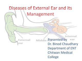
  • 1. Diseases of External Ear and its Management Presented by Dr. Binod Chaudhary Department of ENT Chitwan Medical College
  • 4. Disease of external ear Divided into 1. Disease of the pinna a. Congenital disorders b. Trauma to the auricle c. Inflammatory disorders d. Tumors
  • 5. 2. Disease of external auditory canal a. Congenital disorders b. Trauma c. Inflammation d. Tumors e. Miscellaneous condition
  • 6. Congenital disorders of pinna • Due to developmental abnormalities • May be minor variations or major abnormalities • Minor anomalies – Accessory auricular tag – Bat ear – Lop ear – Preauricular sinus
  • 7. • Major anomalies – Anotia – Microtia
  • 8. Anotia Complete absence of pinna and lobule Usually forms the part of first arch syndrome
  • 9. Microtia • A major developmental anomaly • Frequently associated with anomalies of EAC, middle ear and inner ear • Hearing loss- frequent • Degree of microtia may vary
  • 10. – Grade I microtia: Small external ear and a small but present external ear canal – Grade II microtia: A partially developed ear (usu. top portion underdeveloped) with a closed external ear canal producing a CHL – Grade III microtia: Most common form of microtia with an absent external ear and small peanut-like vestige structure and canal atresia – Grade IV microtia: aka anotia, complete absence of the external ear with canal atresia
  • 11. Treatment options • Do nothing • Surgery – Ear reconstruction using own body’s skin and cartilage – Ear reconstruction using MEDPOR implant • Prosthesis
  • 12. Macrotia Lop/cup ear Preauricular tag Bat ear Congenital anomalies of pinna
  • 13. Preauricular sinus • A depression in front of the crus of helix or above tragus • Is an epithelial track and is due to incomplete fusion of tubercles. • Gets repeatedly infected causing purulent discharge, may form abscess. • Treatment of choice: surgical excision of the track.
  • 14.
  • 15. Trauma to the auricle Hematoma of auricle • Due to injury leading to collection of blood and serum between the auricular cartilage and its perichondrium. • Extravasated blood may clot and then, organize, resulting in a typical deformity called cauliflower ear.
  • 16. • Clinical features – Commonly seen on the anterior surface – Swelling, bluish and tender auricle – If left untreated, necrosis of cartilage and scarring of the auricle – Superadded infection results perichondritis ad abscess formation.
  • 17. Treatment • Aspiration of the hematoma under aseptic condition and pressure dressing to prevent re-accumulation • Incision and drainage if aspiration fails. • All cases should receive antibiotic prophylaxis.
  • 18.
  • 19. Laceration of auricle • The auricle may be cut through-and-through or avulsed partially or totally in RTA, knife injuries, etc. • Treatment includes repairment as early as possible. • Broad spectrum antibiotics given for 1 week.
  • 20.
  • 21. Inflammatory disorders Perichondritis • Is infection of perichondrium of the auricular cartilage • Results from infection secondary to lacerations, hematoma or surgical incisions. • Commonly caused by Pseudomonas aeruginosa
  • 22. Clinical features • Pain, swelling and tender to touch • Patient often has fever • Necrosis of the cartilage, fibrosis and scarring if not treated immediately.
  • 23. Treatment • Should be prompt and vigorous • High dose ciprofloxacin should be used. • Local applicants- magnesium sulfate for soothing • Incision and drainage and C/S if abscess formed. • Surgical debridement to remove unhealthy granulations and necrosed cartilage.
  • 24. Relapsing perichondritis • A rare autoimmune disorder involving cartilage of the ear. • Any cartilage can be involved. • Entire auricle except its lobule becomes inflammed and tender. • External ear canal becomes stenotic. • Treatment consists of high dose of systemic steroids.
  • 26. Chondrodermatitis nodularis chronica helicis • Small painful nodules near the free border of helix. • Tender and patient unable to sleep on the affected side • Treatment is surgical excision of the nodule with its skin and cartilage.
  • 28. Tumors Benign tumors • Hemangioma • Sebaceous cyst • Dermoid cyst • Papilloma • Keratoacanthoma • Neurofibroma Malignant tumors • Squamous cell carcinoma • Basal cell carcinoma • Melanoma
  • 29. Hemangioma • Benign tumors of blood vessels • Congenital tumor commonly seen in children • Bleeds frequently • May get infected • Treatment: surgical excision
  • 30. Dermoid cyst • Developmental cyst • Presents as round mass over the upper part of mastoid behind the pinna • Treatment: surgical excision
  • 31. Sebaceous cyst • Cysts of sebaceous glands • Contains cheesy materials • Common site is postauricular sulcus or below and behind the ear lobule • Treatment: total surgical excision
  • 32. Papilloma • May present as a tufted growth or flat grey plaque and is rough to feel • Viral in origin • Treatment: surgical excision or curettage with cauterization of its base.
  • 33. Squamous cell carcinoma • It can arise anywhere in the external ear, commonly helix. • May present as a painless nodule or an ulcer with raised everted edges and indurated base. • Grows rapidly, invades the surrounding bone and spreads through lymphatics • Treatment: – Small lesions with no nodal metastasis- local excision with 1 cm of external auditory canal – Lesions with nodal metastasis- total amputation of the pinna, often with en bloc removal of parotid gland and cervical lymph nodes.
  • 35. Basal cell carcinoma • Commonly seen over helix and tragus • More common in men beyond 50 yrs • Presents as nodule with central crust, removal of which results in bleeding. • Ulcer has a raised or beaded edge • Lesion often extends circumferentially into the skin, may penetrate deeper to cartilage or bone • Treatment: – Superficial lesion not involving cartilage- irradiation and avoidance of cosmetic deformity – Lesions involving cartilage- surgical excision as in SCC
  • 37. Diseases of External Auditary Canal • Congenital disorders • Trauma • Inflammation • Tumors • Miscellaneous conditions
  • 38. Congenital anomalies Congenital atresia of the EAC • May be either complete or incomplete . • Due to failure of canalization of the ectodermal core that fills the dorsal part of the first brachial cleft. • The outer meatus is obliterated with fibrous tissue or bone while deep meatus and TM are normal • Usually a/w anotia, middle ear and/or inner ear deformities • Hearing loss may be CHL, SNHL or mixed.
  • 39. Treatment • Reconstructive surgery for atresia of EAC (unsatisfactory results) • BAHA or Bonebridge implant system to achieve good hearing system.
  • 41. Collaural fistula • An abnormality of the first branchial cleft. • The fistula has 2 openings: one situated in the neck just below and behind the angle of mandible and the other in the external canal or the middle ear.
  • 42. • Treatment: surgical exploration and excision
  • 43. Trauma to the ear canal • May range from minor laceration of EAC wall to fracture of the bony wall • Foreign body in the EAC may cause trauma to the wall of ear canal. • There may be pain together with bleeding due to laceration. • May get infected if treatment is delayed
  • 44.
  • 45. • Treatment: – Minor injuries require no treatment. – Oral and local antibiotics in more severe cases – Ribbon gauze soaked in 10% ichthyol in glycerine as an ear pack – Reduction of fracture only necessary if the fracture produces occlusion of the EAC
  • 46. Inflammations of EAC May be divided into 1. Infective group – Bacterial • Localized otitis externa (furuncle) • Diffuse otitis externa • Malignant otitis externa – Fungal • otomycosis – Viral • Herpes zoster oticus • Otitis externa hemorrhagica
  • 47. 2. Reactive group – Eczematous otitis externa – Seborrhoeic otitis externa – neurodermatitis
  • 48. Furuncle (Localized otitis externa) • Infection of hair follicle in the outer cartilaginous part of the EAC • Commonly caused by Staphylococcus aureus. • Follows trauma like scratching or cleaning of the EAC by matchsticks, cotton buds, hair clips, nails, etc. • Symptoms – Earache – Swelling/abscess – Discharge – Hearing loss
  • 49. • Signs – Inflamed skin and swelling – Tenderness: tragal tenderness and also with movement of auricle – Discharge – Granulations – Hearing loss • Furuncle at posterior meatal wall causes oedema over the mastoid with obliteration of the retroauricular groove. • Preauricular l.n. may be enlarged and tender
  • 50. • Treatment – Analgesics and local heat – Antibiotics (cloxacillin) – Ear packing with 10% ichthyol in glycerine or other medicated wick – Incision and drainage if abscess formed
  • 51. Diffuse otitis externa • Diffuse inflammation of meatal skin which may spread to involve the pinna and epidermal layer of TM • Commonly seen in hot and humid climate and in swimmers. • Most common factors are – Trauma to the meatal skin – Invasion by pathogenic organisms (S. aureus, P. pyocyaneus) • Clinical features very similar to localized otitis externa.
  • 52. • Differentiating features from localized form are: – Entire EAC is uniformly inflamed and swollen and there is discharge. – No abscess formation – No swelling in the areas adjoining the EAC in diffuse otitis externa
  • 53. • Treatment – Ear toilet – Medicated wicks: aluminium acetate(8%) or silver nitrate(3%) – Antibiotics: cloxacillin or flucloxacillin or ciprofloxacin together with pseudomonas coverage – analgesics
  • 54. Malignant otitis externa • Aka necrotizing otitis externa • It is an inflammatory condition caused by Pseudomonas infection usually in the elderly diabetics, or in those on immunosuppressive drugs. • Early manifestation resemble diffuse otitis externa but there is excruciating pain and appearance of granulations in the ear canal. • Facial paralysis is common • Infection may spread to the skull base and jugular foramen causing multiple cranial nerve palsies.
  • 55. Diagnosis • Severe otalgia in an diabetic patient with granulation tissue in the EAC. • CT scan may show bony destructions • Gallium -67 scan • Technetium 99 bone scan
  • 56. Treatment • Control of diabetics • Toilet of ear canal. Remove discharge, debris and granulations or any dead tissue or bone and send for culture sensitivity. • Antibiotic treatment continued for 6-8 weeks – Gentamicin combined with ticarcillin – Third generation cephalosporins: ceftriaxone 1-2 g/day iv or ceftazidime 1-2 g/day iv combined with aminoglycosides – Quinolones are also effective: combined with rifampin
  • 57. Otomycosis • Is a fungal infection of the EAC caused either by Candida albicans or Aspergillus niger. • Seen in hot and humid climate. • Occurs commonly after entry of water in the EAC, after putting oil and after prolonged use of topical antibiotic eardrops. • Commonly occurs together with CSOM which is actively discharging
  • 58. Clinical features • Itching • Aural fullness • Discomfort and pain • Discharge • Tenderness (in severe case) Examined with otoscope, A. niger appears as black headed filamentous growth and C. albicans appear as yellowish deposit.
  • 59. Treatment: • Thorough cleaning • Broad spectrum topical antifungals (clotrimazole for 10 days) • If there is discharging COM, treat COM as well.
  • 60. Herpes zoster oticus • Aka Ramsay Hunt Syndrome • An infection caused by Varicella Zoster virus. • Usually disease of adults • Characterized by formation of vesicles on the tympanic membrane, meatal skin, concha and postauricular groove. • Patient is ill, complains of severe earache and may have fever. • May involve CN VII and VIII • Triad of SNHL, vertigo and facial palsy
  • 61. Treatment • Oral acyclovir along with high dose steroids. • Labrynthine sedatives for vertigo
  • 62. Otitis externa hemorrhagica • Viral in origin and may be seen in influenzae epidemics. • Characterized by formation of hemorrhagic bullae on the tympanic membrane and deep meatus. • Severe pain and bloody discharge when bullae rupture
  • 63. Treatment • Analgesics for pain relief • Antibiotics for secondary infection of the middle ear if the bulla has ruptured.
  • 64. Eczematous otitis externa • Result of hypersensitivity to infective organisms or topical ear drops such as chloromycetin or neomycin. • Characterized by intense irritation, vesicle formation, oozing and crusting in the canal. • Treatment is withdrawal of causative agent and application of steroids.
  • 65. Miscellaneous conditions Wax • Secreted by sebaceous gland. • Two types: hard and soft. • Seen when self-cleansing mechanism of the ear is disturbed. • So, cleaning with cotton buds, hair clips, matchsticks should be avoided.
  • 66. Clinical features • Discomfort/itching • Feeling of blocked ear • Hearing loss • Tinnitus • Cough • vertigo
  • 67. Treatment • Wax without pain Suctioning Removal by jobson horne probe Syringing
  • 68. • Wax with pain (a/w otitis externa) – Antibiotic ear drops followed by wax softners. – Oil based antibiotic eardrop such as chloramphenicol is preferred to other antibiotic because it also softens the wax to some extent. – Then removed as described above.
  • 69. Foreign body • Common in children than adults. • Common foreign bodies in the EAC Children  Inanimate FB- pieces of paper, eraser, sponge, lead of pencil, etc  Vegetable matter- beans, seeds, etc  Insects- flea, tick, housefly, maggots, etc Adults  Inanimate FB- cotton wool  Insects- flea, tick, housefly, maggots, etc
  • 70. Clinical features • Children often do not tell their parents that they have put a FB in the ear due to fear; incidental finding • Small inanimate FB- no symptoms • Mild hearing loss • Intense pain – live insects • Examination reveals FB in the EAC
  • 71.
  • 72. Treatment Live insects should be first killed by putting oil or water and then only be removed • Methods of removal 1. Removal under microscope
  • 73. 2. Removal by ear syringing
  • 74. 3. Use of head mirror and ear instruments
  • 75. Keratosis obturans • A condition characterized by excess accumulation of hard whitish- yellow debris consisting of desquamated epithelium in the bony part of EAC. • Eventually cause pressure on the bony walls of the EAC and cause resorption of the bone • Clinical features – Blocked ear – Pain and discharge if a/w otitis externa – Cholesteatoma like mass (pearly white hard debris covered by wax) – Automastoidectomy
  • 76. Treatment • Removal as done for wax • General anesthesia may be required because of the pain • Recurrent collection of desquamated epithelium, so regular follow up required.