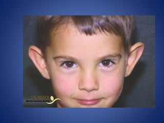
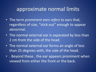
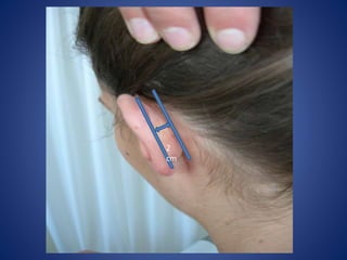
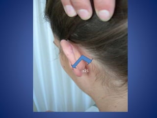
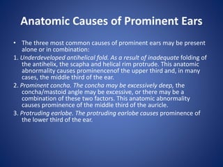
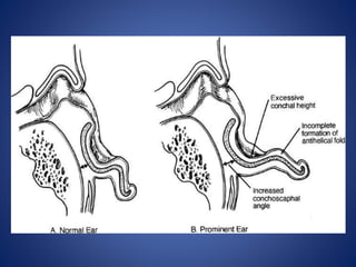
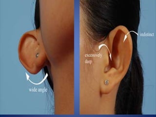
3. approximate normal limits
• The term prominent ears refers to ears that,
regardless of size, “stick out” enough to appear
abnormal.
• The normal external ear is separated by less than
2 cm from the side of the head.
• The normal external ear forms an angle of less
than 25 degrees with, the side of the head.
• . Beyond these , the ear appears prominent when
viewed from either the front or the back.
6. Anatomic Causes of Prominent Ears
• The three most common causes of prominent ears may be present
alone or in combination:
1. Underdeveloped antihelical fold. As a result of inadequate folding of
the antihelix, the scapha and helical rim protrude. This anatomic
abnormality causes prominencenof the upper third and, in many
cases, the middle third of the ear.
2. Prominent concha. The concha may be excessively deep, the
concha/mastoid angle may be excessive, or there may be a
combination of these two factors. This anatomic abnormality
causes prominence of the middle third of the auricle.
3. Protruding earlobe. The protruding earlobe causes prominence of
the lower third of the ear.
7.
8.
9. examples of abnormally shaped
ears that may also be prominent
• Macrotia: refers to excessively large ears that, in addition to
being large, may be“prominent.”
• Constricted ears are abnormally small but tend to appear
“prominent” because the circumference of the helical rim
is inadequate, causing the auricle to cup forward.
• The Stahl’s ear deformity consists of a third crus, in addition
to the crura of the triangular fossa, which traverses the
scapha.
• Cryptotia describes the auricle in which the upper pole of
the helix is buried beneath the temporal skin.
• Question mark ears earn their name because deficiency
ofthe supralobular region gives the ear the shape of a
question mark.
13. Goals of Otoplasty
1. Front view. When viewed from the front the helical rim should be
visible, not set back so far that it is hidden behind the antihelical
fold.
2. Rear view. When viewed from behind, the helical rim should be
straight, not bent like a “C” or a “hockey stick.” If the helical rim is
straight, the setback will be harmonious; that is, the upper, middle,
and lower thirds of the ear will be setback in correct proportion to
each other. If, for example, the middle third is set back too much
relative to the upper and lower thirds, the helical rim will form a
“C” when viewed from behind, creating the so-called telephone
deformity. Similarly, if the earlobe is insufficiently set back, the rear
view will reveal a hockey stick appearance to the helical rim
contour.
3. Lateral view. The contours should be soft and natural, not sharp and
“human-made.”
14. Timing of Otoplasty
• There is no absolute rule about when
otoplasty should be performed.
• In young children with extremely prominent
ears, a reasonable age is approximately 4
years.
• In cases of macrotia associated with
prominence, the procedure performed as
early as age 2 years, hoping that any
restriction of growth is an advantage.
15. Operative Procedure
• The techniques described below are used
alone or in combination depending on the
anatomic deformity and the choice of the
surgeon:
1. Antihelical Fold Manipulation.
2. Conchal Alteration.
3. Correction of Earlobe Prominence.
16. Antihelical Fold Manipulation
• Suturing of cartilage: Mattress sutures are placed from the scapha
and/or triangular fossa to the concha, as described by Mustarde,
and are tied with sufficient tension to increase the definition of the
antihelical fold, thereby setting back the helical rim and scapha.
• Stenstrom technique of anterior abrasion: The anterior surface of
the antihelical fold cartilage is abraded, causing the cartilage to
bend away from the abraded side (principle of Gibson) toward the
side of intact perichondrium.
• Full-thickness incisions :A single full-thickness incision along the
desired curvature of the antihelix permits folding with slight force,
creating an antihelical fold (Luckett procedure). Because the fold is
sharp and unnatural this single-incision technique was modified.
• Converse/Wood-Smith technique, a pair of incisions is made,
parallel to the desired antihelical fold, and tubing sutures are placed
to create a more defined fold.
17. Conchal Alteration
■ Suturing: The angle between the concha and the mastoid skull can be
decreased by placing sutures between the concha and the mastoid
fascia as described by Furnas.
■ Conchal excision: From either an anterior or posterior approach, a
full-thickness crescent of cartilage is removed from the posterior
wall of the concha (taking care not to violate or deform the
antihelical fold), thereby reducing the conchal height. The conchal
defect is meticulously closed with sutures to avoid a visible ridge
within the concha. The excision is designed so that the eventual
closure will lie at the junction of the floor and posterior wall of the
concha where it is least conspicuous and causes the least distortion
of the normal auricular contours.
■ A combination of Furnas suture and Conchal excision technique.
18. Correction of Earlobe Prominence
• Earlobe prominence is not corrected by the
above maneuvers.
• In fact, these maneuvers may increase the
prominence of the earlobe, making earlobe
repositioning the most difficult and neglected
part of the procedure.
19. Correction of Earlobe Prominence
The following technique commonly areused:
• Webster technique: suturing the tail of the helical cartilage
to the concha will correct earlobe prominence.
Unfortunately the tail of the helix does not extend into the
lobule and setting it back does not reliably set back the
earlobe.
• skin excision and sutures between the fibrofatty tissue of
the lobule and the tissues of the neck.
• The best technique is the technique described by Gosain,
or a variation thereof, in which skin is excised on the medial
surface of the earlobe. When this defect is closed with
sutures, a bite of the undersurface of the concha is taken,
which pulls the earlobe toward the head.
20. Alteration of the Position of the
Upper Auricular Pole
• Depending on the degree of prominence
preoperatively of the upper third of the ear,
the antihelical fold creation may be
inadequate to correct the position of the
helical rim, near the root. An additional
mattress suture between the helical rim and
the temporal fascia may be required.
21. a Mustarde scapha-conchal suture
conchal
resection with
primary closure
Furnas conchal-mastoid
suture
22. Other Deformities
• Macrotia: To reduce the size of the ears, the incision is
made on the lateral surface of the ear, just inside the helical
rim. The scapha is reduced and a segment of helical rim is
excised and closed primarily to avoid redundancy.
• Constricted ear : In mild cases, the crus of the helix is
advanced out of the concha and into the helical rim and
standard otoplasty techniques are used in addition . In
severe cases, the cartilage is discarded and a complete
auricular reconstruction performed as in microtia.
• Stahl ear: Various techniques have been described to excise
the extra crus. None is perfect.
23. • Cryptotia: The superior aspect of the auricular cartilage
is pulled out from under the scalp an incision is made
around the now-visible helical rim, and the medial
surface of the freed cartilage is resurfaced with a graft
or flap. In some cases, the buried cartilage is quite
normal, and in other cases, it is markedly abnormal.
• Question mark ear: The deficiency is variable and is
usually treated with a cartilage graft and a V-Y
advancement flap from the retroauricular skin. Often
there is excess in the upper third of the ear requiring
reduction. In severe cases, the entire ear is
reconstructed as in microtia.
24. Postoperative Care
• A bulky, noncompressive dressing is placed for a few
days. Excessive pressure from the dressing will cause
pain, increase swelling, and may lead to abrasion or
even necrosis of auricular skin.
• When the dressing is removed, the patient wears a
loose headband at night for 6 weeks. Again, the
headband should only be tight enough that it does not
fall off. The purpose is to prevent the corrected ear
from being pulled forward when the patient rolls over
in bed. A tight headband can erode the lateral surface
of the ear, creating an open wound.
25. Nonoperative Technique in Infants
• During the early weeks of infancy, the auricular
cartilage has unusual plasticity, attributed to circulating
maternal estrogens.
• During this privileged period, prominent ears and
related deformities can be corrected permanently by
molding the ears into the correct shape with tape and
soft dental compound. The process is continued for
several months or until there is no further
improvement in auricular contour. It is not clear how
long cartilage retains this “moldability” and therefore it
is not clear when infants are too old to have this
technique attempted.
26. Complications
• Hematoma.
• Infection :Cellulitis is rare after otoplasty but is treated aggressively
with intravenous antibiotics in an attempt to avoid chondritis. The
latter may require debridement and leave the ear permanently
disfigured.
• Suture complications : By far the most common complication of
otoplasty is related to suture extrusion in the retroauricular sulcus.
Such sutures are easily removed but may be associated with
unattractive and/or painful granulomas. The use of absorbable
sutures might eliminate this complication. permanent sutures
Monofilament sutures are more likely to protrude but are less likely
to create granulomas. Braided sutures are the opposite: Less likely
to protrude but more likely to be associated with granulomas.
• Overcorrection/unnatural contours :The most common significant
complication of otoplasty is overcorrection.