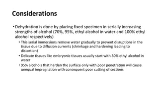
This document provides an overview of decalcification in histopathology. It discusses the need to decalcify bony tissue specimens to make them thin enough for microscopic examination. The key aspects of decalcification covered include the criteria for good decalcifying agents, factors that affect the process, common techniques and decalcifiers used, potential artifacts, and assessing the endpoint of decalcification. The techniques described aim to remove calcium from bone while minimizing damage to tissue morphology and antigenicity.
Histopathology is examination of tissues for presence or absence of changes in their structure due to disease processes. We go through various steps in the process of converting gross sample to microscopic slides.
Decalcification is a process used to remove mineral content from calcified tissues like bone and teeth to allow for microscopic examination. It involves selecting an appropriate decalcifying agent based on factors like the tissue, required staining, and urgency. Common decalcifying agents include acids like nitric acid, formic acid, and chelating agents. The decalcification process must be monitored and the tissue properly processed, sectioned, and stained afterwards to examine it microscopically. Undecalcified sections can also be prepared to examine mineralized and non-mineralized bone ratios.
Tissue processing involves fixing, dehydrating, clearing, infiltrating, and embedding tissue samples to produce diagnostic microscope slides. It stabilizes tissues and removes water, replacing it with paraffin wax. Efficient agitation, heat up to 45°C, low viscosity reagents, and vacuum up to 50.79 kPa can reduce processing time. Stages include fixation with formalin, dehydration using graded alcohols, clearing with xylene or toluene, infiltration with paraffin wax, and embedding for microtomy. Proper orientation during embedding is important for diagnosis.
FIXATIVES in Pathology for Postgraduate and DMLTjenishJebadurai1
This document discusses various fixatives used in histology and cytology techniques. It begins by defining fixation as the process of preserving cells and tissue using physical or chemical methods. Commonly used fixatives include formaldehyde, glutaraldehyde, osmium tetroxide, mercury salts, picric acid, and alcohols. An ideal fixative would be non-toxic, low-cost, and effectively preserve tissue morphology, antigens, and nucleic acids while allowing for long-term storage. Factors like temperature, pH, concentration, and duration impact fixation quality. Proper fixation is important for minimizing artifacts and ensuring high quality staining.
Preparation & stability of large & small volume parentralsROHIT
This document discusses parenteral formulations, including definitions, advantages, disadvantages, and classifications. It provides details on the preparation of small volume parenterals and large volume parenterals, including vehicles, buffers, preservatives, and other excipients used. It also covers the stability considerations for parenteral formulations and factors that influence syringeability, injectability, clogging, drainage, resuspendibility, and sedimentation of suspensions.
This document discusses the process of tissue processing, which involves fixing, dehydrating, clearing, and embedding tissue samples in paraffin wax to allow for thin sectioning. The key stages are fixation using chemicals like formalin to preserve tissue structure, dehydration using increasing concentrations of alcohol, clearing using solvents like xylene to make tissues transparent, and embedding in paraffin wax for sectioning. Automated and manual methods are described. Tissue microarrays allow evaluation of multiple tissue samples on a single slide by arranging small cores in a recipient paraffin block.
Tissue processing involves removing water from tissue and replacing it with paraffin wax to provide rigidity for microscopic examination. The main steps are fixation, dehydration using increasing concentrations of alcohol, clearing with xylene to remove alcohol, and impregnation with molten paraffin wax. Automated tissue processors complete this process overnight using different stations for each step. Factors like tissue size, agitation, heat, and vacuum pressure influence effective processing. Ethyl alcohol is most commonly used for dehydration, while xylene is used for clearing prior to paraffin wax impregnation and embedding.
This document discusses the formulation of sterile solutions, including small volume and large volume parenteral solutions. It covers various types of vehicles that can be used, including aqueous vehicles like water for injection and non-aqueous vehicles like oils. It also discusses additives that can be included, like solubilizing agents, buffers, antioxidants, tonicity adjusting agents, and antimicrobial agents. The document provides details on the preparation of water for injection, considerations for the route of administration, selection of vehicles, and supporting studies required for formulation of sterile solutions.
techniques used for preparing serial sections using microtomes include dehydrating agents and clearing agents ,this slide includes some details on dehydrating and clearing agents
This document provides information about tissue processing for medical laboratory technology (MLT) students. It discusses the basic steps in manual and automatic tissue processing, which are dehydration, clearing, and impregnation. Dehydration removes water from tissue using graded alcohols like ethanol. Clearing removes dehydrating fluids using xylene or other agents. Impregnation infiltrates tissue with paraffin wax. Automatic processors complete the steps more quickly through constant agitation in multiple chambers containing processing chemicals. The goal of tissue processing is to remove water from tissues and replace it with paraffin wax for embedding and microscopic examination.
This document discusses various fixatives used for small biopsy specimens and their applications. It describes the ideal properties of a fixative and the mechanisms of different types of fixatives including aldehydes, mercurials, dichromates, picric acid and alcohol-containing fixatives. For each fixative, the document outlines their composition, principle of action, tissues they are suitable for, fixation time and advantages and disadvantages. It also provides recommendations for fixation of specific tissue biopsies like renal, muscle and liver biopsies. The conclusion emphasizes the importance of using an appropriate fixative to ensure optimal specimen evaluation and diagnosis.
This document discusses various techniques for dehydrating and clearing biological specimens for preservation and mounting, including:
- Using a series of solutions with progressively decreasing water concentration and increasing concentrations of dehydrating agents like ethanol or acetone to remove water from cells and tissues.
- Clearing agents like xylene, chloroform, or clove oil are used to remove alcohol after dehydration and make tissues transparent before embedding in wax or mounting.
- Different agents are suitable for different types of delicate materials, and proper timing in each solution is important to prevent tissues from becoming too brittle or damaged.
MICROTECHNIQUE Killing and fixation moduleAbdulsalm
This document provides instructions for preparing biological specimens for light microscopy. It discusses the key steps of sample collection, killing and fixation, dehydration, clearing, paraffin embedding, microtomy, staining, and observation. Specific fixation fluids, dehydration reagents, clearing agents, and staining methods are described. The goal is to preserve specimens while modifying properties like refractive index to allow examination under a light microscope.
This document discusses various extraction methods for extracting medicinally active compounds from plant materials. It begins with an introduction on the importance of understanding a plant's phytochemicals when selecting an extraction method and solvent. Key methods discussed include maceration, infusion, Soxhlet extraction, hydrodistillation, and microwave-assisted extraction. Each method has advantages and suitabilities depending on the material and desired compound. Selecting the appropriate extraction technique is important to efficiently separate and obtain compounds of interest without degradation.
The document outlines the basic steps in processing biological tissues for microscopic examination: fixation, dehydration, embedding, sectioning, and staining. Chemical fixatives like formalin are used to preserve tissue structure. Dehydration removes water and replaces it with ethanol or methanol. Embedding surrounds tissue with wax or other materials. Sectioning cuts very thin slices using a microtome. Staining adds color to tissue structures using dyes like hematoxylin and eosin for contrast.
This document provides instructions for several histopathology staining techniques, including:
- Periodic Acid Schiff (PAS) staining for polysaccharides and basement membranes in magenta.
- Gram-Twort modification for staining bacteria in paraffin sections in blue-black (Gram positive) and pink (Gram negative).
- Ziehl-Neelsen technique for staining acid-fast bacilli like Mycobacterium tuberculosis red against a blue background.
It also describes the Periodic Acid Schiff/Alcian Blue dual stain to differentiate acid mucins (blue) from neutral mucins and carbohydrates (magenta). Precise protocols and reagent preparations are provided for accurate histological analysis.
WASHING, DRYING AND STERILIZATION OF GLASSWARES.pptxAVINASH K
Good laboratory technique demands clean glassware, because the most carefully executed piece of work may give an erroneous result if dirty glassware is used.
In all instances, glassware must be physically clean; it must be chemically clean; and in many cases, it must be bacteriologic ally clean or sterile.
All glassware must be absolutely grease-free.
This document provides an overview of the tissue processing techniques used to prepare tissue samples for microscopic examination. It describes the main steps in tissue processing as fixation, dehydration, clearing, infiltration/embedding. Dehydration involves removing water from tissues using a series of increasing concentrations of ethanol or other solvents to prevent damage. Clearing replaces the dehydrating fluid with a solvent miscible with both the dehydrating fluid and paraffin wax. The goal is to embed tissues in paraffin wax for microtomy, as it provides sufficient rigidity while being soft enough for thin sectioning without harming tissues or knives. Factors like tissue type, fixation, and desired detail influence processing parameters.
This document discusses the process of preparing histological specimens. It involves fixing tissues to prevent degradation, processing them through dehydration, clearing and embedding in paraffin wax. Tissues are then sectioned using a microtome and stained, commonly with hematoxylin and eosin, to visualize cells and structures under a microscope. The staining highlights nucleic acids, ribosomes and other components to aid examination and study of tissue structure and organization.
Lecture (5) processing of tissue in histopathology laboratoryHafsa Hussein
This document discusses the principles and steps of tissue processing for microscopic examination, including dehydration, clearing, and infiltration/impregnation with paraffin wax.
The key steps are:
1) Dehydration using graded alcohols to remove water from tissues, allowing infiltration with wax. Xylene is commonly used for clearing to remove alcohols.
2) Clearing with xylene or other agents to make tissues transparent before wax infiltration.
3) Infiltration and impregnation with molten paraffin wax provides rigidity to tissues for thin sectioning and microscopic examination. Proper processing is important for examining tissue structures.
1. The document discusses the various steps involved in tissue processing for microscopic examination, which includes fixation, processing, embedding, sectioning and staining of tissues.
2. Key steps include fixation of tissues using formalin to preserve structure, dehydration using increasing concentrations of alcohol, clearing with xylene, impregnation and embedding in paraffin wax.
3. Thin sections are then cut from the paraffin blocks using a microtome and stained, usually with hematoxylin and eosin, for microscopic examination.
This document discusses various extraction methods used to separate medicinally active compounds from plant materials. It begins with an introduction on selecting extraction methods based on compound characteristics and solvent properties. Several conventional methods are described in detail, including maceration, infusion, Soxhlet extraction, and hydrodistillation. Non-conventional methods like ultrasound-assisted extraction and microwave-assisted extraction are also summarized. The document concludes by noting that the appropriate extraction method depends on factors like the target compound and plant material properties.
This document discusses ideal requirements, functions, and commonly used irrigating solutions and intracanal medicaments in endodontic treatment. Sodium hypochlorite and EDTA are the most commonly used irrigants due to their ability to dissolve tissue and remove smear layer. Chlorhexidine and hydrogen peroxide are also discussed. Intracanal medicaments mentioned include eugenol, phenol, camphorated monochlorophenol, formocresol, and calcium hydroxide which are used to disinfect canals and promote healing.
Mechanism of action of various disinfectants.pptxambikaluthra3
The document summarizes various physical and chemical disinfection methods. Physical methods include sunlight, dry heat, moist heat, vibration, radiation, and filtration. Sunlight and heat kill microbes through oxidative effects and protein denaturation. Moist heat is more effective than dry heat. Radiation like UV rays and ionizing radiation damage DNA. Filtration separates but does not kill microbes. Chemical disinfectants include alcohols, aldehydes, phenols, halogens, heavy metals, surfactants, dyes, hydrogen peroxide, ethylene oxide, and beta-propionolactone. They act through processes like dehydration, membrane disruption, protein coagulation, and
Similar to DEHYDRATION - Histopathologic Techniques (20)
Basavarajeeyam is a Sreshta Sangraha grantha (Compiled book ), written by Neelkanta kotturu Basavaraja Virachita. It contains 25 Prakaranas, First 24 Chapters related to Rogas& 25th to Rasadravyas.
Does Over-Masturbation Contribute to Chronic Prostatitis.pptxwalterHu5
In some case, your chronic prostatitis may be related to over-masturbation. Generally, natural medicine Diuretic and Anti-inflammatory Pill can help mee get a cure.
share - Lions, tigers, AI and health misinformation, oh my!.pptxTina Purnat
• Pitfalls and pivots needed to use AI effectively in public health
• Evidence-based strategies to address health misinformation effectively
• Building trust with communities online and offline
• Equipping health professionals to address questions, concerns and health misinformation
• Assessing risk and mitigating harm from adverse health narratives in communities, health workforce and health system
Integrating Ayurveda into Parkinson’s Management: A Holistic ApproachAyurveda ForAll
Explore the benefits of combining Ayurveda with conventional Parkinson's treatments. Learn how a holistic approach can manage symptoms, enhance well-being, and balance body energies. Discover the steps to safely integrate Ayurvedic practices into your Parkinson’s care plan, including expert guidance on diet, herbal remedies, and lifestyle modifications.
Adhd Medication Shortage Uk - trinexpharmacy.comreignlana06
The UK is currently facing a Adhd Medication Shortage Uk, which has left many patients and their families grappling with uncertainty and frustration. ADHD, or Attention Deficit Hyperactivity Disorder, is a chronic condition that requires consistent medication to manage effectively. This shortage has highlighted the critical role these medications play in the daily lives of those affected by ADHD. Contact : +1 (747) 209 – 3649 E-mail : sales@trinexpharmacy.com
Rasamanikya is a excellent preparation in the field of Rasashastra, it is used in various Kushtha Roga, Shwasa, Vicharchika, Bhagandara, Vatarakta, and Phiranga Roga. In this article Preparation& Comparative analytical profile for both Formulationon i.e Rasamanikya prepared by Kushmanda swarasa & Churnodhaka Shodita Haratala. The study aims to provide insights into the comparative efficacy and analytical aspects of these formulations for enhanced therapeutic outcomes.
2. OBJECTIVES
•Describe purpose of dehydration
•Enumerate the different dehydrating agents
•Describe their advantages and disadvantages
3. Principle:
•Removal of intercellular and extracellular water from the tissue
done after fixation and replace them with dehydrating agents
•Done after fixation and after bones and teeth have been
decalcified
4. Considerations
•Dehydration is done by placing fixed specimen in serially increasing
strengths of alcohol (70%, 95%, ethyl alcohol in water and 100% ethyl
alcohol respectively)
• This serial immersions remove water gradually to prevent disruptions in the
tissue due to diffusion currents (shrinkage and hardening leading to
distortion)
• Delicate tissues like embryonic tissues usually start with 30% ethyl alcohol in
water
• 95% alcohols that harden the surface only with poor penetration will cause
unequal impregnation with consequent poor cutting of sections
5. Considerations
•Dehydration times should be as brief as possible to minimize the risk
of extracting cellular constituents
• Prolonged storage in alcohol causes maceration, especially at lower strengths
below 70%
•Storage of specimen may be done at 70-80% alcohol but not for
extended periods due to interference in their staining properties
• Dehydrating agent must not be less than 10x the volume of the tissue for
proper penetration
6. Considerations
•For urgent examinations, temperature may be increased to 37°C
•For complete dehydration with alcohol:
• A layer of anhydrous copper sulfate about ¼ inch is placed at the bottom of
the container and covered with filter paper
• Accelerates dehydration by removing water from dehydrating fluid
• Blue discoloration of copper sulfate crystals indicate full saturation of
dehydrating fluids with water- alcohol should be discarded and changed with
a fresh solutioin
8. A. Alcohol
• used in increasing concentration (70% to 95% ) to prevent
shrinkage and hardening of tissue which may lead to distortion.
• Note:
• Too low alcohol concentration: leads to tissue maceration
•concentrated alcohol: tissue hardening
9. A. Alcohol
1. Ethyl alcohol
• best dehydrating agent (fast acting, mixes with water and
penetrates tissues easily)
• Clear colorless, flammable
• Most recommended for routine dehydration
2. Methyl alcohol
• toxic
• used for blood and tissue films and for smear preparations
10. A. Alcohol
3. Butyl alcohol
•Utilized in plant and animal microtechniques
•Slow acting causing less shrinkage and hardening than ethyl
alcohol
•Recommended for tissues which do nor require rapid processing
11. B. Acetone
• Cheap and rapid acting (dehydrates biopsies for ½ to 2 hours)
but penetrates poorly
• Clear colorless fluid that mixes with water, ethanol and most
organic solvents
• More miscible with epoxy resins than alcohol but extremely
volatile and flammable
• Removes most lipids from tissues
• not recommended for routine tissue purposes due to
considerable tissue shrinkage
12. C. Dioxane (Diethylene dioxide)
• excellent dehydrating and clearing agent
• miscible with water, paraffin , alcohol and xylol
• Less tissue shrinkage vs alcohol
• Tissues may be stored for long periods without loss in consistency or
staining properties
• Disadvantages: expensive, toxic vapour, tissues tend to ribbon poorly
• Not routine due to highly toxic effect (use only in well ventilated room)
• Should not be recycled due to the risk of forming explosive peroxides
13. D. Cellosolve (Ethylene glycol
monoethyl ether)
• rapid dehydrating agent,
• Specimen may be transferred from water or normal saline
directly into cellosolve for storage with no hardening or
distortion
• Flammable at 43-49°C, toxic by inhalation, skin contact
and ingestion to the reproductive, fetal, urinary and
blood (Substitute: Propylene based ethylene glycos)
14. D. Triethyl phosphate
• soluble in water, alcohol, ether, benzene chloroform,
acetone and xylene
• removes water from tissues readily with little distortion
and hardening of tissues.
15. D. Tetrahydrofuran
• clearing and dehydrating agent since it is miscible in both water and
paraffin.
• causes less shrinkage, easier cutting of sections with fewer artifacts
• Improves most staining procedures
• Toxic on ingestion and inhalation, offensive odor (use in well
ventilated room)
• Eye irritant- prolonged exposure (up to 6 mos) may cause
conjunctival irritation
•Skin irritant- no practical way to protect skin except for Teflon
gloves