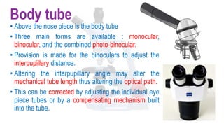
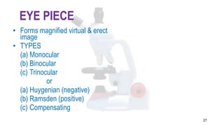
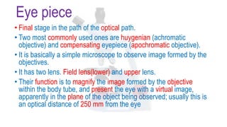
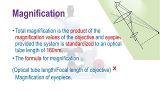
Download to read offline


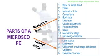
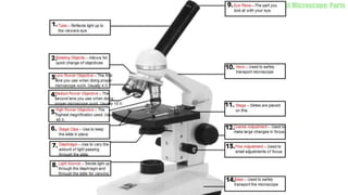
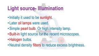
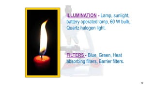
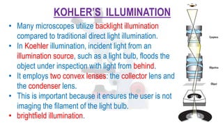
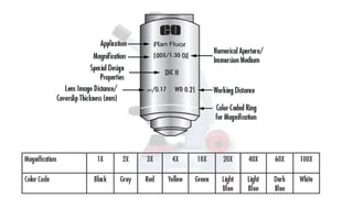

The document describes the key components and functioning of a light microscope. It discusses the light source, condenser, stage, objectives of different magnifications, body tube, eyepiece, and how their interaction allows for magnification and imaging of small specimens. The document also covers advantages such as cost-effectiveness and simple sample preparation, and disadvantages like limited resolution and need for staining.


![PERI-PROSTHETIC FRACTURE NAIL-PLATE CONSTRUCT [NPC].pptx](https://cdn.slidesharecdn.com/ss_thumbnails/drarunkumardrmohamedashrafperiprostheticfrasturenail-plateconstructnpc-260209164459-7e9d15a1-thumbnail.jpg?width=640&height=640&fit=bounds)


