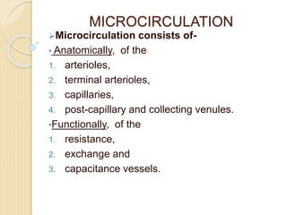
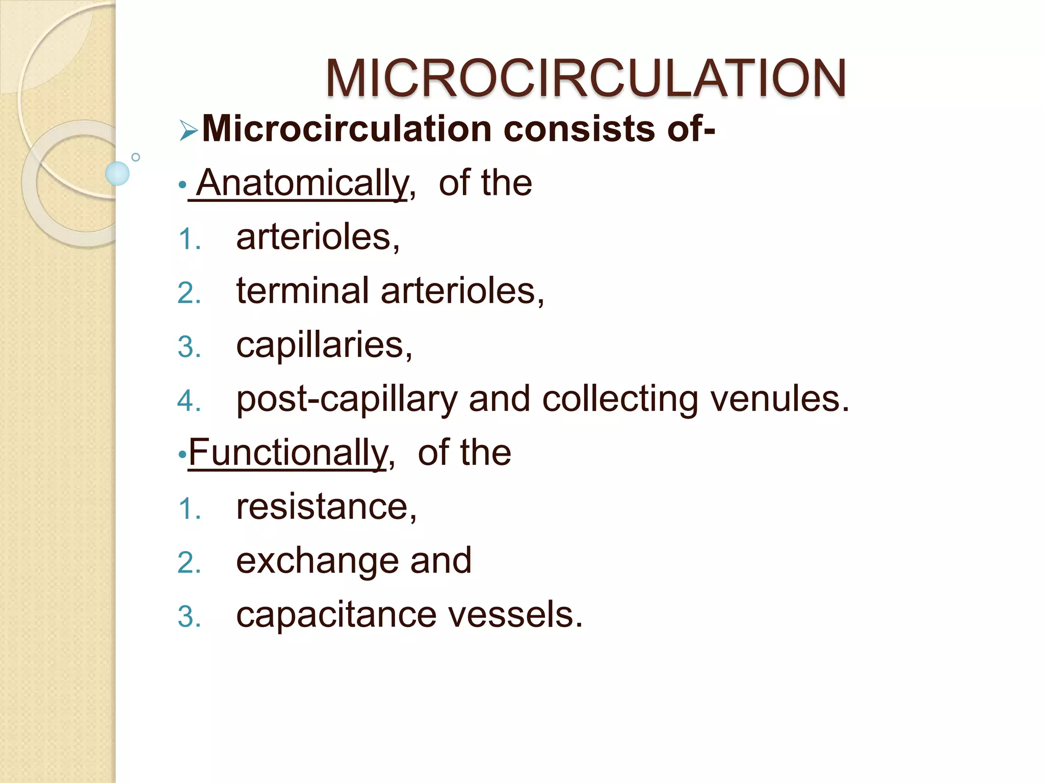
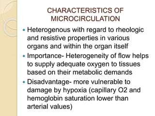
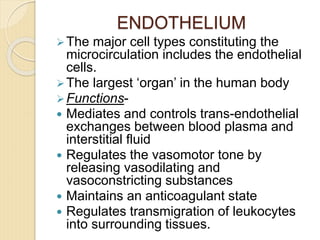
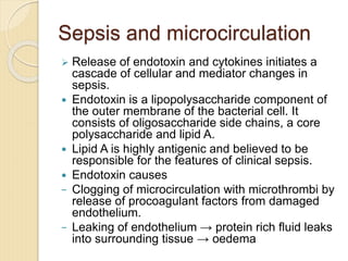
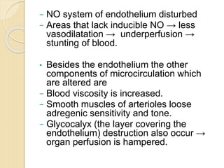
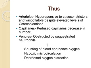
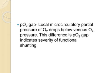
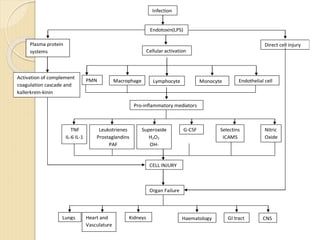
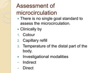
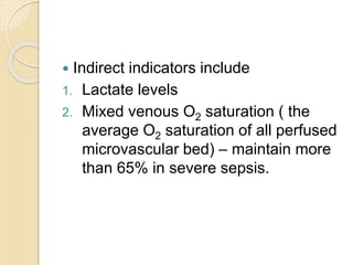
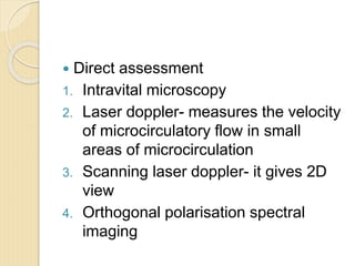
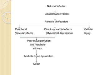
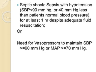
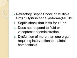
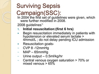
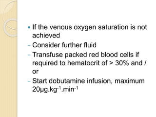
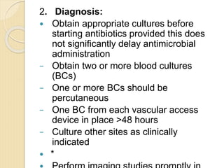
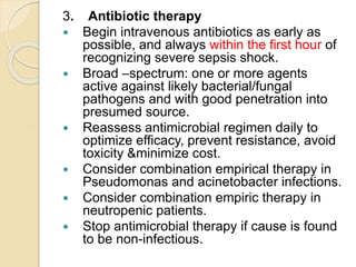
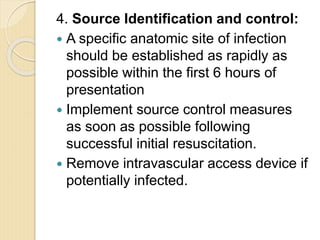
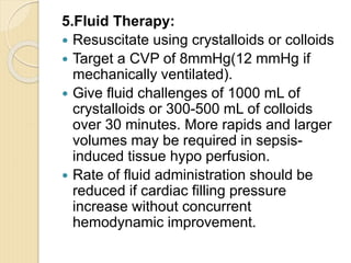
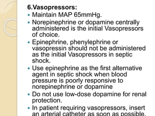
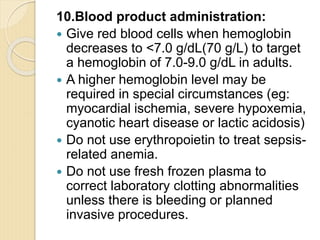
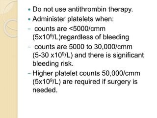
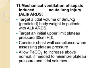
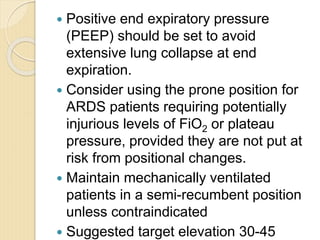
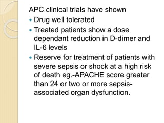
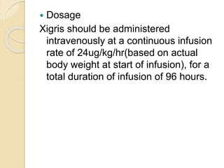
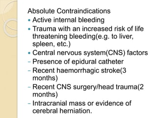
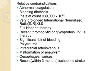
MICROCIRCULATION
Microcirculation consists of-
• Anatomically, of the
1. arterioles,
2. terminal arterioles,
3. capillaries,
4. post-capillary and collecting venules.
•Functionally, of the
1. resistance,
2. exchange and
3. capacitance vessels.
Resistance vessels (Arterioles)
• <100-200 mm in diameter)
• final branch of the arterial system
• marks the beginning of microcirculation.
Exchange vessels (Capillaries)
 lined by a single layer of endothelium
 contains no smooth muscle cells
 cannot change their diameter actively
CAPILLARY RECRUITMENT- The body
can cope up with increased metabolic
demands by increasing the number of
capillaries → increasing the exchange
surface area.
Capacitance vessels (Venules)
 diameter greater than 30 mm
 start acquiring smooth muscles cells
 termed ‘capacitance vessels’ since
they hold almost 70% of the total
circulating blood volume
CHARACTERISTICS OF
MICROCIRCULATION
 Heterogenous with regard to rheologic
and resistive properties in various
organs and within the organ itself
 Importance- Heterogeneity of flow helps
to supply adequate oxygen to tissues
based on their metabolic demands
 Disadvantage- more vulnerable to
damage by hypoxia (capillary O2 and
hemoglobin saturation lower than
arterial values)
ENDOTHELIUM
The major cell types constituting the
microcirculation includes the endothelial
cells.
The largest ‘organ’ in the human body
Functions-
 Mediates and controls trans-endothelial
exchanges between blood plasma and
interstitial fluid
 Regulates the vasomotor tone by
releasing vasodilating and
vasoconstricting substances
 Maintains an anticoagulant state
 Regulates transmigration of leukocytes
into surrounding tissues.
Sepsis and microcirculation
 Release of endotoxin and cytokines initiates a
cascade of cellular and mediator changes in
sepsis.
 Endotoxin is a lipopolysaccharide component of
the outer membrane of the bacterial cell. It
consists of oligosaccharide side chains, a core
polysaccharide and lipid A.
 Lipid A is highly antigenic and believed to be
responsible for the features of clinical sepsis.
 Endotoxin causes
− Clogging of microcirculation with microthrombi by
release of procoagulant factors from damaged
endothelium.
− Leaking of endothelium → protein rich fluid leaks
into surrounding tissue → oedema
− NO system of endothelium disturbed
− Areas that lack inducible NO → less
vasodilatation → underperfusion →
stunting of blood.
• Besides the endothelium the other
components of microcirculation which
are altered are
− Blood viscosity is increased.
− Smooth muscles of arterioles loose
adregenic sensitivity and tone.
− Glycocalyx (the layer covering the
endothelium) destruction also occur →
organ perfusion is hampered.
Thus
 Arterioles- Hyporeponsive to vasoconstrictors
and vasodilators despite elevated levels of
Catecholamines.
 Capillaries- Perfused capillaries decrease in
number.
 Venules- Obstructed by sequestrated
neutrophils
↓
Shunting of blood and hence oxygen
Hypoxic microcirculation
Decreased oxygen extraction
 pO2 gap- Local microcirculatory partial
pressure of O2 drops below venous O2
pressure. This difference is pO2 gap
indicates severity of functional
shunting.
Infection
Endotoxin(LPS)
Plasma protein
systems
Activation of complement
coagulation cascade and
kallerkrein-kinin
Direct cell injury
CELL INJURY
Organ Failure
Lungs Heart and
Vasculature
Kidneys Haematology GI tract CNS
Cellular activation
PMN Macrophage Lymphocyte Monocyte Endothelial cell
Pro-inflammatory mediators
TNF
IL-6 IL-1
Leukotrienes
Prostaglandins
PAF
Superoxide
H2O2
OH-
G-CSF Selectins
ICAMS
Nitric
Oxide
Assessment of
microcirculation
 There is no single goal standard to
assess the microcirculation.
 Clinically by
1. Colour
2. Capillary refill
3. Temperature of the distal part of the
body.
 Investigational modalities
− Indirect
− Direct
 Indirect indicators include
1. Lactate levels
2. Mixed venous O2 saturation ( the
average O2 saturation of all perfused
microvascular bed) – maintain more
than 65% in severe sepsis.
 Direct assessment
1. Intravital microscopy
2. Laser doppler- measures the velocity
of microcirculatory flow in small
areas of microcirculation
3. Scanning laser doppler- it gives 2D
view
4. Orthogonal polarisation spectral
imaging
Nidus of infection
Bloodstream invasion
Release of mediators
Peripheral Direct myocardial effects Cellular
Vascular effects (Myocardial depression) Injury
Poor tissue perfusion
and metabolic
acidosis
Multiple organ dysfunction
Death
Sepsis and its management
 Bacteremia: Presence of bacteria in blood as
evidenced by positive blood cultures.
 Septicemia: Presence of microbes or their
toxins in blood.
 Systemic inflammatory response
syndrome(SIRS): Two or more of the
following conditions:
1. Fever(oral temp>38°C) or
hypothermia(<36°C)
2. Tachypnoea(>24 bpm)
3. Tachcardia(HR>90)
4. Leucocytosis(>12,000),
Leucopenia(<4000),or >10%bands;
SIRS may have non infectious etiology.
 Sepsis: SIRS of microbial etiology
 Severe sepsis/ sepsis syndrome: Sepsis with
one or more signs of organ dysfunction- eg
1. Cardiovascular: SBP<=90, MAP<=70 mm Hg.
That responds to administrations of i.v fluids.
2. Renal: Urine output <0.5 ml/kg/hr for 1 hr
despite adequate fluid resuscitation.
3. Respiratory: PaO2/FIO2<=250 or if the lung is
the only affected organ, <=200
4. Haematologic: Platelet count<80,000/µL or
50% decrease from the highest value recorded
over previous 3 days.
5. Unexplained metabolic acidosis: pH <=7.30 or
base deficit >=5.0 mEq/L & plasma lactate level
>1.5 times the normal limit for the reporting lab.
6. Adequate fluid resuscitation: PAWP>=12 or
CVP >=8 mmHg.
 Septic shock: Sepsis with hypotension
(SBP<90 mm hg, or 40 mm Hg less
than patients normal blood pressure)
for at least 1 hr despite adequate fluid
resuscitation:
Or
Need for Vasopressors to maintain SBP
>=90 mm Hg or MAP >=70 mm Hg.
 Refractory Septic Shock or Multiple
Organ Dysfunction Syndrome(MODS):
1. Septic shock that lasts for >1 hr,
2. Does not respond to fluid or
vasopressor administration,
3. Dysfuntion of more than one organ
requiring intervention to maintain
homeostasis.
Surviving Sepsis
Campaign(SSC):
In 2004 the first set of guidelines were given, which
were further modified in 2008.
2008 guidelines1:
1. Initial resuscitation:(first 6 hrs)
 Begin resuscitation immediately in patients with
hypotension or elevated serum lactate >
4mmol/L ; do not delay pending ICU admission
 Resuscitation goals-
− CVP 8 -12mmHg
− MAP – 65mmHg
− Urine output – 0.5ml/kg/hr
− Central venous oxygen saturation > 70% or
mixed venous > 65%
 If the venous oxygen saturation is not
achieved
− Consider further fluid
− Transfuse packed red blood cells if
required to hematocrit of > 30% and /
or
− Start dobutamine infusion, maximum
20μg.kg-1.min-1
2. Diagnosis:
 Obtain appropriate cultures before
starting antibiotics provided this does
not significantly delay antimicrobial
administration
− Obtain two or more blood cultures
(BCs)
− One or more BCs should be
percutaneous
− One BC from each vascular access
device in place >48 hours
− Culture other sites as clinically
indicated
 *
3. Antibiotic therapy
 Begin intravenous antibiotics as early as
possible, and always within the first hour of
recognizing severe sepsis shock.
 Broad –spectrum: one or more agents
active against likely bacterial/fungal
pathogens and with good penetration into
presumed source.
 Reassess antimicrobial regimen daily to
optimize efficacy, prevent resistance, avoid
toxicity &minimize cost.
 Consider combination empirical therapy in
Pseudomonas and acinetobacter infections.
 Consider combination empiric therapy in
neutropenic patients.
 Stop antimicrobial therapy if cause is found
to be non-infectious.
4. Source Identification and control:
 A specific anatomic site of infection
should be established as rapidly as
possible within the first 6 hours of
presentation
 Implement source control measures
as soon as possible following
successful initial resuscitation.
 Remove intravascular access device if
potentially infected.
5.Fluid Therapy:
 Resuscitate using crystalloids or colloids
 Target a CVP of 8mmHg(12 mmHg if
mechanically ventilated).
 Give fluid challenges of 1000 mL of
crystalloids or 300-500 mL of colloids
over 30 minutes. More rapids and larger
volumes may be required in sepsis-
induced tissue hypo perfusion.
 Rate of fluid administration should be
reduced if cardiac filling pressure
increase without concurrent
hemodynamic improvement.
6.Vasopressors:
 Maintain MAP 65mmHg.
 Norepinephrine or dopamine centrally
administered is the initial Vasopressors
of choice.
 Epinephrine, phenylephrine or
vasopressin should not be administered
as the initial Vasopressors in septic
shock.
 Use epinephrine as the first alternative
agent in septic shock when blood
pressure is poorly responsive to
norepinephrine or dopamine
 Do not use low-dose dopamine for renal
protection.
 In patient requiring vasopressors, insert
7.Inotropic therapy:
 Use dobutamine in patients with
myocardial dysfunction as evidenced
by elevated cardiac filling pressure
and low cardiac output.
 Do not increase cardiac index to
predetermined supranormal levels.
8.Steroids:
 Consider intravenous hydrocortisone for
adult septic shock when hypotension
responds poorly to adequate fluid
resuscitation and Vasopressors.
 Hydrocortisone is preferred to
dexamethasone.
 Steroid therapy may be weaned once
vasopressors are no longer required.
 Hydrocortisone dose should be
300mg/day.
 Do not use corticosteroids to treat sepsis
in the absence of shock unless the
patient’s endocrine or corticosteroid
history warrants it.
9.Recombinant Human Activated
Protein C(rhAPC):
 Consider rhAPC in adult patients with
sepsis-induced organ dysfunction with
clinical assessment of high risk of
death (typically APACHE II >25 or
multiple organ failure) if there are no
contraindications.
 Adult patients with severe sepsis and
low risk of death(eg: APACHE II <20
or one organ failure) should not
receive rhAPC.
10.Blood product administration:
 Give red blood cells when hemoglobin
decreases to <7.0 g/dL(70 g/L) to target
a hemoglobin of 7.0-9.0 g/dL in adults.
 A higher hemoglobin level may be
required in special circumstances (eg:
myocardial ischemia, severe hypoxemia,
cyanotic heart disease or lactic acidosis)
 Do not use erythropoietin to treat sepsis-
related anemia.
 Do not use fresh frozen plasma to
correct laboratory clotting abnormalities
unless there is bleeding or planned
invasive procedures.
 Do not use antithrombin therapy.
 Administer platelets when:
− counts are <5000/cmm
(5x10⁹/L)regardless of bleeding
− counts are 5000 to 30,000/cmm
(5-30 x109/L) and there is significant
bleeding risk.
− Higher platelet counts 50,000/cmm
(5x10⁹/L) are required if surgery is
needed.
11.Mechanical ventilation of sepsis
induced acute lung injury
(ALI)/ ARDS:
 Target a tidal volume of 6mL/kg
(predicted) body weight in patients
with ALI/ ARDS.
 Target an initial upper limit plateau
pressure 30cm H20.
 Consider chest wall compliance when
assessing plateau pressure
 Allow PaCO2 to increase above
normal, if needed to minimize plateau
pressure and tidal volumes.
 Positive end expiratory pressure
(PEEP) should be set to avoid
extensive lung collapse at end
expiration.
 Consider using the prone position for
ARDS patients requiring potentially
injurious levels of FiO2 or plateau
pressure, provided they are not put at
risk from positional changes.
 Maintain mechanically ventilated
patients in a semi-recumbent position
unless contraindicated
 Suggested target elevation 30-45
 Noninvasive ventilation may be
considered in the minority of
ALI/ARDS patients with mild-moderate
hypoxemic respiratory failure. The
patients need to be hemodynamically
stable, comfortable, easily arousable,
able to protect/clear their airway, and
expected to recover rapidly.
 Use a weaning protocol and a
spontaneous breathing trial (SBT)
regularly to elevate the potential for
discontinuing mechanical ventilation.
 SBT options include a low level of
pressure support with continuous
positive airway pressure 5 cm H2O or
a T-piece.
 Before the SBT, patients should be
− be arousable
− be hemodynamically stable without
Vasopressors
− have no new potentially serious
conditions
− require FiO2 levels that can be safely
delivered with a face mask or nasal
cannula.
 Use either intermittent bolus sedation
or continuous infusion sedation to
predetermined end points (sedation
scales), with daily
interruption/lightening to produce
awakening. Re-titrate if necessary.
 Avoid neuromuscular blockers where
possible. Monitor depth of block with
train-of-four when using continuous
infusions.
12.Control of blood glucose:
 As per the guidelines of June 2009,
recommendation is against
intravenous insulin therapy titrated to
keep the blood glucose in the normal
range (80-110mg/dl) in patients with
severe sepsis.
 Consider initiating insulin therapy
when blood glucose levels exceed
180mg/dl with a target blood glucose
<=180mg/dl.
13.Renal replacement:
 Intermittent hemodialysis and
continuous veno-venous hemofiltration
14.Bicarbonate therapy:
Do not use bicarbonate theory for the
purpose of improving haemodynamics or
reducing vasopressor requirement when
treating hypoperfusion-induced lactic
acidemia with pH> 7.15.
15.Deep vein thrombosis prophylaxis:
 * Use either low-dose unfractionated
heparin(UFH) or low molecular weight
heparin(LMWH), unless contraindicated.
 Use a mechanical prophylactic device,
such as compression stockings or an
intermittent compression device , when
heparin is contraindicated.
16.Stress ulcer prophylaxis:
 Provide stress ulcer prophylaxis using
H2 blocker or proton pump inhibitor.
 Benefits of prevention of upper GI
bleed must be weighed against the
potential for development of
ventilator0acquired pneumonia.
Xigris
 Xigris is a drotrecogin alfa activated, a
recombinanat form of the natural
protein activated protein C (APC)
 Inhibits INFLAMMATION
− Decreases level of tumor necrosis
factor (TNF) and interleukin-6 (IL-6)
− Blocks leukocyte adhesion to selectins
− Limits the inflammatory response
induced by thrombin
 Inhibits COAGULATION
− Inhibits Factors Va and VIIIa
− Thrombin generation
− Prothrombin F1.2, and thrombin-
antithrombin(TAT) complexes
 Promotes FIBRINOLYSIS
− Decreases levels of plasminogen
activator inhibirtor-1(PAI-1)
− Decreases generation of thrombin-
activatable fibrinolysis-inhibitor(TAFI)
APC clinical trials have shown
 Drug well tolerated
 Treated patients show a dose
dependant reduction in D-dimer and
IL-6 levels
 Reserve for treatment of patients with
severe sepsis or shock at a high risk
of death eg.-APACHE score greater
than 24 or two or more sepsis-
associated organ dysfunction.
 Dosage
Xigris should be administered
intravenously at a continuous infusion
rate of 24ug/kg/hr(based on actual
body weight at start of infusion), for a
total duration of infusion of 96 hours.
Absolute Contraindications
 Active internal bleeding
 Trauma with an increased risk of life
threatening bleeding(e.g. to liver,
spleen, etc.)
 Central nervous system(CNS) factors
− Presence of epidural catheter
− Recent haemorrhagic stroke(3
months)
− Recent CNS surgery/head trauma(2
months)
− Intracranial mass or evidence of
cerebral herniation.
Relative contraindications
 Abnormal coagulation
− Bleeding diathesis
− Platelet count <30,000 x 106/l
− Very prolonged International Normalized
Ratio(INR)>3.0
− Full Heparin therapy
− Recent thrombolytic or glycoprotein IIb/IIIa
therapy
 Significant risk of bleeding
− Polytrauma
− Intracranial arteriovenous
− Malformation or aneurysm
− Oesophageal varices
 Recent(within 3 months) ischaemic stroke
Thank you

Microcirculation.ppt

  • 1.
    MICROCIRCULATION Microcirculation consists of- •Anatomically, of the 1. arterioles, 2. terminal arterioles, 3. capillaries, 4. post-capillary and collecting venules. •Functionally, of the 1. resistance, 2. exchange and 3. capacitance vessels.
  • 2.
    Resistance vessels (Arterioles) •<100-200 mm in diameter) • final branch of the arterial system • marks the beginning of microcirculation. Exchange vessels (Capillaries)  lined by a single layer of endothelium  contains no smooth muscle cells  cannot change their diameter actively CAPILLARY RECRUITMENT- The body can cope up with increased metabolic demands by increasing the number of capillaries → increasing the exchange surface area.
  • 3.
    Capacitance vessels (Venules) diameter greater than 30 mm  start acquiring smooth muscles cells  termed ‘capacitance vessels’ since they hold almost 70% of the total circulating blood volume
  • 4.
    CHARACTERISTICS OF MICROCIRCULATION  Heterogenouswith regard to rheologic and resistive properties in various organs and within the organ itself  Importance- Heterogeneity of flow helps to supply adequate oxygen to tissues based on their metabolic demands  Disadvantage- more vulnerable to damage by hypoxia (capillary O2 and hemoglobin saturation lower than arterial values)
  • 5.
    ENDOTHELIUM The major celltypes constituting the microcirculation includes the endothelial cells. The largest ‘organ’ in the human body Functions-  Mediates and controls trans-endothelial exchanges between blood plasma and interstitial fluid  Regulates the vasomotor tone by releasing vasodilating and vasoconstricting substances  Maintains an anticoagulant state  Regulates transmigration of leukocytes into surrounding tissues.
  • 6.
    Sepsis and microcirculation Release of endotoxin and cytokines initiates a cascade of cellular and mediator changes in sepsis.  Endotoxin is a lipopolysaccharide component of the outer membrane of the bacterial cell. It consists of oligosaccharide side chains, a core polysaccharide and lipid A.  Lipid A is highly antigenic and believed to be responsible for the features of clinical sepsis.  Endotoxin causes − Clogging of microcirculation with microthrombi by release of procoagulant factors from damaged endothelium. − Leaking of endothelium → protein rich fluid leaks into surrounding tissue → oedema
  • 7.
    − NO systemof endothelium disturbed − Areas that lack inducible NO → less vasodilatation → underperfusion → stunting of blood. • Besides the endothelium the other components of microcirculation which are altered are − Blood viscosity is increased. − Smooth muscles of arterioles loose adregenic sensitivity and tone. − Glycocalyx (the layer covering the endothelium) destruction also occur → organ perfusion is hampered.
  • 8.
    Thus  Arterioles- Hyporeponsiveto vasoconstrictors and vasodilators despite elevated levels of Catecholamines.  Capillaries- Perfused capillaries decrease in number.  Venules- Obstructed by sequestrated neutrophils ↓ Shunting of blood and hence oxygen Hypoxic microcirculation Decreased oxygen extraction
  • 9.
     pO2 gap-Local microcirculatory partial pressure of O2 drops below venous O2 pressure. This difference is pO2 gap indicates severity of functional shunting.
  • 10.
    Infection Endotoxin(LPS) Plasma protein systems Activation ofcomplement coagulation cascade and kallerkrein-kinin Direct cell injury CELL INJURY Organ Failure Lungs Heart and Vasculature Kidneys Haematology GI tract CNS Cellular activation PMN Macrophage Lymphocyte Monocyte Endothelial cell Pro-inflammatory mediators TNF IL-6 IL-1 Leukotrienes Prostaglandins PAF Superoxide H2O2 OH- G-CSF Selectins ICAMS Nitric Oxide
  • 11.
    Assessment of microcirculation  Thereis no single goal standard to assess the microcirculation.  Clinically by 1. Colour 2. Capillary refill 3. Temperature of the distal part of the body.  Investigational modalities − Indirect − Direct
  • 12.
     Indirect indicatorsinclude 1. Lactate levels 2. Mixed venous O2 saturation ( the average O2 saturation of all perfused microvascular bed) – maintain more than 65% in severe sepsis.
  • 13.
     Direct assessment 1.Intravital microscopy 2. Laser doppler- measures the velocity of microcirculatory flow in small areas of microcirculation 3. Scanning laser doppler- it gives 2D view 4. Orthogonal polarisation spectral imaging
  • 14.
    Nidus of infection Bloodstreaminvasion Release of mediators Peripheral Direct myocardial effects Cellular Vascular effects (Myocardial depression) Injury Poor tissue perfusion and metabolic acidosis Multiple organ dysfunction Death
  • 15.
    Sepsis and itsmanagement  Bacteremia: Presence of bacteria in blood as evidenced by positive blood cultures.  Septicemia: Presence of microbes or their toxins in blood.  Systemic inflammatory response syndrome(SIRS): Two or more of the following conditions: 1. Fever(oral temp>38°C) or hypothermia(<36°C) 2. Tachypnoea(>24 bpm) 3. Tachcardia(HR>90) 4. Leucocytosis(>12,000), Leucopenia(<4000),or >10%bands; SIRS may have non infectious etiology.
  • 16.
     Sepsis: SIRSof microbial etiology  Severe sepsis/ sepsis syndrome: Sepsis with one or more signs of organ dysfunction- eg 1. Cardiovascular: SBP<=90, MAP<=70 mm Hg. That responds to administrations of i.v fluids. 2. Renal: Urine output <0.5 ml/kg/hr for 1 hr despite adequate fluid resuscitation. 3. Respiratory: PaO2/FIO2<=250 or if the lung is the only affected organ, <=200 4. Haematologic: Platelet count<80,000/µL or 50% decrease from the highest value recorded over previous 3 days. 5. Unexplained metabolic acidosis: pH <=7.30 or base deficit >=5.0 mEq/L & plasma lactate level >1.5 times the normal limit for the reporting lab. 6. Adequate fluid resuscitation: PAWP>=12 or CVP >=8 mmHg.
  • 17.
     Septic shock:Sepsis with hypotension (SBP<90 mm hg, or 40 mm Hg less than patients normal blood pressure) for at least 1 hr despite adequate fluid resuscitation: Or Need for Vasopressors to maintain SBP >=90 mm Hg or MAP >=70 mm Hg.
  • 18.
     Refractory SepticShock or Multiple Organ Dysfunction Syndrome(MODS): 1. Septic shock that lasts for >1 hr, 2. Does not respond to fluid or vasopressor administration, 3. Dysfuntion of more than one organ requiring intervention to maintain homeostasis.
  • 19.
    Surviving Sepsis Campaign(SSC): In 2004the first set of guidelines were given, which were further modified in 2008. 2008 guidelines1: 1. Initial resuscitation:(first 6 hrs)  Begin resuscitation immediately in patients with hypotension or elevated serum lactate > 4mmol/L ; do not delay pending ICU admission  Resuscitation goals- − CVP 8 -12mmHg − MAP – 65mmHg − Urine output – 0.5ml/kg/hr − Central venous oxygen saturation > 70% or mixed venous > 65%
  • 20.
     If thevenous oxygen saturation is not achieved − Consider further fluid − Transfuse packed red blood cells if required to hematocrit of > 30% and / or − Start dobutamine infusion, maximum 20μg.kg-1.min-1
  • 21.
    2. Diagnosis:  Obtainappropriate cultures before starting antibiotics provided this does not significantly delay antimicrobial administration − Obtain two or more blood cultures (BCs) − One or more BCs should be percutaneous − One BC from each vascular access device in place >48 hours − Culture other sites as clinically indicated  *
  • 22.
    3. Antibiotic therapy Begin intravenous antibiotics as early as possible, and always within the first hour of recognizing severe sepsis shock.  Broad –spectrum: one or more agents active against likely bacterial/fungal pathogens and with good penetration into presumed source.  Reassess antimicrobial regimen daily to optimize efficacy, prevent resistance, avoid toxicity &minimize cost.  Consider combination empirical therapy in Pseudomonas and acinetobacter infections.  Consider combination empiric therapy in neutropenic patients.  Stop antimicrobial therapy if cause is found to be non-infectious.
  • 23.
    4. Source Identificationand control:  A specific anatomic site of infection should be established as rapidly as possible within the first 6 hours of presentation  Implement source control measures as soon as possible following successful initial resuscitation.  Remove intravascular access device if potentially infected.
  • 24.
    5.Fluid Therapy:  Resuscitateusing crystalloids or colloids  Target a CVP of 8mmHg(12 mmHg if mechanically ventilated).  Give fluid challenges of 1000 mL of crystalloids or 300-500 mL of colloids over 30 minutes. More rapids and larger volumes may be required in sepsis- induced tissue hypo perfusion.  Rate of fluid administration should be reduced if cardiac filling pressure increase without concurrent hemodynamic improvement.
  • 25.
    6.Vasopressors:  Maintain MAP65mmHg.  Norepinephrine or dopamine centrally administered is the initial Vasopressors of choice.  Epinephrine, phenylephrine or vasopressin should not be administered as the initial Vasopressors in septic shock.  Use epinephrine as the first alternative agent in septic shock when blood pressure is poorly responsive to norepinephrine or dopamine  Do not use low-dose dopamine for renal protection.  In patient requiring vasopressors, insert
  • 26.
    7.Inotropic therapy:  Usedobutamine in patients with myocardial dysfunction as evidenced by elevated cardiac filling pressure and low cardiac output.  Do not increase cardiac index to predetermined supranormal levels.
  • 27.
    8.Steroids:  Consider intravenoushydrocortisone for adult septic shock when hypotension responds poorly to adequate fluid resuscitation and Vasopressors.  Hydrocortisone is preferred to dexamethasone.  Steroid therapy may be weaned once vasopressors are no longer required.  Hydrocortisone dose should be 300mg/day.  Do not use corticosteroids to treat sepsis in the absence of shock unless the patient’s endocrine or corticosteroid history warrants it.
  • 28.
    9.Recombinant Human Activated ProteinC(rhAPC):  Consider rhAPC in adult patients with sepsis-induced organ dysfunction with clinical assessment of high risk of death (typically APACHE II >25 or multiple organ failure) if there are no contraindications.  Adult patients with severe sepsis and low risk of death(eg: APACHE II <20 or one organ failure) should not receive rhAPC.
  • 29.
    10.Blood product administration: Give red blood cells when hemoglobin decreases to <7.0 g/dL(70 g/L) to target a hemoglobin of 7.0-9.0 g/dL in adults.  A higher hemoglobin level may be required in special circumstances (eg: myocardial ischemia, severe hypoxemia, cyanotic heart disease or lactic acidosis)  Do not use erythropoietin to treat sepsis- related anemia.  Do not use fresh frozen plasma to correct laboratory clotting abnormalities unless there is bleeding or planned invasive procedures.
  • 30.
     Do notuse antithrombin therapy.  Administer platelets when: − counts are <5000/cmm (5x10⁹/L)regardless of bleeding − counts are 5000 to 30,000/cmm (5-30 x109/L) and there is significant bleeding risk. − Higher platelet counts 50,000/cmm (5x10⁹/L) are required if surgery is needed.
  • 31.
    11.Mechanical ventilation ofsepsis induced acute lung injury (ALI)/ ARDS:  Target a tidal volume of 6mL/kg (predicted) body weight in patients with ALI/ ARDS.  Target an initial upper limit plateau pressure 30cm H20.  Consider chest wall compliance when assessing plateau pressure  Allow PaCO2 to increase above normal, if needed to minimize plateau pressure and tidal volumes.
  • 32.
     Positive endexpiratory pressure (PEEP) should be set to avoid extensive lung collapse at end expiration.  Consider using the prone position for ARDS patients requiring potentially injurious levels of FiO2 or plateau pressure, provided they are not put at risk from positional changes.  Maintain mechanically ventilated patients in a semi-recumbent position unless contraindicated  Suggested target elevation 30-45
  • 33.
     Noninvasive ventilationmay be considered in the minority of ALI/ARDS patients with mild-moderate hypoxemic respiratory failure. The patients need to be hemodynamically stable, comfortable, easily arousable, able to protect/clear their airway, and expected to recover rapidly.  Use a weaning protocol and a spontaneous breathing trial (SBT) regularly to elevate the potential for discontinuing mechanical ventilation.
  • 34.
     SBT optionsinclude a low level of pressure support with continuous positive airway pressure 5 cm H2O or a T-piece.  Before the SBT, patients should be − be arousable − be hemodynamically stable without Vasopressors − have no new potentially serious conditions − require FiO2 levels that can be safely delivered with a face mask or nasal cannula.
  • 35.
     Use eitherintermittent bolus sedation or continuous infusion sedation to predetermined end points (sedation scales), with daily interruption/lightening to produce awakening. Re-titrate if necessary.  Avoid neuromuscular blockers where possible. Monitor depth of block with train-of-four when using continuous infusions.
  • 36.
    12.Control of bloodglucose:  As per the guidelines of June 2009, recommendation is against intravenous insulin therapy titrated to keep the blood glucose in the normal range (80-110mg/dl) in patients with severe sepsis.  Consider initiating insulin therapy when blood glucose levels exceed 180mg/dl with a target blood glucose <=180mg/dl. 13.Renal replacement:  Intermittent hemodialysis and continuous veno-venous hemofiltration
  • 37.
    14.Bicarbonate therapy: Do notuse bicarbonate theory for the purpose of improving haemodynamics or reducing vasopressor requirement when treating hypoperfusion-induced lactic acidemia with pH> 7.15. 15.Deep vein thrombosis prophylaxis:  * Use either low-dose unfractionated heparin(UFH) or low molecular weight heparin(LMWH), unless contraindicated.  Use a mechanical prophylactic device, such as compression stockings or an intermittent compression device , when heparin is contraindicated.
  • 38.
    16.Stress ulcer prophylaxis: Provide stress ulcer prophylaxis using H2 blocker or proton pump inhibitor.  Benefits of prevention of upper GI bleed must be weighed against the potential for development of ventilator0acquired pneumonia.
  • 39.
    Xigris  Xigris isa drotrecogin alfa activated, a recombinanat form of the natural protein activated protein C (APC)  Inhibits INFLAMMATION − Decreases level of tumor necrosis factor (TNF) and interleukin-6 (IL-6) − Blocks leukocyte adhesion to selectins − Limits the inflammatory response induced by thrombin
  • 40.
     Inhibits COAGULATION −Inhibits Factors Va and VIIIa − Thrombin generation − Prothrombin F1.2, and thrombin- antithrombin(TAT) complexes  Promotes FIBRINOLYSIS − Decreases levels of plasminogen activator inhibirtor-1(PAI-1) − Decreases generation of thrombin- activatable fibrinolysis-inhibitor(TAFI)
  • 41.
    APC clinical trialshave shown  Drug well tolerated  Treated patients show a dose dependant reduction in D-dimer and IL-6 levels  Reserve for treatment of patients with severe sepsis or shock at a high risk of death eg.-APACHE score greater than 24 or two or more sepsis- associated organ dysfunction.
  • 42.
     Dosage Xigris shouldbe administered intravenously at a continuous infusion rate of 24ug/kg/hr(based on actual body weight at start of infusion), for a total duration of infusion of 96 hours.
  • 43.
    Absolute Contraindications  Activeinternal bleeding  Trauma with an increased risk of life threatening bleeding(e.g. to liver, spleen, etc.)  Central nervous system(CNS) factors − Presence of epidural catheter − Recent haemorrhagic stroke(3 months) − Recent CNS surgery/head trauma(2 months) − Intracranial mass or evidence of cerebral herniation.
  • 44.
    Relative contraindications  Abnormalcoagulation − Bleeding diathesis − Platelet count <30,000 x 106/l − Very prolonged International Normalized Ratio(INR)>3.0 − Full Heparin therapy − Recent thrombolytic or glycoprotein IIb/IIIa therapy  Significant risk of bleeding − Polytrauma − Intracranial arteriovenous − Malformation or aneurysm − Oesophageal varices  Recent(within 3 months) ischaemic stroke
  • 45.

Editor's Notes

  • #22 * 1-3 beta D glucan assay, anti mannan and mannan antibody assay if available and invasive candidiasis is in diff diagnosis as cause of infection.
  • #26 *changed in 2012 protocol..
  • #31 *changed
  • #38 * CHANGED