SlideShare a Scribd company logo
CARDIAC OUTPUT
MONITORING
Dr. Ananya Nanda
SECTIONS…
• Definition
• Features of an ideal monitor
• Types of monitoring CO
• Fick s principle and its application
• Thermodilution and dye dilution techniques
• Minimally invasive methods – PICCO, LIDCO, FLOTRAC, TEE, OD
• Non invasive methods NICO, TTE, MODELFLOW, BIOIMPEDANCE
• Summary
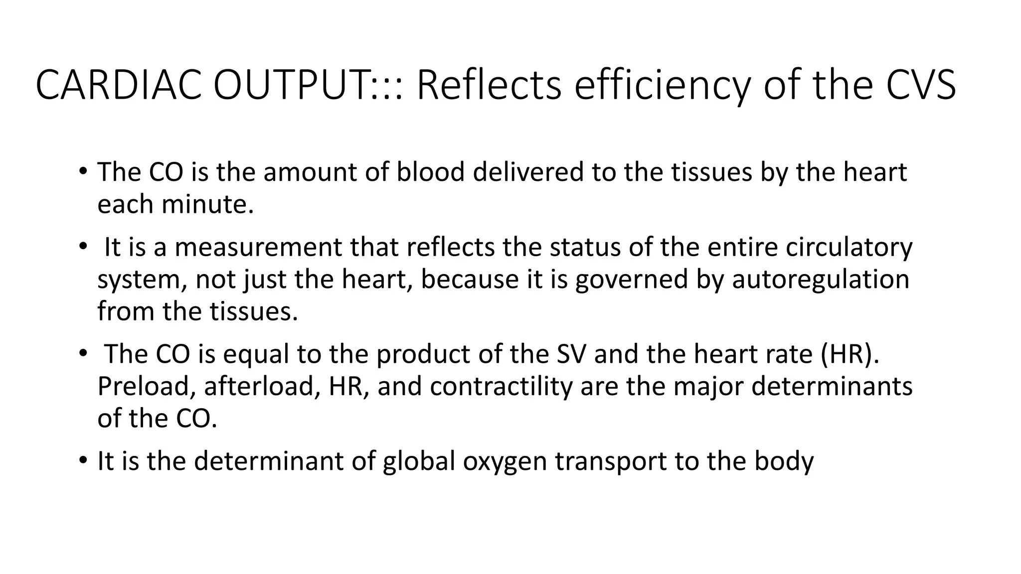
CARDIAC OUTPUT::: Reflects efficiency of the CVS
• The CO is the amount of blood delivered to the tissues by the heart
each minute.
• It is a measurement that reflects the status of the entire circulatory
system, not just the heart, because it is governed by autoregulation
from the tissues.
• The CO is equal to the product of the SV and the heart rate (HR).
Preload, afterload, HR, and contractility are the major determinants
of the CO.
• It is the determinant of global oxygen transport to the body
WHY and WHEN should we measure?
• In critically ill
• High risk surgical patients (in whom large fluid shifts are
expected) with bleeding and hemodynamic instability
Low Cardiac Output Leads To Significant Morbidity And
Mortality
• Allows us to assess the blood flow to the tissues, and provides
information on how to best support a failing circulation ins a
goal directed therapy
• Clinical Assessment of cardiac output is unreliable/ inaccurate
An Ideal Cardiac Output Monitor
• Safe, accurate and repetitive - Reliable during various physiological
states
• Quick and easy to use both in terms of set-up and interpretation of
information
• Operator independent
Methods of CO Monitoring
INVASIVE MINIMALLY INVASIVE NON INVASIVE
1. PA Catheter
• Dye dilution
technique
• Thermodilution
technique
1. Lithium dilution CO
(LiDCO)
2. Pulse contour analysis
(PiCCO)
3. FloTrac
4. Esophgeal Doppler (ED)
5. Transesophgeal echo
(TEE);
1. Partial gas rebreathing
(NICO)
2. Thoracic bioimpedance/
bioreactance
3. endotracheal cardiac output
monitor (ECOM)
4. TTE
5. Photoelectric
plethysmography
FICK s METHOD: gold standard
PRINCIPLE - the total
uptake (or) release of a
substance by an organ is
the product of the blood
flow through the organ
and the arteriovenous
concentration difference
of the substance .
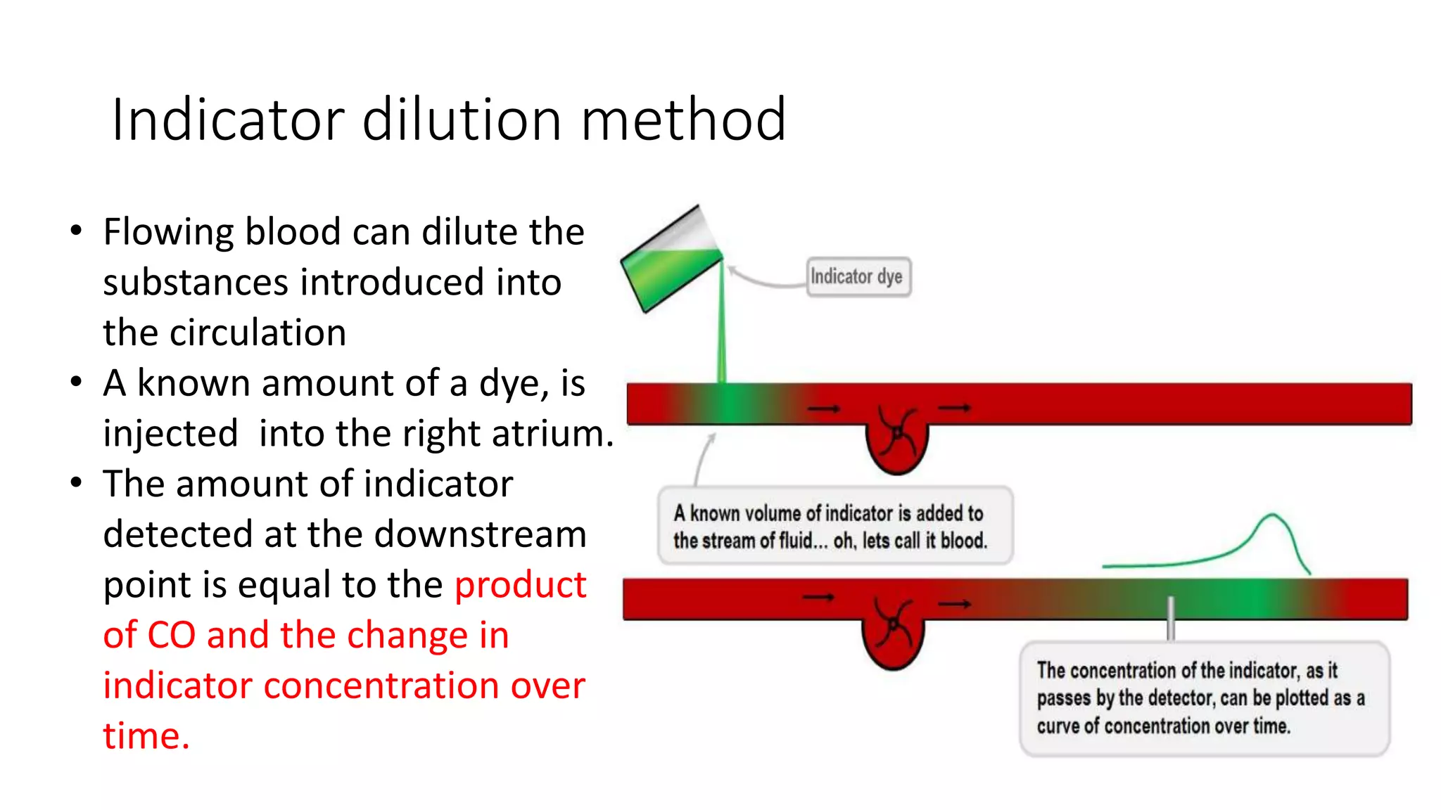
Indicator dilution method
• Flowing blood can dilute the
substances introduced into
the circulation
• A known amount of a dye, is
injected into the right atrium.
• The amount of indicator
detected at the downstream
point is equal to the product
of CO and the change in
indicator concentration over
time.
Indicator dilution method
• The concentration of the dye is recorded as the dye passes through
one of the peripheral arteries, giving a curve.
Stewart–Hamilton equation
• Cold Saline
• ICG
• Lithium Ions
Thermodilution Method
• the indicator- cold
saline/dextrose.
• Affected by the phases of
respiration.
• The thermistor records
the change in blood
temperature with time
and uses it to records and
displays a
temperature-time curve/
Thermo dilution curve
• Variations in the
speed of cold water
injection can result in
altered measurement.
AUC area under
thermodilution curve
m0 = amount (or mass)
of injected cold saline
Modified Stewart-Hamilton Conservation Of Heat
Equation
• The degree of change in temperature is inversely proportional to cardiac
output.
• The higher the cardiac output, the faster he blood flow and the shorter and
steeper the thermodilution curve.
• In low cardiac output, the curve is slurred and lazy.
• Even more so in tricuspid regurgitation.
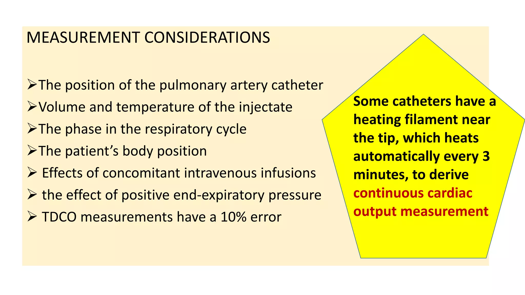
MEASUREMENT CONSIDERATIONS
The position of the pulmonary artery catheter
Volume and temperature of the injectate
The phase in the respiratory cycle
The patient’s body position
 Effects of concomitant intravenous infusions
 the effect of positive end-expiratory pressure
 TDCO measurements have a 10% error
Some catheters have a
heating filament near
the tip, which heats
automatically every 3
minutes, to derive
continuous cardiac
output measurement
Method
Principle Advantages Disadvantages
Invasive
Pulmonary
artery
catheter
(PAC)
Stewart-Hamilton equation
: the rate of blood flow is
inversely proportional to the
change in temperature over
time.
•Very accurate
•Clinical benefit in
monitoring multi-
factorial shock
states and cardiac
cases
Risk of:
•Dysrhythmias
•Cardiac perforation
•Tamponade
•Pneumothorax
•Valve damage
•Infection
•Emboli
PACMAN trial showed 10% incidence of complications, ESCAPE trial 5%.
ESCAPE trial demonstrated functional improvement with PAC guided therapy used in patients
with congestive heart failure.
The evidence
• No effect on mortality, LOS, or cost of care in either general ICU or high risk surgical patients
• No effect on surgical outcomes when used preoperatively to optimise haemodynamics.
Minimally Invasive Techniques
• Lithium dilution CO (LiDCO) - lithium dilution
• Pulse contour analysis CO - transpulmonary thermodilution
• FloTrac®/Vigileo® system and MostCare® require no external
calibration.
• Esophgeal Doppler (ED),
• transesophgeal echocardiography (TEE)
Mechanism
All of these methods are based on the Morphology Of The Arterial
Pressure Curve.
It is therefore important to obtain a precise curve morphology.
Pulse Contour Analysis - Principle
t [s]
P [mm Hg]
Area under
pressure curve
Shape of
pressure curve
PCCO = cal • HR •


Systole
P(t)
SVR
+ C(p) •
dP
dt
( ) dt
Aortic
compliance
Heart
rate
Patient-specific calibration factor
(determined by thermodilution)
• Arterial pressure waveform determined by interaction of
stroke volume and SVR.
• based on the principle that area under the systolic part
of the arterial pressure waveform is proportional to the
SV.
Limitations
Values can be affected by:
• Buffering of the arterial curve
• insufficient zeroing
• Furthermore, the analysis of pulse pressure is of limited accuracy
during periods of hemodynamic instability, as for example in the rapid
changes in vascular resistance found in septic patients and in cases of
liver dysfunction
• Arrhythmias
• IABP
• Severe aortic regurgitation
Lithium Dilution Cardiac Output system (LiDCO plus)
• Uses a peripheral lithium indicator sensor
• acceptable accuracy----- frequent calibrations
• less invasive that the PiCCO system, since it requires no central venous access
• CO is calculated based on Li dose and area according to the concentration time
circulation.
• A bolus of lithium chloride is injected into venous line and arterial concentration is
measured by withdrawing blood across disposable lithium sensitive sensor
containing an ionophor selectively permeable to Li.
• Contraindications
• chronic lithium use,
• recent NDNMB,
• early pregnancy
Can calculate a number of derived parameters:
• systolic pressure variation,
• pulse pressure variation,
• cardiac index,
• stroke volume,
• stroke volume index,
• stroke volume variation,
• systemic vascular resistance and
• systemic vascular resistance index.
PICCO system
• It requires both central venous (femoral or internal jugular) and arterial
cannulation (femoral/radial).
• it combines pulse contour analysis with the transpulmonary
thermodilution CO measurement
• It requires manual calibration every 8 h and hourly during
hemodynamic instability.
• can calculate pulse pressure variation (PPV) stroke volume variation (SVV)
• very sensitive preload parameters
• indicate the point of the patient on the Frank–Starling curve
• SVV value of 9.5% or more, will increase SV by at least 5% in response to a
100-ml volume load, with a sensitivity of 79% and specificity of 93%.'
Volemic Status Of
Ventilated Patients.
Extravascular Lung Water
(EVLW)- Lung Edema And
Vascular Permeability
Optimize Use Of
Vasoactive Drugs, Diuretics
Or Dialysis
ERNEST
STARLING
OTTO FRANK
FloTrac / VIGILEO
system (2005)
• pulse contour analysis device 2005
• no external calibration, operator independent
and easy to use.
• Good arterial waveform quality is a prerequisite.
• Various studies have validated the efficacy of
FloTrac with PAC and find good correlation
• Accuracy is affected in patients with significant arrhythmias, IABP or morbid
obesity.
• In patients with low SVR, undergoing liver transplantation or septicemia it is
not found as accurate as PAC
• When the cardiac index (CI) was <2.2, the data demonstrated that the FV
was outperformed by PAC in both empty (30.8%, n = 13 vs. 57.1%, n = 14,
respectively) and PSF states (66%, n = 3 vs. 50%, n = 4, respectively).
Pressure recording analytic method(PRAM)
• a new, less invasive technique allowing beat-by-beat stroke volume
monitoring from the pressure signals recorded in femoral or radial
arteries
TEE (Trans Esophageal Echocardiography)
• Doppler technique is used to measure CO by Simpson’s rule measuring
SV multiplied by HR.
• Flow is measured by area under the Doppler velocity waveform that
gives VTI
• Measurement can be done at the level of pulmonary artery, mitral or
aortic valve.
• TEE views used for measurement are
• midesophageal aortic long axis view and
• deep transgastric long axis view with pulsed and continuous wave Doppler respectively
Trans Esophageal Echocardiography (TEE)
• an important tool for the
assessment of cardiac structures,
filling status and cardiac
contractility
• by measuring both the velocity
and the cross-sectional area of
blood flow in the LVOT or aorta or
PA.
• Flow = CSA X Velocity
• SV= flow X ET ( Systolic Ejection
time)
• CO=SV X HR
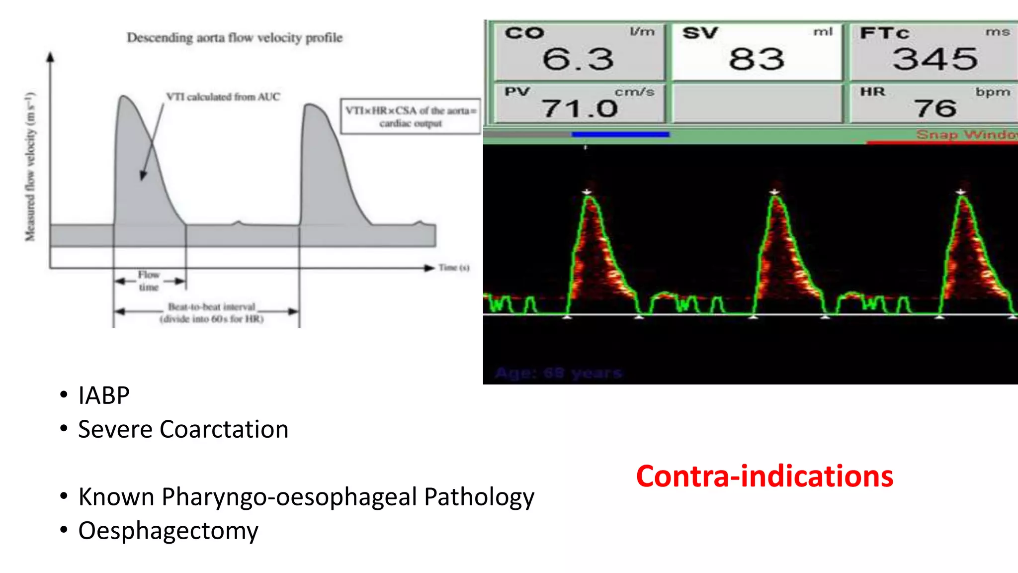
Esophageal Doppler
FLOW= CSA X Vti
• Major limiting factor is that it measures flow only in
descending thoracic aorta which is 70% of total flow.
• Values calculated from these are stroke volume (SV)
flow-time corrected (FTc) and cardiac output (CO).
• IT provides
• Heart Rate (HR), Stroke Distance (SD), Maximum Acceleration
(MA),
• Flow-time (FT) Peak Velocity (PV)
Using manual input of age, weight and height; body surface area (BSA) and body mass
index (BMI), cardiac index (CI) and stroke volume index (SVI or SI) can be calculated.
• IABP
• Severe Coarctation
• Known Pharyngo-oesophageal Pathology
• Oesphagectomy
Contra-indications
NON INVASIVE METHODS
• Partial Gas Rebreathing
• Thoracic Bioimpedance
• Bioreactance
• The Modelflow- FINGER PROBE Based CO Monitor
• TTE/ USG
NON INVASIVE METHODS :Partial gas rebreathing
 known as the NICO system
uses indirect Fick’s principle to calculate CO.
At steady state, the amount of CO2 entering the lungs via the pulmonary
artery is proportional to the CO and equals the amount exiting the lungs
via expiration and pulmonary veins.
CO2 Produced per min
CO= -------------------------------------
PvCO2- PaCO2
It is used in intubated patients under mechanical ventilation.
THORACIC BIOIMPEDANCE
Based on the hypothesis by considering thorax as a cylinder perfused
with fluid with specific resistivity.
 Electrodes six in number are placed (two on either side of neck and
four in lower thorax) on the patient and the resistance to current
flowing from the outermost to innermost electrodes is measured.
A low-grade electrical current, from 2 - 4 mA is emitted, and
received by the adjacent electrodes
 Impedance to the current flow produces a waveform
 Changes in CO will change the amount of aortic blood and will be
reflected in a change TEB
BioReactance
Method used by the NICOM®
Analyzes the changes in amplitude and frequency of the electrical
impulses as they course through the chest.
 Advantages
• significant reduction of effect of electrical interferences, patient
movements or positioning, or displacement of the electrodes, which
can give rise to data error.
The Modelflow- FINGER PROBE based CO monitor
• Modelflow-Nexfin® system
• analyzes pulse pressure noninvasively using photoelectric plethysmography
in combination with an inflatable finger cuff.
PRINCIPLE
Cardiac output is calculated through continuous monitorization of arterial
pressure and analysis of pulse wave morphology, based on the study of the
area of the systolic pressure wave and on the Windkessel triple elements
model individualized for each patient (Modelflow method).
• The measurements obtained include continuous CO, SV, SVR and left
ventricle contractility index.
TTE
• can be used to estimate cardiac output by
direct visualisation of the contracting heart in
real time.
• safe and most reliable cardiac output
monitors in the critically ill.
• Using transthoracic echocardiography four
views are obtained (parasternal long axis,
parasternal short axis, apical, and subcostal
• Helps in assessment of ventricular function
and size of cardiac chambers.
Summary
• The choice of one device or other should be determined by the
experience of operator, facility of use and interpretation of the
results, the precision of the system, and its cost-effectiveness.
• Monitoring of the critical patient must be global, with
multiparametric monitoring combining the hemodynamic parameters
and the metabolic data regards to oxygen transport and consumption,
with the purpose of optimizing tissue perfusion and improving
survival of the critically ill patient.
Thank you
• PAC-MAN trial failed to show any benefit or harm with the use
of PAC.
• ESCAPE TRIAL IN heart failure patients--- Addition of the PAC to
careful clinical assessment increased anticipated adverse events, but
did not affect overall mortality and hospitalization.

More Related Content

What's hot

Patients with pacemaker anaesthetic implications
Patients with pacemaker anaesthetic implicationsPatients with pacemaker anaesthetic implications
Patients with pacemaker anaesthetic implications
Gowri Shankar
 
Flotrac
FlotracFlotrac
Flotrac
Vijay Anand
 
Advanced haemodynamics
Advanced haemodynamicsAdvanced haemodynamics
Advanced haemodynamics
Lambert Winston
 
Basic haemodynamic monitoring
Basic haemodynamic monitoringBasic haemodynamic monitoring
Basic haemodynamic monitoring
Deepankar Mishra
 
Pulmonary artery catheter
Pulmonary artery catheterPulmonary artery catheter
Pulmonary artery catheter
Arun Aru
 
Capnography in icu
Capnography  in icuCapnography  in icu
Capnography in icu
Eman Mahmoud
 
Cardiopulmonary bypass
Cardiopulmonary bypassCardiopulmonary bypass
Cardiopulmonary bypass
Abeer Nakera
 
Which cardiac output monitoring?
Which cardiac output monitoring?Which cardiac output monitoring?
Which cardiac output monitoring?
International Fluid Academy
 
Advanced hemodynamic monitoring
Advanced hemodynamic monitoringAdvanced hemodynamic monitoring
Advanced hemodynamic monitoring
Ghaleb Almekhlafi
 
Ecmo
EcmoEcmo
MEASUREMENT OF CARDIAC OUTPUT
MEASUREMENT OF CARDIAC OUTPUTMEASUREMENT OF CARDIAC OUTPUT
MEASUREMENT OF CARDIAC OUTPUT
AmudhaLakshmi1
 
Flo trac
Flo tracFlo trac
Recruitment maneuvers in ards
Recruitment maneuvers in ardsRecruitment maneuvers in ards
Recruitment maneuvers in ards
Anusha Jahagirdar
 
Ventilator setting
Ventilator settingVentilator setting
Ventilator setting
HINDUJACON
 
Deep hypothermic circulatory arrest in pediatric cardiac sur
Deep hypothermic circulatory arrest in pediatric cardiac surDeep hypothermic circulatory arrest in pediatric cardiac sur
Deep hypothermic circulatory arrest in pediatric cardiac sur
Manu Jacob
 
Haemodynamic monitoring-Minati
Haemodynamic monitoring-MinatiHaemodynamic monitoring-Minati
Haemodynamic monitoring-Minati
Minati Choudhury
 
Module 3.2 Hemodynamic Monitoring
Module 3.2 Hemodynamic Monitoring Module 3.2 Hemodynamic Monitoring
Module 3.2 Hemodynamic Monitoring
Hannah Nelson
 
Static and dynamic indices of hemodynamic monitoring
Static and dynamic indices of hemodynamic monitoringStatic and dynamic indices of hemodynamic monitoring
Static and dynamic indices of hemodynamic monitoring
Bhargav Mundlapudi
 
Picco
PiccoPicco
Picco
gnivri1666
 
One lung ventilation
One lung ventilationOne lung ventilation
One lung ventilation
DR SHADAB KAMAL
 

What's hot (20)

Patients with pacemaker anaesthetic implications
Patients with pacemaker anaesthetic implicationsPatients with pacemaker anaesthetic implications
Patients with pacemaker anaesthetic implications
 
Flotrac
FlotracFlotrac
Flotrac
 
Advanced haemodynamics
Advanced haemodynamicsAdvanced haemodynamics
Advanced haemodynamics
 
Basic haemodynamic monitoring
Basic haemodynamic monitoringBasic haemodynamic monitoring
Basic haemodynamic monitoring
 
Pulmonary artery catheter
Pulmonary artery catheterPulmonary artery catheter
Pulmonary artery catheter
 
Capnography in icu
Capnography  in icuCapnography  in icu
Capnography in icu
 
Cardiopulmonary bypass
Cardiopulmonary bypassCardiopulmonary bypass
Cardiopulmonary bypass
 
Which cardiac output monitoring?
Which cardiac output monitoring?Which cardiac output monitoring?
Which cardiac output monitoring?
 
Advanced hemodynamic monitoring
Advanced hemodynamic monitoringAdvanced hemodynamic monitoring
Advanced hemodynamic monitoring
 
Ecmo
EcmoEcmo
Ecmo
 
MEASUREMENT OF CARDIAC OUTPUT
MEASUREMENT OF CARDIAC OUTPUTMEASUREMENT OF CARDIAC OUTPUT
MEASUREMENT OF CARDIAC OUTPUT
 
Flo trac
Flo tracFlo trac
Flo trac
 
Recruitment maneuvers in ards
Recruitment maneuvers in ardsRecruitment maneuvers in ards
Recruitment maneuvers in ards
 
Ventilator setting
Ventilator settingVentilator setting
Ventilator setting
 
Deep hypothermic circulatory arrest in pediatric cardiac sur
Deep hypothermic circulatory arrest in pediatric cardiac surDeep hypothermic circulatory arrest in pediatric cardiac sur
Deep hypothermic circulatory arrest in pediatric cardiac sur
 
Haemodynamic monitoring-Minati
Haemodynamic monitoring-MinatiHaemodynamic monitoring-Minati
Haemodynamic monitoring-Minati
 
Module 3.2 Hemodynamic Monitoring
Module 3.2 Hemodynamic Monitoring Module 3.2 Hemodynamic Monitoring
Module 3.2 Hemodynamic Monitoring
 
Static and dynamic indices of hemodynamic monitoring
Static and dynamic indices of hemodynamic monitoringStatic and dynamic indices of hemodynamic monitoring
Static and dynamic indices of hemodynamic monitoring
 
Picco
PiccoPicco
Picco
 
One lung ventilation
One lung ventilationOne lung ventilation
One lung ventilation
 

Similar to cardiac output pptx

Minimally invasive Cardiac output Monitoring
Minimally invasive Cardiac output MonitoringMinimally invasive Cardiac output Monitoring
Minimally invasive Cardiac output Monitoring
JeanPaulDushime1
 
cardiacoutputmonitoring-190708221224.pdf
cardiacoutputmonitoring-190708221224.pdfcardiacoutputmonitoring-190708221224.pdf
cardiacoutputmonitoring-190708221224.pdf
aishabajwa8081
 
HEMODYNMAMICS MONITORING IN CRITICAL CARE NURSING
HEMODYNMAMICS MONITORING IN CRITICAL CARE NURSINGHEMODYNMAMICS MONITORING IN CRITICAL CARE NURSING
HEMODYNMAMICS MONITORING IN CRITICAL CARE NURSING
GraceTaiwo3
 
satya sai 2018 cardiac output.pdf
satya sai 2018 cardiac output.pdfsatya sai 2018 cardiac output.pdf
satya sai 2018 cardiac output.pdf
CutiePie71
 
BASIC ANAESTHETIC MONITORING
BASIC ANAESTHETIC MONITORING BASIC ANAESTHETIC MONITORING
BASIC ANAESTHETIC MONITORING
HARITHANALLABOTHULA1
 
Hemodynamic Monitoring .pptx
Hemodynamic Monitoring  .pptxHemodynamic Monitoring  .pptx
Hemodynamic Monitoring .pptx
anesthesia2023
 
REVIEW OF HEMODYNAMIC MONITORING
REVIEW OF HEMODYNAMIC MONITORINGREVIEW OF HEMODYNAMIC MONITORING
REVIEW OF HEMODYNAMIC MONITORING
Ghaleb Almekhlafi
 
hemodynamic monitoring
hemodynamic monitoringhemodynamic monitoring
hemodynamic monitoring
gagan brar
 
Hemodynamic monitoring
Hemodynamic  monitoringHemodynamic  monitoring
Hemodynamic monitoring
TarunChandra13
 
Echocardiography
EchocardiographyEchocardiography
Echocardiography
Dr.Debmalya Saha
 
cardiac output measurment and monitoring ppt-1.pptx
cardiac output measurment and monitoring ppt-1.pptxcardiac output measurment and monitoring ppt-1.pptx
cardiac output measurment and monitoring ppt-1.pptx
shekinah41
 
MONITORING IN ANAESTHESIA.pptx
MONITORING IN ANAESTHESIA.pptxMONITORING IN ANAESTHESIA.pptx
MONITORING IN ANAESTHESIA.pptx
Amin Badamosi
 
Monitoring of critically ill
Monitoring of critically illMonitoring of critically ill
Monitoring of critically ill
StudyTricksByIshu
 
physiological monitoring of a surgical patient.pptx
physiological monitoring of a surgical patient.pptxphysiological monitoring of a surgical patient.pptx
physiological monitoring of a surgical patient.pptx
kiogakimathi
 
Hemodynamic parameters & fluid therapy Asim
Hemodynamic parameters &  fluid therapy AsimHemodynamic parameters &  fluid therapy Asim
Hemodynamic parameters & fluid therapy Asim
Muhammad Asim Rana
 
3. CVS monitoring.pptx
3. CVS monitoring.pptx3. CVS monitoring.pptx
3. CVS monitoring.pptx
FLOWERSOFPAKISTAN
 
Monitoring_New (1).pptx
Monitoring_New (1).pptxMonitoring_New (1).pptx
Monitoring_New (1).pptx
NehaMasarkar1
 
Hemodynamic monitoring
Hemodynamic monitoringHemodynamic monitoring
Hemodynamic monitoring
Pratik Tantia
 
The menagerie of monitoring tools by Professor Jean-Louis Teboul
The menagerie of monitoring tools by Professor Jean-Louis TeboulThe menagerie of monitoring tools by Professor Jean-Louis Teboul
The menagerie of monitoring tools by Professor Jean-Louis Teboul
CICM 2019 Annual Scientific Meeting
 
Cardiac catheterisation
Cardiac catheterisationCardiac catheterisation
Cardiac catheterisation
Kunwar Saurabh
 

Similar to cardiac output pptx (20)

Minimally invasive Cardiac output Monitoring
Minimally invasive Cardiac output MonitoringMinimally invasive Cardiac output Monitoring
Minimally invasive Cardiac output Monitoring
 
cardiacoutputmonitoring-190708221224.pdf
cardiacoutputmonitoring-190708221224.pdfcardiacoutputmonitoring-190708221224.pdf
cardiacoutputmonitoring-190708221224.pdf
 
HEMODYNMAMICS MONITORING IN CRITICAL CARE NURSING
HEMODYNMAMICS MONITORING IN CRITICAL CARE NURSINGHEMODYNMAMICS MONITORING IN CRITICAL CARE NURSING
HEMODYNMAMICS MONITORING IN CRITICAL CARE NURSING
 
satya sai 2018 cardiac output.pdf
satya sai 2018 cardiac output.pdfsatya sai 2018 cardiac output.pdf
satya sai 2018 cardiac output.pdf
 
BASIC ANAESTHETIC MONITORING
BASIC ANAESTHETIC MONITORING BASIC ANAESTHETIC MONITORING
BASIC ANAESTHETIC MONITORING
 
Hemodynamic Monitoring .pptx
Hemodynamic Monitoring  .pptxHemodynamic Monitoring  .pptx
Hemodynamic Monitoring .pptx
 
REVIEW OF HEMODYNAMIC MONITORING
REVIEW OF HEMODYNAMIC MONITORINGREVIEW OF HEMODYNAMIC MONITORING
REVIEW OF HEMODYNAMIC MONITORING
 
hemodynamic monitoring
hemodynamic monitoringhemodynamic monitoring
hemodynamic monitoring
 
Hemodynamic monitoring
Hemodynamic  monitoringHemodynamic  monitoring
Hemodynamic monitoring
 
Echocardiography
EchocardiographyEchocardiography
Echocardiography
 
cardiac output measurment and monitoring ppt-1.pptx
cardiac output measurment and monitoring ppt-1.pptxcardiac output measurment and monitoring ppt-1.pptx
cardiac output measurment and monitoring ppt-1.pptx
 
MONITORING IN ANAESTHESIA.pptx
MONITORING IN ANAESTHESIA.pptxMONITORING IN ANAESTHESIA.pptx
MONITORING IN ANAESTHESIA.pptx
 
Monitoring of critically ill
Monitoring of critically illMonitoring of critically ill
Monitoring of critically ill
 
physiological monitoring of a surgical patient.pptx
physiological monitoring of a surgical patient.pptxphysiological monitoring of a surgical patient.pptx
physiological monitoring of a surgical patient.pptx
 
Hemodynamic parameters & fluid therapy Asim
Hemodynamic parameters &  fluid therapy AsimHemodynamic parameters &  fluid therapy Asim
Hemodynamic parameters & fluid therapy Asim
 
3. CVS monitoring.pptx
3. CVS monitoring.pptx3. CVS monitoring.pptx
3. CVS monitoring.pptx
 
Monitoring_New (1).pptx
Monitoring_New (1).pptxMonitoring_New (1).pptx
Monitoring_New (1).pptx
 
Hemodynamic monitoring
Hemodynamic monitoringHemodynamic monitoring
Hemodynamic monitoring
 
The menagerie of monitoring tools by Professor Jean-Louis Teboul
The menagerie of monitoring tools by Professor Jean-Louis TeboulThe menagerie of monitoring tools by Professor Jean-Louis Teboul
The menagerie of monitoring tools by Professor Jean-Louis Teboul
 
Cardiac catheterisation
Cardiac catheterisationCardiac catheterisation
Cardiac catheterisation
 

More from ananya nanda

pulmonaryPFTfunc Tests.pdf
pulmonaryPFTfunc Tests.pdfpulmonaryPFTfunc Tests.pdf
pulmonaryPFTfunc Tests.pdf
ananya nanda
 
Anaesthetic Implications Of Lung Resection (3).ppt
Anaesthetic Implications Of Lung Resection (3).pptAnaesthetic Implications Of Lung Resection (3).ppt
Anaesthetic Implications Of Lung Resection (3).ppt
ananya nanda
 
ALTERNATIVES TO double lumen tubes IN ONE LUNG VENTILATION.pptx
ALTERNATIVES TO double lumen tubes IN ONE LUNG VENTILATION.pptxALTERNATIVES TO double lumen tubes IN ONE LUNG VENTILATION.pptx
ALTERNATIVES TO double lumen tubes IN ONE LUNG VENTILATION.pptx
ananya nanda
 
PHYSIOLOGY OF One lung ventilation.pptx
PHYSIOLOGY OF One lung ventilation.pptxPHYSIOLOGY OF One lung ventilation.pptx
PHYSIOLOGY OF One lung ventilation.pptx
ananya nanda
 
Spinal anaesthesia basics
Spinal anaesthesia  basicsSpinal anaesthesia  basics
Spinal anaesthesia basics
ananya nanda
 
acute surgical pain management
acute surgical pain managementacute surgical pain management
acute surgical pain management
ananya nanda
 
Anatomy of brachial plexus
Anatomy of brachial plexusAnatomy of brachial plexus
Anatomy of brachial plexus
ananya nanda
 
Principles of icu ventilators
Principles of icu ventilatorsPrinciples of icu ventilators
Principles of icu ventilators
ananya nanda
 

More from ananya nanda (8)

pulmonaryPFTfunc Tests.pdf
pulmonaryPFTfunc Tests.pdfpulmonaryPFTfunc Tests.pdf
pulmonaryPFTfunc Tests.pdf
 
Anaesthetic Implications Of Lung Resection (3).ppt
Anaesthetic Implications Of Lung Resection (3).pptAnaesthetic Implications Of Lung Resection (3).ppt
Anaesthetic Implications Of Lung Resection (3).ppt
 
ALTERNATIVES TO double lumen tubes IN ONE LUNG VENTILATION.pptx
ALTERNATIVES TO double lumen tubes IN ONE LUNG VENTILATION.pptxALTERNATIVES TO double lumen tubes IN ONE LUNG VENTILATION.pptx
ALTERNATIVES TO double lumen tubes IN ONE LUNG VENTILATION.pptx
 
PHYSIOLOGY OF One lung ventilation.pptx
PHYSIOLOGY OF One lung ventilation.pptxPHYSIOLOGY OF One lung ventilation.pptx
PHYSIOLOGY OF One lung ventilation.pptx
 
Spinal anaesthesia basics
Spinal anaesthesia  basicsSpinal anaesthesia  basics
Spinal anaesthesia basics
 
acute surgical pain management
acute surgical pain managementacute surgical pain management
acute surgical pain management
 
Anatomy of brachial plexus
Anatomy of brachial plexusAnatomy of brachial plexus
Anatomy of brachial plexus
 
Principles of icu ventilators
Principles of icu ventilatorsPrinciples of icu ventilators
Principles of icu ventilators
 

Recently uploaded

CHEMOTHERAPY_RDP_CHAPTER 4_ANTI VIRAL DRUGS.pdf
CHEMOTHERAPY_RDP_CHAPTER 4_ANTI VIRAL DRUGS.pdfCHEMOTHERAPY_RDP_CHAPTER 4_ANTI VIRAL DRUGS.pdf
CHEMOTHERAPY_RDP_CHAPTER 4_ANTI VIRAL DRUGS.pdf
rishi2789
 
How STIs Influence the Development of Pelvic Inflammatory Disease.pptx
How STIs Influence the Development of Pelvic Inflammatory Disease.pptxHow STIs Influence the Development of Pelvic Inflammatory Disease.pptx
How STIs Influence the Development of Pelvic Inflammatory Disease.pptx
FFragrant
 
Chapter 11 Nutrition and Chronic Diseases.pptx
Chapter 11 Nutrition and Chronic Diseases.pptxChapter 11 Nutrition and Chronic Diseases.pptx
Chapter 11 Nutrition and Chronic Diseases.pptx
Earlene McNair
 
Journal Article Review on Rasamanikya
Journal Article Review on RasamanikyaJournal Article Review on Rasamanikya
Journal Article Review on Rasamanikya
Dr. Jyothirmai Paindla
 
Top 10 Best Ayurvedic Kidney Stone Syrups in India
Top 10 Best Ayurvedic Kidney Stone Syrups in IndiaTop 10 Best Ayurvedic Kidney Stone Syrups in India
Top 10 Best Ayurvedic Kidney Stone Syrups in India
Swastik Ayurveda
 
Integrating Ayurveda into Parkinson’s Management: A Holistic Approach
Integrating Ayurveda into Parkinson’s Management: A Holistic ApproachIntegrating Ayurveda into Parkinson’s Management: A Holistic Approach
Integrating Ayurveda into Parkinson’s Management: A Holistic Approach
Ayurveda ForAll
 
The Electrocardiogram - Physiologic Principles
The Electrocardiogram - Physiologic PrinciplesThe Electrocardiogram - Physiologic Principles
The Electrocardiogram - Physiologic Principles
MedicoseAcademics
 
share - Lions, tigers, AI and health misinformation, oh my!.pptx
share - Lions, tigers, AI and health misinformation, oh my!.pptxshare - Lions, tigers, AI and health misinformation, oh my!.pptx
share - Lions, tigers, AI and health misinformation, oh my!.pptx
Tina Purnat
 
CHEMOTHERAPY_RDP_CHAPTER 2 _LEPROSY.pdf1
CHEMOTHERAPY_RDP_CHAPTER 2 _LEPROSY.pdf1CHEMOTHERAPY_RDP_CHAPTER 2 _LEPROSY.pdf1
CHEMOTHERAPY_RDP_CHAPTER 2 _LEPROSY.pdf1
rishi2789
 
Netter's Atlas of Human Anatomy 7.ed.pdf
Netter's Atlas of Human Anatomy 7.ed.pdfNetter's Atlas of Human Anatomy 7.ed.pdf
Netter's Atlas of Human Anatomy 7.ed.pdf
BrissaOrtiz3
 
Top Effective Soaps for Fungal Skin Infections in India
Top Effective Soaps for Fungal Skin Infections in IndiaTop Effective Soaps for Fungal Skin Infections in India
Top Effective Soaps for Fungal Skin Infections in India
SwisschemDerma
 
8 Surprising Reasons To Meditate 40 Minutes A Day That Can Change Your Life.pptx
8 Surprising Reasons To Meditate 40 Minutes A Day That Can Change Your Life.pptx8 Surprising Reasons To Meditate 40 Minutes A Day That Can Change Your Life.pptx
8 Surprising Reasons To Meditate 40 Minutes A Day That Can Change Your Life.pptx
Holistified Wellness
 
Local Advanced Lung Cancer: Artificial Intelligence, Synergetics, Complex Sys...
Local Advanced Lung Cancer: Artificial Intelligence, Synergetics, Complex Sys...Local Advanced Lung Cancer: Artificial Intelligence, Synergetics, Complex Sys...
Local Advanced Lung Cancer: Artificial Intelligence, Synergetics, Complex Sys...
Oleg Kshivets
 
Promoting Wellbeing - Applied Social Psychology - Psychology SuperNotes
Promoting Wellbeing - Applied Social Psychology - Psychology SuperNotesPromoting Wellbeing - Applied Social Psychology - Psychology SuperNotes
Promoting Wellbeing - Applied Social Psychology - Psychology SuperNotes
PsychoTech Services
 
ABDOMINAL TRAUMA in pediatrics part one.
ABDOMINAL TRAUMA in pediatrics part one.ABDOMINAL TRAUMA in pediatrics part one.
ABDOMINAL TRAUMA in pediatrics part one.
drhasanrajab
 
Ear and its clinical correlations By Dr. Rabia Inam Gandapore.pptx
Ear and its clinical correlations By Dr. Rabia Inam Gandapore.pptxEar and its clinical correlations By Dr. Rabia Inam Gandapore.pptx
Ear and its clinical correlations By Dr. Rabia Inam Gandapore.pptx
Dr. Rabia Inam Gandapore
 
Light House Retreats: Plant Medicine Retreat Europe
Light House Retreats: Plant Medicine Retreat EuropeLight House Retreats: Plant Medicine Retreat Europe
Light House Retreats: Plant Medicine Retreat Europe
Lighthouse Retreat
 
A Classical Text Review on Basavarajeeyam
A Classical Text Review on BasavarajeeyamA Classical Text Review on Basavarajeeyam
A Classical Text Review on Basavarajeeyam
Dr. Jyothirmai Paindla
 
REGULATION FOR COMBINATION PRODUCTS AND MEDICAL DEVICES.pptx
REGULATION FOR COMBINATION PRODUCTS AND MEDICAL DEVICES.pptxREGULATION FOR COMBINATION PRODUCTS AND MEDICAL DEVICES.pptx
REGULATION FOR COMBINATION PRODUCTS AND MEDICAL DEVICES.pptx
LaniyaNasrink
 
TEST BANK For Community Health Nursing A Canadian Perspective, 5th Edition by...
TEST BANK For Community Health Nursing A Canadian Perspective, 5th Edition by...TEST BANK For Community Health Nursing A Canadian Perspective, 5th Edition by...
TEST BANK For Community Health Nursing A Canadian Perspective, 5th Edition by...
Donc Test
 

Recently uploaded (20)

CHEMOTHERAPY_RDP_CHAPTER 4_ANTI VIRAL DRUGS.pdf
CHEMOTHERAPY_RDP_CHAPTER 4_ANTI VIRAL DRUGS.pdfCHEMOTHERAPY_RDP_CHAPTER 4_ANTI VIRAL DRUGS.pdf
CHEMOTHERAPY_RDP_CHAPTER 4_ANTI VIRAL DRUGS.pdf
 
How STIs Influence the Development of Pelvic Inflammatory Disease.pptx
How STIs Influence the Development of Pelvic Inflammatory Disease.pptxHow STIs Influence the Development of Pelvic Inflammatory Disease.pptx
How STIs Influence the Development of Pelvic Inflammatory Disease.pptx
 
Chapter 11 Nutrition and Chronic Diseases.pptx
Chapter 11 Nutrition and Chronic Diseases.pptxChapter 11 Nutrition and Chronic Diseases.pptx
Chapter 11 Nutrition and Chronic Diseases.pptx
 
Journal Article Review on Rasamanikya
Journal Article Review on RasamanikyaJournal Article Review on Rasamanikya
Journal Article Review on Rasamanikya
 
Top 10 Best Ayurvedic Kidney Stone Syrups in India
Top 10 Best Ayurvedic Kidney Stone Syrups in IndiaTop 10 Best Ayurvedic Kidney Stone Syrups in India
Top 10 Best Ayurvedic Kidney Stone Syrups in India
 
Integrating Ayurveda into Parkinson’s Management: A Holistic Approach
Integrating Ayurveda into Parkinson’s Management: A Holistic ApproachIntegrating Ayurveda into Parkinson’s Management: A Holistic Approach
Integrating Ayurveda into Parkinson’s Management: A Holistic Approach
 
The Electrocardiogram - Physiologic Principles
The Electrocardiogram - Physiologic PrinciplesThe Electrocardiogram - Physiologic Principles
The Electrocardiogram - Physiologic Principles
 
share - Lions, tigers, AI and health misinformation, oh my!.pptx
share - Lions, tigers, AI and health misinformation, oh my!.pptxshare - Lions, tigers, AI and health misinformation, oh my!.pptx
share - Lions, tigers, AI and health misinformation, oh my!.pptx
 
CHEMOTHERAPY_RDP_CHAPTER 2 _LEPROSY.pdf1
CHEMOTHERAPY_RDP_CHAPTER 2 _LEPROSY.pdf1CHEMOTHERAPY_RDP_CHAPTER 2 _LEPROSY.pdf1
CHEMOTHERAPY_RDP_CHAPTER 2 _LEPROSY.pdf1
 
Netter's Atlas of Human Anatomy 7.ed.pdf
Netter's Atlas of Human Anatomy 7.ed.pdfNetter's Atlas of Human Anatomy 7.ed.pdf
Netter's Atlas of Human Anatomy 7.ed.pdf
 
Top Effective Soaps for Fungal Skin Infections in India
Top Effective Soaps for Fungal Skin Infections in IndiaTop Effective Soaps for Fungal Skin Infections in India
Top Effective Soaps for Fungal Skin Infections in India
 
8 Surprising Reasons To Meditate 40 Minutes A Day That Can Change Your Life.pptx
8 Surprising Reasons To Meditate 40 Minutes A Day That Can Change Your Life.pptx8 Surprising Reasons To Meditate 40 Minutes A Day That Can Change Your Life.pptx
8 Surprising Reasons To Meditate 40 Minutes A Day That Can Change Your Life.pptx
 
Local Advanced Lung Cancer: Artificial Intelligence, Synergetics, Complex Sys...
Local Advanced Lung Cancer: Artificial Intelligence, Synergetics, Complex Sys...Local Advanced Lung Cancer: Artificial Intelligence, Synergetics, Complex Sys...
Local Advanced Lung Cancer: Artificial Intelligence, Synergetics, Complex Sys...
 
Promoting Wellbeing - Applied Social Psychology - Psychology SuperNotes
Promoting Wellbeing - Applied Social Psychology - Psychology SuperNotesPromoting Wellbeing - Applied Social Psychology - Psychology SuperNotes
Promoting Wellbeing - Applied Social Psychology - Psychology SuperNotes
 
ABDOMINAL TRAUMA in pediatrics part one.
ABDOMINAL TRAUMA in pediatrics part one.ABDOMINAL TRAUMA in pediatrics part one.
ABDOMINAL TRAUMA in pediatrics part one.
 
Ear and its clinical correlations By Dr. Rabia Inam Gandapore.pptx
Ear and its clinical correlations By Dr. Rabia Inam Gandapore.pptxEar and its clinical correlations By Dr. Rabia Inam Gandapore.pptx
Ear and its clinical correlations By Dr. Rabia Inam Gandapore.pptx
 
Light House Retreats: Plant Medicine Retreat Europe
Light House Retreats: Plant Medicine Retreat EuropeLight House Retreats: Plant Medicine Retreat Europe
Light House Retreats: Plant Medicine Retreat Europe
 
A Classical Text Review on Basavarajeeyam
A Classical Text Review on BasavarajeeyamA Classical Text Review on Basavarajeeyam
A Classical Text Review on Basavarajeeyam
 
REGULATION FOR COMBINATION PRODUCTS AND MEDICAL DEVICES.pptx
REGULATION FOR COMBINATION PRODUCTS AND MEDICAL DEVICES.pptxREGULATION FOR COMBINATION PRODUCTS AND MEDICAL DEVICES.pptx
REGULATION FOR COMBINATION PRODUCTS AND MEDICAL DEVICES.pptx
 
TEST BANK For Community Health Nursing A Canadian Perspective, 5th Edition by...
TEST BANK For Community Health Nursing A Canadian Perspective, 5th Edition by...TEST BANK For Community Health Nursing A Canadian Perspective, 5th Edition by...
TEST BANK For Community Health Nursing A Canadian Perspective, 5th Edition by...
 

cardiac output pptx

  • 2. SECTIONS… • Definition • Features of an ideal monitor • Types of monitoring CO • Fick s principle and its application • Thermodilution and dye dilution techniques • Minimally invasive methods – PICCO, LIDCO, FLOTRAC, TEE, OD • Non invasive methods NICO, TTE, MODELFLOW, BIOIMPEDANCE • Summary
  • 3. CARDIAC OUTPUT::: Reflects efficiency of the CVS • The CO is the amount of blood delivered to the tissues by the heart each minute. • It is a measurement that reflects the status of the entire circulatory system, not just the heart, because it is governed by autoregulation from the tissues. • The CO is equal to the product of the SV and the heart rate (HR). Preload, afterload, HR, and contractility are the major determinants of the CO. • It is the determinant of global oxygen transport to the body
  • 4. WHY and WHEN should we measure? • In critically ill • High risk surgical patients (in whom large fluid shifts are expected) with bleeding and hemodynamic instability Low Cardiac Output Leads To Significant Morbidity And Mortality • Allows us to assess the blood flow to the tissues, and provides information on how to best support a failing circulation ins a goal directed therapy • Clinical Assessment of cardiac output is unreliable/ inaccurate
  • 5. An Ideal Cardiac Output Monitor • Safe, accurate and repetitive - Reliable during various physiological states • Quick and easy to use both in terms of set-up and interpretation of information • Operator independent
  • 6. Methods of CO Monitoring INVASIVE MINIMALLY INVASIVE NON INVASIVE 1. PA Catheter • Dye dilution technique • Thermodilution technique 1. Lithium dilution CO (LiDCO) 2. Pulse contour analysis (PiCCO) 3. FloTrac 4. Esophgeal Doppler (ED) 5. Transesophgeal echo (TEE); 1. Partial gas rebreathing (NICO) 2. Thoracic bioimpedance/ bioreactance 3. endotracheal cardiac output monitor (ECOM) 4. TTE 5. Photoelectric plethysmography
  • 7. FICK s METHOD: gold standard PRINCIPLE - the total uptake (or) release of a substance by an organ is the product of the blood flow through the organ and the arteriovenous concentration difference of the substance .
  • 8. Indicator dilution method • Flowing blood can dilute the substances introduced into the circulation • A known amount of a dye, is injected into the right atrium. • The amount of indicator detected at the downstream point is equal to the product of CO and the change in indicator concentration over time.
  • 9. Indicator dilution method • The concentration of the dye is recorded as the dye passes through one of the peripheral arteries, giving a curve. Stewart–Hamilton equation • Cold Saline • ICG • Lithium Ions
  • 10.
  • 11. Thermodilution Method • the indicator- cold saline/dextrose. • Affected by the phases of respiration. • The thermistor records the change in blood temperature with time and uses it to records and displays a temperature-time curve/ Thermo dilution curve
  • 12. • Variations in the speed of cold water injection can result in altered measurement. AUC area under thermodilution curve m0 = amount (or mass) of injected cold saline
  • 14. • The degree of change in temperature is inversely proportional to cardiac output. • The higher the cardiac output, the faster he blood flow and the shorter and steeper the thermodilution curve. • In low cardiac output, the curve is slurred and lazy. • Even more so in tricuspid regurgitation.
  • 15. MEASUREMENT CONSIDERATIONS The position of the pulmonary artery catheter Volume and temperature of the injectate The phase in the respiratory cycle The patient’s body position  Effects of concomitant intravenous infusions  the effect of positive end-expiratory pressure  TDCO measurements have a 10% error Some catheters have a heating filament near the tip, which heats automatically every 3 minutes, to derive continuous cardiac output measurement
  • 16. Method Principle Advantages Disadvantages Invasive Pulmonary artery catheter (PAC) Stewart-Hamilton equation : the rate of blood flow is inversely proportional to the change in temperature over time. •Very accurate •Clinical benefit in monitoring multi- factorial shock states and cardiac cases Risk of: •Dysrhythmias •Cardiac perforation •Tamponade •Pneumothorax •Valve damage •Infection •Emboli PACMAN trial showed 10% incidence of complications, ESCAPE trial 5%. ESCAPE trial demonstrated functional improvement with PAC guided therapy used in patients with congestive heart failure. The evidence • No effect on mortality, LOS, or cost of care in either general ICU or high risk surgical patients • No effect on surgical outcomes when used preoperatively to optimise haemodynamics.
  • 17. Minimally Invasive Techniques • Lithium dilution CO (LiDCO) - lithium dilution • Pulse contour analysis CO - transpulmonary thermodilution • FloTrac®/Vigileo® system and MostCare® require no external calibration. • Esophgeal Doppler (ED), • transesophgeal echocardiography (TEE)
  • 18. Mechanism All of these methods are based on the Morphology Of The Arterial Pressure Curve. It is therefore important to obtain a precise curve morphology.
  • 19. Pulse Contour Analysis - Principle t [s] P [mm Hg] Area under pressure curve Shape of pressure curve PCCO = cal • HR •   Systole P(t) SVR + C(p) • dP dt ( ) dt Aortic compliance Heart rate Patient-specific calibration factor (determined by thermodilution) • Arterial pressure waveform determined by interaction of stroke volume and SVR. • based on the principle that area under the systolic part of the arterial pressure waveform is proportional to the SV.
  • 20. Limitations Values can be affected by: • Buffering of the arterial curve • insufficient zeroing • Furthermore, the analysis of pulse pressure is of limited accuracy during periods of hemodynamic instability, as for example in the rapid changes in vascular resistance found in septic patients and in cases of liver dysfunction • Arrhythmias • IABP • Severe aortic regurgitation
  • 21. Lithium Dilution Cardiac Output system (LiDCO plus) • Uses a peripheral lithium indicator sensor • acceptable accuracy----- frequent calibrations • less invasive that the PiCCO system, since it requires no central venous access • CO is calculated based on Li dose and area according to the concentration time circulation. • A bolus of lithium chloride is injected into venous line and arterial concentration is measured by withdrawing blood across disposable lithium sensitive sensor containing an ionophor selectively permeable to Li. • Contraindications • chronic lithium use, • recent NDNMB, • early pregnancy
  • 22. Can calculate a number of derived parameters: • systolic pressure variation, • pulse pressure variation, • cardiac index, • stroke volume, • stroke volume index, • stroke volume variation, • systemic vascular resistance and • systemic vascular resistance index.
  • 23. PICCO system • It requires both central venous (femoral or internal jugular) and arterial cannulation (femoral/radial). • it combines pulse contour analysis with the transpulmonary thermodilution CO measurement • It requires manual calibration every 8 h and hourly during hemodynamic instability.
  • 24. • can calculate pulse pressure variation (PPV) stroke volume variation (SVV) • very sensitive preload parameters • indicate the point of the patient on the Frank–Starling curve • SVV value of 9.5% or more, will increase SV by at least 5% in response to a 100-ml volume load, with a sensitivity of 79% and specificity of 93%.' Volemic Status Of Ventilated Patients. Extravascular Lung Water (EVLW)- Lung Edema And Vascular Permeability Optimize Use Of Vasoactive Drugs, Diuretics Or Dialysis
  • 26. FloTrac / VIGILEO system (2005) • pulse contour analysis device 2005 • no external calibration, operator independent and easy to use. • Good arterial waveform quality is a prerequisite. • Various studies have validated the efficacy of FloTrac with PAC and find good correlation
  • 27. • Accuracy is affected in patients with significant arrhythmias, IABP or morbid obesity. • In patients with low SVR, undergoing liver transplantation or septicemia it is not found as accurate as PAC • When the cardiac index (CI) was <2.2, the data demonstrated that the FV was outperformed by PAC in both empty (30.8%, n = 13 vs. 57.1%, n = 14, respectively) and PSF states (66%, n = 3 vs. 50%, n = 4, respectively).
  • 28. Pressure recording analytic method(PRAM) • a new, less invasive technique allowing beat-by-beat stroke volume monitoring from the pressure signals recorded in femoral or radial arteries
  • 29. TEE (Trans Esophageal Echocardiography) • Doppler technique is used to measure CO by Simpson’s rule measuring SV multiplied by HR. • Flow is measured by area under the Doppler velocity waveform that gives VTI • Measurement can be done at the level of pulmonary artery, mitral or aortic valve. • TEE views used for measurement are • midesophageal aortic long axis view and • deep transgastric long axis view with pulsed and continuous wave Doppler respectively
  • 30. Trans Esophageal Echocardiography (TEE) • an important tool for the assessment of cardiac structures, filling status and cardiac contractility • by measuring both the velocity and the cross-sectional area of blood flow in the LVOT or aorta or PA. • Flow = CSA X Velocity • SV= flow X ET ( Systolic Ejection time) • CO=SV X HR
  • 31. Esophageal Doppler FLOW= CSA X Vti • Major limiting factor is that it measures flow only in descending thoracic aorta which is 70% of total flow. • Values calculated from these are stroke volume (SV) flow-time corrected (FTc) and cardiac output (CO). • IT provides • Heart Rate (HR), Stroke Distance (SD), Maximum Acceleration (MA), • Flow-time (FT) Peak Velocity (PV) Using manual input of age, weight and height; body surface area (BSA) and body mass index (BMI), cardiac index (CI) and stroke volume index (SVI or SI) can be calculated.
  • 32. • IABP • Severe Coarctation • Known Pharyngo-oesophageal Pathology • Oesphagectomy Contra-indications
  • 33. NON INVASIVE METHODS • Partial Gas Rebreathing • Thoracic Bioimpedance • Bioreactance • The Modelflow- FINGER PROBE Based CO Monitor • TTE/ USG
  • 34. NON INVASIVE METHODS :Partial gas rebreathing  known as the NICO system uses indirect Fick’s principle to calculate CO. At steady state, the amount of CO2 entering the lungs via the pulmonary artery is proportional to the CO and equals the amount exiting the lungs via expiration and pulmonary veins. CO2 Produced per min CO= ------------------------------------- PvCO2- PaCO2 It is used in intubated patients under mechanical ventilation.
  • 35. THORACIC BIOIMPEDANCE Based on the hypothesis by considering thorax as a cylinder perfused with fluid with specific resistivity.  Electrodes six in number are placed (two on either side of neck and four in lower thorax) on the patient and the resistance to current flowing from the outermost to innermost electrodes is measured. A low-grade electrical current, from 2 - 4 mA is emitted, and received by the adjacent electrodes  Impedance to the current flow produces a waveform  Changes in CO will change the amount of aortic blood and will be reflected in a change TEB
  • 36. BioReactance Method used by the NICOM® Analyzes the changes in amplitude and frequency of the electrical impulses as they course through the chest.  Advantages • significant reduction of effect of electrical interferences, patient movements or positioning, or displacement of the electrodes, which can give rise to data error.
  • 37.
  • 38. The Modelflow- FINGER PROBE based CO monitor • Modelflow-Nexfin® system • analyzes pulse pressure noninvasively using photoelectric plethysmography in combination with an inflatable finger cuff. PRINCIPLE Cardiac output is calculated through continuous monitorization of arterial pressure and analysis of pulse wave morphology, based on the study of the area of the systolic pressure wave and on the Windkessel triple elements model individualized for each patient (Modelflow method). • The measurements obtained include continuous CO, SV, SVR and left ventricle contractility index.
  • 39. TTE • can be used to estimate cardiac output by direct visualisation of the contracting heart in real time. • safe and most reliable cardiac output monitors in the critically ill. • Using transthoracic echocardiography four views are obtained (parasternal long axis, parasternal short axis, apical, and subcostal • Helps in assessment of ventricular function and size of cardiac chambers.
  • 40.
  • 41. Summary • The choice of one device or other should be determined by the experience of operator, facility of use and interpretation of the results, the precision of the system, and its cost-effectiveness. • Monitoring of the critical patient must be global, with multiparametric monitoring combining the hemodynamic parameters and the metabolic data regards to oxygen transport and consumption, with the purpose of optimizing tissue perfusion and improving survival of the critically ill patient.
  • 43. • PAC-MAN trial failed to show any benefit or harm with the use of PAC. • ESCAPE TRIAL IN heart failure patients--- Addition of the PAC to careful clinical assessment increased anticipated adverse events, but did not affect overall mortality and hospitalization.

Editor's Notes

  1. •An important component of goal directed therapy (GDT), i.e., when a monitor is used in conjunction with administration of fluids and vasopressors to achieve set therapeutic endpoints thereby improving patient care and outcome
  2. based on the concept that oxygen consumed by the tissues per unit time is equal to the amount of oxygen extracted per unit time from the circulation. The oxygen extracted from the circulation is the product of the arteriovenous oxygen content difference and the CO.
  3. A cold solution of D/W 5% or normal saline (temperature 0°C) is injected into the right atrium from a proximal catheter port •This solution causes a decrease in blood temperature in right heart and flows to the pulmonary artery where the temperature is measured by a thermistor placed in the pulmonary artery catheter . The thermistor records the change in blood temperature with time and sends this information to an electronic instrument that records and displays a temperature-time curve
  4. Temperature change is minimal if there is a high blood flow but Temperature change high if blood flow is low. lot of variability. You should take measurements in expiration. You have to take a mean of 3 measurements. The mean has to be 15% different to the previous mean, otherwise it is within the margin of error. The thermodilution cardiac output can vary by 10% from measurement to measurement without any change in the condition of the patient Too much injected cold stuff causes underestimation of cardiac output. Too little injected cold stuff causes overestimation of cardiac output.
  5. Complications • Associated with CVL (arterial puncture, nerve injury, embolism, pneumothorax) • Associated with catheterisation (arrhythmias, RV rupture) • Associated with prolonged catheter insertion (PA rupture, pulmonary infarction, thrombosis, stenosis) •
  6. The dose of lithium needed (0.15–0.3 mmol for an average adult) is very small and has no known pharmacological effects It is contraindicated in patients on Li therapy and calibration is also affected by neuromuscular blockers as quaternary ammonium residue causes electrode to drift.
  7. Indicator solution injected via central venous cannula and blood temperature changes are detected by a thermistor tip catheter placed in the artery.
  8. distal oesophagus measures the blood flow in the descending aorta at midthoracic level. aorta is considered as a cylinder.
  9. Hemodynamic monitoring aims to is reduce mortality in the critically ill patient.