Phage Therapy against mutildrug-resistant Pseudomonas aeruginosa using a murine model
•
1 like•260 views

Presented by Meshack Tweya Omwega, John Maingi, Anthony Kebira and Atunga Nyachieo at the Kenya One Health Online Conference, 6-8 December 2021

Recommended
More Related Content
What's hot
What's hot (20)
Similar to Phage Therapy against mutildrug-resistant Pseudomonas aeruginosa using a murine model
Similar to Phage Therapy against mutildrug-resistant Pseudomonas aeruginosa using a murine model (20)
More from ILRI
More from ILRI (20)
Recently uploaded
Recently uploaded (20)
Phage Therapy against mutildrug-resistant Pseudomonas aeruginosa using a murine model
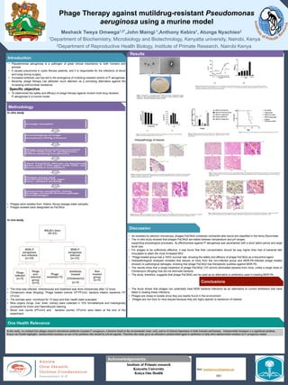
- 1. Phage Therapy against mutildrug-resistant Pseudomonas aeruginosa using a murine model Meshack Tweya Omwega1,2*,John Maingi 1,Anthony Kebira1, Atunga Nyachieo2 K 1Department of Biochemistry, Microbiology and Biotechnology, Kenyatta university, Nairobi, Kenya 2Department of Reproductive Health Biology, Institute of Primate Research, Nairobi Kenya • Pseudomonas aeruginosa is a pathogen of great clinical importance to both humans and animals • It causes pneumonia in cystic fibrosis patients, and it is responsible for the infections of blood and lungs during surgery . • Increased antibiotic use has led to the emergence of multidrug-resistant strains of P. aeruginosa. • Recently, phage therapy has attracted much attention as a promising alternative against the increasing antimicrobial resistance. Specific objective • To determined the safety and efficacy of phage therapy against virulent multi-drug resistant P. aeruginosa in a murine model. Introduction Add your information, graphs and imkk Figure 1. Morphology of P aeruginosa phage PaCIKb2: (a) Electron micrographs of phage PaCIKb2 at 1500× magnification.(b) Electron micrograph of phage Pa/CI/Kb2 at 60000× magnification. (c) Plaque morphology of phage Pa/CI/Kb2 Results In vitro study • Phages were isolated from Kibera, Kenya sewage water samples. • Phages isolated were designated as PaCIKb2. In vivo study • The mice was infected intravenously and treatment was done intravenosly after 12 hours. • Clindamycin dose (8mg/kg), Phage treated viremia 108 PFU/ml, bacteria infetion bactemia 108 CFU/ml. • The animals were monitored for 10 days and their health state evaluated. • Mice organs (lungs, liver, brain, kidney) were collected in 10% formaldehyde and histologically processed for Eosin and Haematoxylin staining. • Blood viral counts (PFU/ml) and bacteria counts( CFU/ml) were taken at the end of the experiment. Methodology Institute of Primate research Kenyatta University Kenya One Health Add your information, graphs and images to this section. One Health Relevance • As revealed by electron microscopy, phages PaCIKb2 contained contractile tails hence are classified in the famiy Myoviridae. • The in-vitro study showed that phages PaCIKb2 are stable between temperature and pH ranges, supporting physiological processes. Its effectiveness against P. aeruginosa was ascertained with a short latent period and large burst size. • For phages to be sufficiently effective, it was found that their concentration should be way higher than that of bacterial titer inoculated to attain the most increased MOI. • Phage treated group had a 100% survival rate, showing the safety and efficacy of phage PaCIKb2 as a biocontrol agent. • Histopathological analyses revealed that tissues of mice from the non-infected group and MDR-PA-infected phage treated showed no pathological damages, showing that phage PaCIkb2 has therapeutic qualities against MDR-PA. • The results show that a single treatment of phage PaCIKb2 (108 pfu/ml) eliminated bacteria from mice, unlike a single dose of Clindamycin (8mg/kg) that did not eliminate bacteria. • The study, therefore, suggests that phage PaCIKb2 can be used as an alternative to antibiotics used in treating MDR-PA. • The study shows that phages can potentially treat MDR bacteria infections as an alternative to current antibiotics that have failed in treating these infections. • Phages are cheap to isolate since they are readily found in the environment. • Phages are non toxic to mice tissues because they are highy specific to bacterium of interest. Discussion Histopathology of tissues Figure 2 (a) Phage PaCIKb2 one-step growth curve. Bacteriophages showed a latent period of about 15 min and a burst size of 316 viral particles per infected cell. (b) The lytic ability of phage PaCIKb2 in vitro. P. aeruginosa (2 × 10⁸CFU mL-1) was infected with the phage at different multiplicities of infection. *p<0.05 statistically significant between test assays and control (MOI=0). Figure 3. The Stability of phage PaCIKb2 at different (a) Temperatures or (b) pH. *p<0.05 statistically significant between test assays and controls (+4°C or pH 7) Figure 4. Bacteremia and viremia levels from infected mice, MDR-PA received no treatment, and phage control had no bacteria. Each group had five mice. Significance levels* p < 0.05 and **p<0.001 Figure 5. Survival rates of mice treated after 12 hours of bacterial inoculation (p<0.001) Figure 6: Hematoxylin and eosin-stained Brain tissues; (a) Non-infected group, (b) Phage infected, (c) MDR-PA non treated group B- Lymphocytic infiltration (inflammation), (d) MDR-PA Phage treated group (e) MDR-PA antibiotic (Clindamycin) treated group B- Lymphocytic infiltration. Magnification= ×400 Figure 7: Hematoxylin and eosin-stained Liver tissues; (a) Non-infected group, (b) Phage infected, (c) MDR-PA non treated group M- Perivascular lymphocytic infiltration (inflammation), W- Severe congestion (d) MDR-PA Phage treated group (e) MDR-PA antibiotic (Clindamycin) treated group W- Severe congestion Magnification= ×400 Figure 8. Hematoxylin and eosin-stained kidney tissues; (a) Non-infected group, (b) Phage infected, (c) MDR-PA non treated group I- Inflammatory cell infiltrate. (d) MDR-PA Phage treated group (e) MDR-PA antibiotic (Clindamycin) treated group I- mild inflammatory cell infiltrate. Magnification= ×400 Figure 9. Hematoxylin and eosin-stained Lung tissues; (a) Non-infected group, (b) Phage infected, (c) MDR-PA non-treated group; Y- lymphocytic infiltrated septae. (d) MDR-PA Phage treated group (e) MDR-PA antibiotic (Clindamycin) treated group; S- Perivascular fibrosis. Magnification= ×400 Conclusions Acknowledgements: Mail: meshacktweya2@gmail.com 2021 In this study, we isolated lytic phages aimed to decolonize antibiotic-resistant P. aeruginosa, a bacteria found in the environment( water, soil), and its of clinical importance to both Animals and humans. Antimicrobial resistance is a significant problem. Kenya one Health highlights antimicrobial resistance as one of the problems that should be solved urgently. Therefore this study gives an alternative antimicrobial agent to antibiotics to help solve antimicrobial resiatnce in P. aeruginosa strains.