MEIOSIS
(THE REDUCTION DIVISION)
By
PB Mallikharjuna PhD
GFGC Yelahanka Bangalore
7th and 8th June 2021
July 13, 2021 GFGC Yelahanka 1
Introduction : Meiosis is a very important and compulsory
biological process of all eukaryotic organisms including plants.
• It is a component of the true sexual reproduction in order to
produce the haploid gametes (gametogenesis) or the spores
(sporogenesis).
• Therefore, meiosis ensures the production of haploid phase in
the life cycle of sexually reproducing organisms, whereas
fertilization restores the diploid phase.
July 13, 2021 GFGC Yelahanka 2
 Meiosis is the specialized cell division that mainly occurs in the
reproductive or sex cells such as the pollen mother cells (PMC)
and megaspore mother cells(MMC) in higher plants. Thereby, the
haploid microspore or megaspore tetrads are produced.
 Meiocytes are the cells which undergo meiosis.
 The term Meiosis was coined by Farmer(1905).
 The process of meiosis was first described by Oscar Hertwig, who
observed it in sea urchin eggs. Later Edouard Van Beneden expanded it.
 It can be defined as the special type of cell division where a single
diploid reproductive cell(2N) is divided into four haploid gamete
cells(N).
July 13, 2021 GFGC Yelahanka 3
• The key features of meiosis involves two sequential cycles of nuclear and
cell division called meiosis I and meiosis II, but only a single cycle of DNA
replication. Meiosis I is initiated after the parental chromosomes have
replicated to produce identical sister chromatids at the S phase.
• Meiosis involves pairing of homologous chromosomes and recombination
between them.
• Four haploid cells are formed at the end of meiosis II.
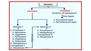
• Meiotic events can be grouped under the following phases:-
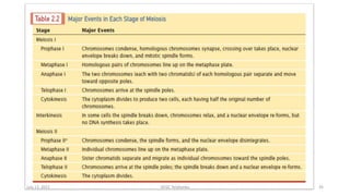
 Meiosis-I ( ): Prophase I, Metaphase
I, Anaphase I, and Telophase I.
 Meiosis-II( ): Prophase II ,
Metaphase II, Anaphase II, and Telophase II
July 13, 2021 GFGC Yelahanka 4
KARYOKINESIS CYTOKINESIS
July 13, 2021 GFGC Yelahanka 5
July 13, 2021 GFGC Yelahanka 6
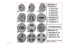
MITOSIS - I
Prophase I
• It is of longest phase of karyokinesis of meiosis which is
about 200 times more complex than that of the prophase of
Mitosis.
• Hence it is further divided into five sub stages i.e.,
 Leptotene,
 Zygotene,
 Pachytene,
 Diplotene, and
 Diakinesis.
July 13, 2021 GFGC Yelahanka 7
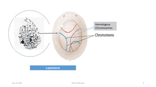
Leptotene/ Leptonema :
(a) Chromosomes are long thread like with chromomeres (beaded) on it.
(b) the volume of nucleus increases.
(c) Half of the chromosomes are from male(paternal) and the remaining half are
from female (maternal)parent.
(d) Chromosome with similar structure are known as homologous chromosomes.
(e) Leptonemal chromosomes have a definite polarization and forms loops whose
ends are attached to the nuclear envelope at points near the centrioles,
contained within an aster. Such peculiar arrangement is termed as
bouquet stage (in animals) and syndet knot (in plants).
(f) E.M. (electron microscope) reveals that chromosomes are composed of paired
chromatids, a dense proteinaceous filament or axial core lies within the groove
between the sister- chromatids of each chromosome.
July 13, 2021 GFGC Yelahanka 8
Leptotene
Homologous
Chromosomes
July 13, 2021 GFGC Yelahanka 9
Synapsis
July 13, 2021 GFGC Yelahanka 10
Zygotene / Zygonema
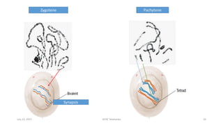
(a) Pairing or “synapsis” of homologous chromosomes takes place in this stage.
(b) Synapsis may be of following types.
 Procentric : Starting at the centromere.
 Proterminal : Starting at the end.
 Localised random : Starting at various points.
(c) Paired chromosomes are called bivalents, which by further molecular packing and
spiralization becomes shorter and thicker.
(d) Pairing of homologous chromosomes in a zipper-fashion. Number of bivalents (paired
homologous chromosomes) is half to total number of chromosomes in a diploid cell. Each
bivalent is formed of one paternal and one maternal chromosome (i.e. one chromosome
derived from each parent). Ex in Allium cepa eight bivalents are present (in PMC/MMC)
(e) Under EM, a filamentous ladder like nucleoproteinaceous complex, called Synaptenemal
Complex between the homologous chromosomes, which is discovered by “Moses” (1953).
July 13, 2021 GFGC Yelahanka 11
Pachytene/Pachynema
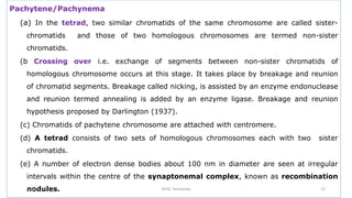
(a) In the tetrad, two similar chromatids of the same chromosome are called sister-
chromatids and those of two homologous chromosomes are termed non-sister
chromatids.
(b Crossing over i.e. exchange of segments between non-sister chromatids of
homologous chromosome occurs at this stage. It takes place by breakage and reunion
of chromatid segments. Breakage called nicking, is assisted by an enzyme endonuclease
and reunion termed annealing is added by an enzyme ligase. Breakage and reunion
hypothesis proposed by Darlington (1937).
(c) Chromatids of pachytene chromosome are attached with centromere.
(d) A tetrad consists of two sets of homologous chromosomes each with two sister
chromatids.
(e) A number of electron dense bodies about 100 nm in diameter are seen at irregular
intervals within the centre of the synaptonemal complex, known as recombination
nodules.
July 13, 2021 GFGC Yelahanka 12
Diplotene/Diplonema
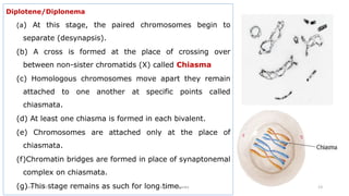
(a) At this stage, the paired chromosomes begin to
separate (desynapsis).
(b) A cross is formed at the place of crossing over
between non-sister chromatids (X) called Chiasma
(c) Homologous chromosomes move apart they remain
attached to one another at specific points called
chiasmata.
(d) At least one chiasma is formed in each bivalent.
(e) Chromosomes are attached only at the place of
chiasmata.
(f)Chromatin bridges are formed in place of synaptonemal
complex on chiasmata.
(g) This stage remains as such for long time.
July 13, 2021 GFGC Yelahanka 13
Diakinesis
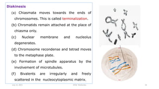
(a) Chiasmata moves towards the ends of
chromosomes. This is called terminalization.
(b) Chromatids remain attached at the place of
chiasma only.
(c) Nuclear membrane and nucleolus
degenerates.
(d) Chromosome recondense and tetrad moves
to the metaphase plate.
(e) Formation of spindle apparatus by the
involvement of microtubules.
(f) Bivalents are irregularly and freely
scattered in the nucleocytoplasmic matrix.
July 13, 2021 GFGC Yelahanka 14
Metaphase I : involves;
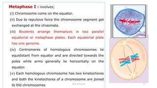
(i) Chromosome come on the equator.
(ii) Due to repulsive force the chromosome segment get
exchanged at the chiasmata.
(iii) Bivalents arrange themselves in two parallel
equatorial or metaphase plates. Each equatorial plate
has one genome.
(iv) Centromeres of homologous chromosomes lie
equidistant from equator and are directed towards the
poles while arms generally lie horizontally on the
equator.
(v) Each homologous chromosome has two kinetochores
and both the kinetochores of a chromosome are joined
to the chromosomes
July 13, 2021 GFGC Yelahanka 15
Anaphase-I
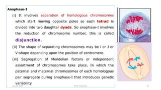
(i) It involves separation of homologous chromosomes
which start moving opposite poles so each tetrad is
divided into two daughter dyads. So anaphase-I involves
the reduction of chromosome number, this is called
disjunction.
(ii) The shape of separating chromosomes may be i or J or
V-shape depending upon the position of centromere.
(iii) Segregation of Mendelian factors or independent
assortment of chromosomes take place. In which the
paternal and maternal chromosomes of each homologous
pair segregate during anaphase-I that introduces genetic
variability.
July 13, 2021 GFGC Yelahanka 16
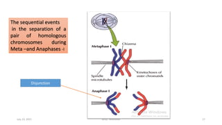
The sequential events
in the separation of a
pair of homologous
chromosomes during
Meta –and Anaphases -I
Disjunction
July 13, 2021 GFGC Yelahanka 17
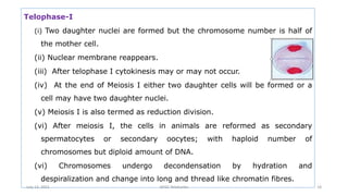
Telophase-I
(i) Two daughter nuclei are formed but the chromosome number is half of
the mother cell.
(ii) Nuclear membrane reappears.
(iii) After telophase I cytokinesis may or may not occur.
(iv) At the end of Meiosis I either two daughter cells will be formed or a
cell may have two daughter nuclei.
(v) Meiosis I is also termed as reduction division.
(vi) After meiosis I, the cells in animals are reformed as secondary
spermatocytes or secondary oocytes; with haploid number of
chromosomes but diploid amount of DNA.
(vi) Chromosomes undergo decondensation by hydration and
despiralization and change into long and thread like chromatin fibres.
July 13, 2021 GFGC Yelahanka 18
Interphase : Generally there is no interphase between meiosis-I and meiosis-II.
A brief interphase called interkinesis, or intermeiotic interphase sometimes may
exist. During this interphase (S phase), there is no replication of chromosomes,
Cytokinesis-I : It may or may not be present. When present, it occurs by cell-
furrow formation in animal cells and cell plate formation in plant cells.
Significance of meiosis-I
(i) It separates the homologous chromosomes to reduce the chromosome
number to the haploid state, a necessity for sexual reproduction.
(ii) It introduces variation by forming new gene combinations through crossing
over and random assortment of paternal and maternal chromosomes.
(iii) Sometimes, it may cause chromosomal mutation by abnormal disjunction.
(iv) It induces the cells to produce gametes for sexual reproduction or spores
for asexual reproduction.
July 13, 2021 GFGC Yelahanka 19
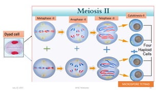
Meiosis-I
Dyad cell
Metaphase -II Anaphase -II Telophase -II
Cytokinesis-II
MICROSPORE TETRAD
July 13, 2021 GFGC Yelahanka 20
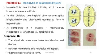
Meiosis-II ( Homotytic or equational division)
• Meiosis-II is exactly like mitosis, so it is also
known as meiotic mitosis.
• In this division, two haploid chromosome splits
longitudinally and distributed equally to form 4
haploid cells.
• It completes in 4 stages - Prophase-II,
Metaphase-II, Anaphase-II, Telophase-II.
Prophase-II:
• The dyad chromosomes becomes shorter and
thicker.
• Nuclear membrane and nucleolus disappear.
• Spindle fiber starts to form.
July 13, 2021 GFGC Yelahanka 21
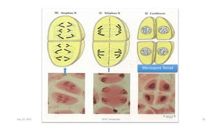
Metaphase-II:
• The dyads chromosomes comes to equatorial plane.
• Spindle fibers organize between poles and attaches to centromere of
chromosome.
Anaphase-II:
• Centromere of each chromosome divides and sister-chromatids separates to
form two daughter chromosomes.
• Spindle fiber contracts and pull the daughter chromosome apart towards
opposite pole.
Telophase-II:
• Chromosome become organize at respective pole into nuclei.
• Chromosome elongates to form thin networks of chromatin.
• Nuclear membrane and nucleolus reappears
July 13, 2021 GFGC Yelahanka 22
Microspore Tetrad
July 13, 2021 GFGC Yelahanka 23
Cytokinesis-II:
• The result of cytokinesis is four haploid daughter cells
(gametes or spores).
• Cytokinesis takes place by cell plate formation in plant cells
• Successive methods: cytokinesis followed by each nuclear
division resulting in 4 haploid cells. E.g., Monocot plants
(Allium cepa)
• Simultaneous methods: cytokinesis occurs only after meiosis-
II to form 4 haploid cells. E.g., Dicot plants
• In animal cells, cytokinesis occurs by furrow formation or
depression.
July 13, 2021 GFGC Yelahanka 24
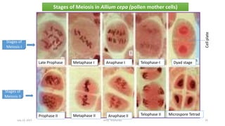
Stages of
Meiosis-I
Stages of
Meiosis II
Stages of Meiosis in Allium cepa (pollen mother cells)
Late Prophase Metaphase I Anaphase I Telophase-I Dyad stage
Cell
plate
Prophase II Metaphase II Anaphase II Telophase II Microspore Tetrad
July 13, 2021 GFGC Yelahanka 25
July 13, 2021 GFGC Yelahanka 26
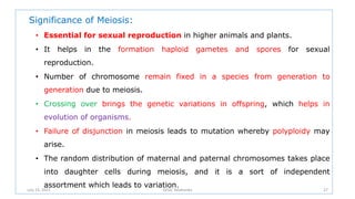
Significance of Meiosis:
• Essential for sexual reproduction in higher animals and plants.
• It helps in the formation haploid gametes and spores for sexual
reproduction.
• Number of chromosome remain fixed in a species from generation to
generation due to meiosis.
• Crossing over brings the genetic variations in offspring, which helps in
evolution of organisms.
• Failure of disjunction in meiosis leads to mutation whereby polyploidy may
arise.
• The random distribution of maternal and paternal chromosomes takes place
into daughter cells during meiosis, and it is a sort of independent
assortment which leads to variation.
July 13, 2021 GFGC Yelahanka 27
Synaptonemal Complex
 The synaptonemal complex (SC) is a protein structure that forms
between homologous chromosomes during meiosis.
 It is a tripartite structure consisting of two parallel lateral regions and a
central element. The fibres of lateral filaments are called synaptomeres.
 This "tripartite structure" is seen during the pachytene stage of the first
meiotic prophase, both in males and in females during gametogenesis.
 It mediates synapsis, recombination and chiasmata formation in
eukaryotes including plants.
 Further, SC functions primarily as a scaffold to allow interacting
chromatids to complete their crossover activities.
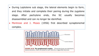
 During Leptotene sub stage, the lateral elements begin to form,
and they initiate and complete their pairing during the zygotene
stage. After pachytene ends, the SC usually becomes
disassembled and can no longer be identified.
 Montrose and J. Moses (1956) first described synaptonemal
complex.
How SC is formed?
 The lateral elements of the synaptonemal complex start
to attach to individual chromosomes during leptotene, but
the central element, which actually joins homologous
chromosomes together, does not form until zygotene.
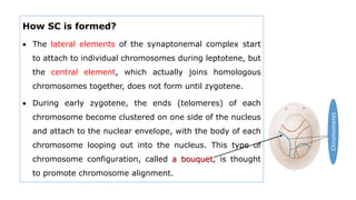
 During early zygotene, the ends (telomeres) of each
chromosome become clustered on one side of the nucleus
and attach to the nuclear envelope, with the body of each
chromosome looping out into the nucleus. This type of
chromosome configuration, called a bouquet, is thought
to promote chromosome alignment.
Chromomeres
 The alignment of similar-sized chromosomes facilitates
formation of synaptonemal complexes, which become fully
developed during pachytene
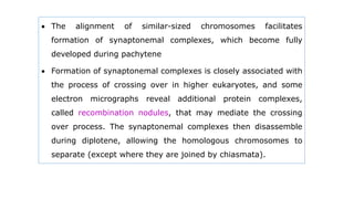
 Formation of synaptonemal complexes is closely associated with
the process of crossing over in higher eukaryotes, and some
electron micrographs reveal additional protein complexes,
called recombination nodules, that may mediate the crossing
over process. The synaptonemal complexes then disassemble
during diplotene, allowing the homologous chromosomes to
separate (except where they are joined by chiasmata).
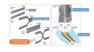
Protein Electron micrograph
Diagrammatic
Pairing like a zipper
The sequential events
of SC in the meiosis-I
Functions of SC
 It helps in the proper alignment of homologous chromosomes
during pairing (Synapsis) in the meiotic division.
 It stabilizes the pairing of homologous chromosomes
 It facilitates the crossing over and thereby genetic
recombination.
 Lack of SC as in male Drosophila no crossing over occurs
(Complete Linkage).
The Cytological Basis of Crossing over
Introduction : It is not necessary that chiasmata should be associated
with exchange of chromosome segments. Therefore, to demonstrate that
crossing over is associated with actual exchange of chromosome segments, special
experiments were devised by C. Stern in Drosophila and by H.S. Creighton
and B. McClintock in corn. These experiments were reported in 1931 and
had utilized chromosomes, whose morphology was altered due to
chromosomal aberrations in order to make it identifiable from its
homologue.
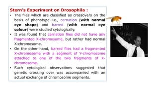
Stern’s Experiment on Drosophila :
• The flies which are classified as crossovers on the
basis of phenotype i.e., carnation (with normal
eye shape) and barred (with normal eye
colour) were studied cytologically.
• It was found that carnation flies did not have any
fragmented X-chromosome, but rather had normal
X-chromosome.
• On the other hand, barred flies had a fragmented
X-chromosome with a segment of Y-chromosome
attached to one of the two fragments of X-
chromosome.
• Such cytological observations suggested that
genetic crossing over was accompanied with an
actual exchange of chromosome segments.
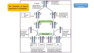
Test cross
Gametogenesis
The flowchart of Stern’s
experiment in Drosophila
Created by X-ray
exposure
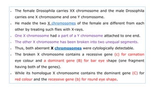
 The female Drosophila carries XX chromosome and the male Drosophila
carries one X chromosome and one Y chromosome.
 He made the two X chromosomes of the female are different from each
other by treating such flies with X-rays.
 One X chromosome had a part of a Y chromosome attached to one end.
 The other X chromosome has been broken into two unequal segments.
 Thus, both aberrant X chromosomes were cytologically detectable.
 The broken X chromosome contains a recessive gene (c) for carnation
eye colour and a dominant gene (B) for bar eye shape (one fragment
having both of the genes).
 While its homologue X chromosome contains the dominant gene (C) for
red colour and the recessive gene (b) for round eye shape.
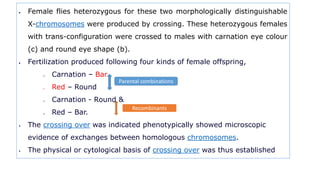
 Female flies heterozygous for these two morphologically distinguishable
X-chromosomes were produced by crossing. These heterozygous females
with trans-configuration were crossed to males with carnation eye colour
(c) and round eye shape (b).
 Fertilization produced following four kinds of female offspring,
o Carnation – Bar
o Red – Round
o Carnation - Round &
o Red – Bar.
 The crossing over was indicated phenotypically showed microscopic
evidence of exchanges between homologous chromosomes.
 The physical or cytological basis of crossing over was thus established
Parental combinations
Recombinants
Experiments by Harriet Creighton and Barbara McClintock on
Maize :
• Harriet Creighton and Barbara McClintock (1931) demonstrated the
correlation between genetic crossing over and the exchange of parts of
homologous chromosomes.
• They analyzed crosses involving two loci on chromosome 9 of maize.
▪ C = Coloured seed
▪ c = colourless seed
▪ Wx – Starchy endosperm, and
▪ wx = waxy endosperm
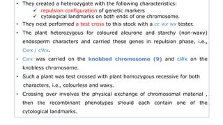
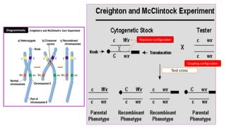
• They made use of a chromosome with a densely stained “knob” at one end and
an extra (translocated) piece of chromosome at the other end.
• They created a heterozygote with the following characteristics:
 repulsion configuration of genetic markers
 cytological landmarks on both ends of one chromosome.
• They next performed a test cross to this stock with a cc wx wx tester.
• The plant heterozygous for coloured aleurone and starchy (non-waxy)
endosperm characters and carried these genes in repulsion phase, i.e.,
Cwx / cWx.
• Cwx was carried on the and cWx on the
knobless chromosome.
• Such a plant was test crossed with plant homozygous recessive for both
characters, i.e., colourless and waxy.
• Crossing over involves the physical exchange of chromosomal material ,
then the recombinant phenotypes should each contain one of the
cytological landmarks.
Test cross
Repulsion configuration
Diagrammatic
Coupling configuration
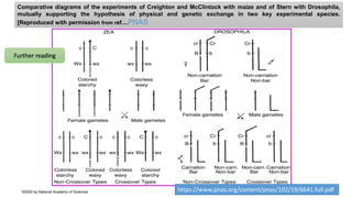
Comparative diagrams of the experiments of Creighton and McClintock with maize and of Stern with Drosophila,
mutually supporting the hypothesis of physical and genetic exchange in two key experimental species.
[Reproduced with permission from ref....PNAS
©2005 by National Academy of Sciences https://www.pnas.org/content/pnas/102/19/6641.full.pdf
Further reading