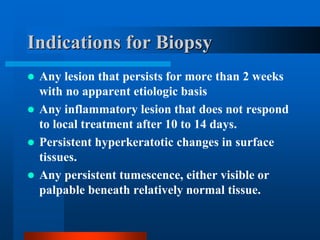
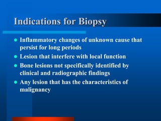
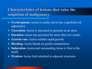
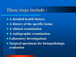
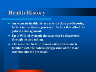
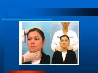
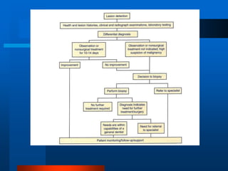
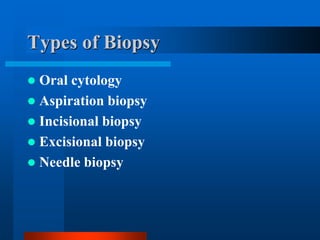
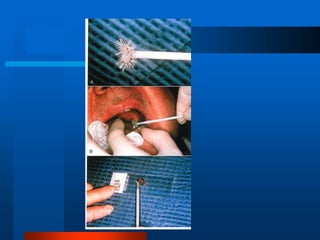
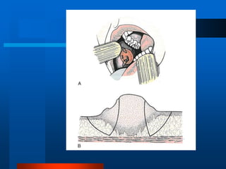
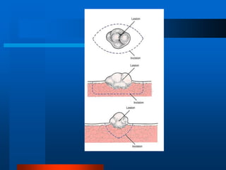
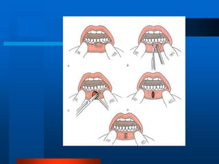
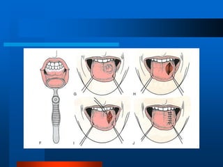
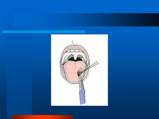
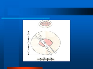
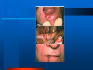
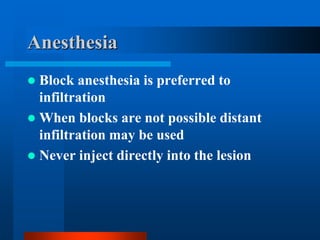
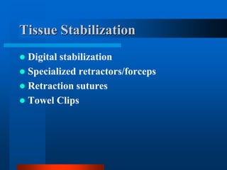
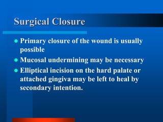
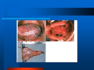
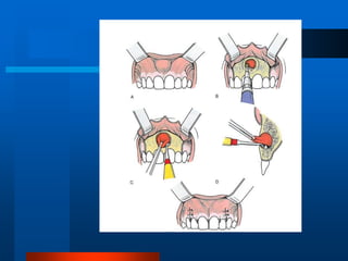
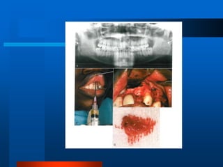
Principles and Techniques of
Biopsy
Reza Tabrizi DMD
Assistant professor of OMFS
Shahid Beheshti University of Medical Sciences
Email:tabmed@gmail.com
Principles and Techniques of
Biopsy
 It is important to develop a systematic
approach in evaluating a patient with a
lesion in the Oral and Maxillofacial
region.
These steps include :
 A detailed health history
 A history of the specific lesion
 A clinical examination
 A radiographic examination
 Laboratory investigations
 Surgical specimens for histopathologic
evaluation
Health History
 An accurate health history may disclose predisposing
factors in the disease process or factors that affect the
patients management.
 Up to 90% of systemic deseases can be discovered
through history taking.
 The same can be true of oral lesions when one is
familiar with the natural progression of the more
common disease processes.
Medical conditions that
warrant special care include:
 Congenital heart defects
 Coagulopathies
 Hypertension
 Poorly controlled diabetics
 Immunocompromised patients
History of the Lesion
Questions to Ask
 Duration of the lesion
 Changes in size and rate of change
 Changes in the character of the lesion.
– Lump to ulcer, etc
 Associated systemic symptoms:
– fever
– nausea
– anorexia
More Questions to Ask
 Pain
 Abnormal sensations
 Anesthesia
 A feeling of swelling
 Bad taste or smell
 Dysphagia
 Swelling or tenderness of adjacent lymph
nodes
 Character of the pain if present
Historical Reasons for the
Lesions:
 Trauma to the area
 Recent toothache
 Habits
Clinical Examination
 The clinical examination should always
include when possible:
– Inspection
– Palpation
– Percussion
– Auscultation
Clinical Evaluation
 The anatomic location of the lesion/mass
 The physical character of the lesion/mass
 The size and shape of the lesion/mass
 Single vs. multiple lesions
 The surface of the lesion
 The color of the lesion
 The sharpness of the boundaries of the lesion
 The consistency of the lesion to palpation
 Presence of pulsation
 Lymph node examination
Radiographic Examination
 The radiographic appearance may provide
clues that will help determine the nature of the
lesion.
 A radiolucency with sharp borders will often be
a cyst
 A ragged radiolucency will often be a more
aggressive lesion
 Radiopaque dyes and instruments can help
differentiate normal anatomy
Laboratory Investigation
 Oral lesions may be manifestations of
systemic disease.
 If a systemic disease is suspected it should
be pursued.
These include:
 Tumor of hyperparathyroidism
 Padgets disease
 Multiple myeloma
 Determination of serum calcium,
phosphorus, and alkaline phosphatase
and protein can be very useful in excluding
certain pathological processes.
Indications for Biopsy
 Any lesion that persists for more than 2 weeks
with no apparent etiologic basis
 Any inflammatory lesion that does not respond
to local treatment after 10 to 14 days.
 Persistent hyperkeratotic changes in surface
tissues.
 Any persistent tumescence, either visible or
palpable beneath relatively normal tissue.
Indications for Biopsy
 Inflammatory changes of unknown cause that
persist for long periods
 Lesion that interfere with local function
 Bone lesions not specifically identified by
clinical and radiographic findings
 Any lesion that has the characteristics of
malignancy
Characteristics of lesions that raise the
suspicion of malignancy.
 Erythroplasia- lesion is totally red or has a speckled red
appearance.
 Ulceration- lesion is ulcerated or presents as an ulcer.
 Duration- lesion has persisted for more than two weeks.
 Growth rate- lesion exhibits rapid growth
 Bleeding- lesion bleeds on gentle manipulation
 Induration- lesion and surrounding tissue is firm to the
touch
 Fixation- lesion feels attached to adjacent structures
What is a Biopsy?
 Biopsy is the removal of tissue for the
purpose of diagnostic examination.
Types of Biopsy
 Oral cytology
 Aspiration biopsy
 Incisional biopsy
 Excisional biopsy
 Needle biopsy
Oral Cytology
 Developed as a diagnostic screening
procedure to monitor large tissue areas
for dysplastic changes.
 Most frequently used to screen for
uterine cervix malignancy
 May be helpful with monitoring
postradiation changes, herpes,
pemphigus.
The Disadvantage of oral cytological
procedures include:
 Not very reliable with many false positives.
 Expertise in oral cytology is not widely
available
 The lesion is repeatedly scraped with a
moistened tongue depressor or spatula type
instrument. The cells obtained are smeared on
a glass slide and immediately fixed with a
fixative spray or solution.
Aspiration Biopsy
 Aspiration biopsy is the use of a needle and syringe
to penetrate a lesion for aspiration if its contents.
 Indications:
– To determine the presents of fluid within a lesion
– To a certain the type of fluid within a lesion
– When exploration of an intraosseous lesion is
indicated
Aspiration
 An 18 gauge needle on a 5 or 10 ml
syringe is inserted into the area under
investigation after anesthesia is obtained.
 The syringe is aspirated and the needle
redirected if necessary to find the fluid
cavity.
Incisional Biopsy
 An incisional biopsy is a biopsy that
samples only a particular portion or
representative part of a lesion.
 If a lesion is large or has different
characteristics in various locations more
than one area may need to be sampled
Incisional Biopsy
 Indications:
– Size limitations
– Hazardous location of the lesion
– Great suspicion of malignancy
 Technique:
– Representative areas are biopsied in a wedge fashion.
– Margins should extend into normal tissue on the deep
surface.
– Necrotic tissue should be avoided.
– A narrow deep specimen is better than a broad shallow
one.
Excisional Biopsy
An excisional biposy implies the complete removal of
the lesion.
 Indications:
– Should be employed with small lesions. Less than 1cm
– The lesion on clinical exam appears benign.
– When complete excision with a margin of normal tissue is
possible without mutilation.
Excisional Biopsy
 Technique:
– The entire lesion with 2 to 3mm of normal
appearing tissue surrounding the lesion is excised
if benign.
Principles of Surgery
Anesthesia
 Block anesthesia is preferred to
infiltration
 When blocks are not possible distant
infiltration may be used
 Never inject directly into the lesion
Tissue Stabilization
 Digital stabilization
 Specialized retractors/forceps
 Retraction sutures
 Towel Clips
Hemostasis
 Suction devices should be avoided
 Gauze compresses are usually adequate
 Gauze wrapped low volume suction may
be used if needed
Incisions
 Incisions should be made with a scalpel.
 They should be converging
 Should extend beyond the suspected depth of the lesion
 They should parallel important structures
 Margins should include 2 to 3mm of normal appearing
tissue if the lesion is thought to be benign.
 5mm or more may be necessary with lesions that appear
malignant, vascular, pigmented, or have diffuse borders.
Handling of the Tissue
Specimen
 Direct handling of the lesion will expose it
to crush injury resulting in alteration the
cellular architecture.
Specimen Care
 The specimen should be immediately
placed in 10% formalin solution, and be
completely immersed.
Margins of the Biopsy
 Margins of the tissue should be identified
to orient the pathologist. A silk suture is
often adequate. Illustrations are also
very helpful and should be included.
Surgical Closure
 Primary closure of the wound is usually
possible
 Mucosal undermining may be necessary
 Elliptical incision on the hard palate or
attached gingiva may be left to heal by
secondary intention.
Biopsy Data Sheet
 A biopsy data sheet should be completed
and the specimen immediately labeled.
All pertinent history and descriptions of
the lesion must be conveyed.
Intraosseous and Hard Tissue
Biopsy
 Intraosseous lesions are most often the
result of problems associated with the
dentition.
Indications for Intraosseous
Biopsy
 Any intraosseous lesion that fails to
respond to routine treatment of the
dentition.
 Any intraosseous lesion that appears
unrelated to the dentition.
 Palpation of the area of the lesion with comparison
to the opposite side.
 Any radiolucent lesion should have an aspiration
biopsy performed prior to surgical exploration.
– Information from the aspiration will provide valuable
information about the lesion.
• Solid
• Fluid Filled
• Vascular
• Without Contents
Clinical Exam
Principles of Surgery
 Mucperiosteal flaps should be designed to allow
adequate access for incisional/excisional biopsy.
 Incisions should be over sound bone
 Cortical perforation must be considered when
designing flaps
 Flaps should be full thickness
 Major neurovascular structures should be avoided
Principles of Surgery
 Osseous windows should be submitted with the
specimen
 Osseous preformations can be enlarged to gain
access
 Avoid roots and neurovascular structures
 The tissue consistency and nature of the lesion will
determine the ease of removal
Principles of Surgery
 Incisional biopsies only require removal of a
section of tissue
 Soft tissue overlying the lesion should be
reapproximated following thorough irrigation
of the operative site.
 The specimen should be handled as previously
described
Biopsy Results: What If ?
 They don’t corroborate your clinical impression
– Repeat the biopsy!!!
– Determine if the tissue was looked at by an Oral
Pathologist
– The results show malignancy
When To Refer For Biopsy
 When the health of the patient requires special management
that the dentist feel unprepared to handle
 The size and surgical difficulty is beyond the level of skill
that the dentist feels he/she possesses
 If the dentist is concerned about the possibility of
malignancy

Principles and Techniques of Biopsy

  • 1.
    Principles and Techniquesof Biopsy Reza Tabrizi DMD Assistant professor of OMFS Shahid Beheshti University of Medical Sciences Email:tabmed@gmail.com
  • 2.
    Principles and Techniquesof Biopsy  It is important to develop a systematic approach in evaluating a patient with a lesion in the Oral and Maxillofacial region.
  • 3.
    These steps include:  A detailed health history  A history of the specific lesion  A clinical examination  A radiographic examination  Laboratory investigations  Surgical specimens for histopathologic evaluation
  • 4.
    Health History  Anaccurate health history may disclose predisposing factors in the disease process or factors that affect the patients management.  Up to 90% of systemic deseases can be discovered through history taking.  The same can be true of oral lesions when one is familiar with the natural progression of the more common disease processes.
  • 5.
    Medical conditions that warrantspecial care include:  Congenital heart defects  Coagulopathies  Hypertension  Poorly controlled diabetics  Immunocompromised patients
  • 6.
  • 7.
    Questions to Ask Duration of the lesion  Changes in size and rate of change  Changes in the character of the lesion. – Lump to ulcer, etc  Associated systemic symptoms: – fever – nausea – anorexia
  • 8.
    More Questions toAsk  Pain  Abnormal sensations  Anesthesia  A feeling of swelling  Bad taste or smell  Dysphagia  Swelling or tenderness of adjacent lymph nodes  Character of the pain if present
  • 9.
    Historical Reasons forthe Lesions:  Trauma to the area  Recent toothache  Habits
  • 10.
    Clinical Examination  Theclinical examination should always include when possible: – Inspection – Palpation – Percussion – Auscultation
  • 11.
    Clinical Evaluation  Theanatomic location of the lesion/mass  The physical character of the lesion/mass  The size and shape of the lesion/mass  Single vs. multiple lesions  The surface of the lesion  The color of the lesion  The sharpness of the boundaries of the lesion  The consistency of the lesion to palpation  Presence of pulsation  Lymph node examination
  • 14.
    Radiographic Examination  Theradiographic appearance may provide clues that will help determine the nature of the lesion.  A radiolucency with sharp borders will often be a cyst  A ragged radiolucency will often be a more aggressive lesion  Radiopaque dyes and instruments can help differentiate normal anatomy
  • 15.
    Laboratory Investigation  Orallesions may be manifestations of systemic disease.  If a systemic disease is suspected it should be pursued.
  • 16.
    These include:  Tumorof hyperparathyroidism  Padgets disease  Multiple myeloma  Determination of serum calcium, phosphorus, and alkaline phosphatase and protein can be very useful in excluding certain pathological processes.
  • 17.
    Indications for Biopsy Any lesion that persists for more than 2 weeks with no apparent etiologic basis  Any inflammatory lesion that does not respond to local treatment after 10 to 14 days.  Persistent hyperkeratotic changes in surface tissues.  Any persistent tumescence, either visible or palpable beneath relatively normal tissue.
  • 19.
    Indications for Biopsy Inflammatory changes of unknown cause that persist for long periods  Lesion that interfere with local function  Bone lesions not specifically identified by clinical and radiographic findings  Any lesion that has the characteristics of malignancy
  • 20.
    Characteristics of lesionsthat raise the suspicion of malignancy.  Erythroplasia- lesion is totally red or has a speckled red appearance.  Ulceration- lesion is ulcerated or presents as an ulcer.  Duration- lesion has persisted for more than two weeks.  Growth rate- lesion exhibits rapid growth  Bleeding- lesion bleeds on gentle manipulation  Induration- lesion and surrounding tissue is firm to the touch  Fixation- lesion feels attached to adjacent structures
  • 21.
    What is aBiopsy?  Biopsy is the removal of tissue for the purpose of diagnostic examination.
  • 24.
    Types of Biopsy Oral cytology  Aspiration biopsy  Incisional biopsy  Excisional biopsy  Needle biopsy
  • 25.
    Oral Cytology  Developedas a diagnostic screening procedure to monitor large tissue areas for dysplastic changes.  Most frequently used to screen for uterine cervix malignancy  May be helpful with monitoring postradiation changes, herpes, pemphigus.
  • 26.
    The Disadvantage oforal cytological procedures include:  Not very reliable with many false positives.  Expertise in oral cytology is not widely available  The lesion is repeatedly scraped with a moistened tongue depressor or spatula type instrument. The cells obtained are smeared on a glass slide and immediately fixed with a fixative spray or solution.
  • 28.
    Aspiration Biopsy  Aspirationbiopsy is the use of a needle and syringe to penetrate a lesion for aspiration if its contents.  Indications: – To determine the presents of fluid within a lesion – To a certain the type of fluid within a lesion – When exploration of an intraosseous lesion is indicated
  • 29.
    Aspiration  An 18gauge needle on a 5 or 10 ml syringe is inserted into the area under investigation after anesthesia is obtained.  The syringe is aspirated and the needle redirected if necessary to find the fluid cavity.
  • 30.
    Incisional Biopsy  Anincisional biopsy is a biopsy that samples only a particular portion or representative part of a lesion.  If a lesion is large or has different characteristics in various locations more than one area may need to be sampled
  • 31.
    Incisional Biopsy  Indications: –Size limitations – Hazardous location of the lesion – Great suspicion of malignancy  Technique: – Representative areas are biopsied in a wedge fashion. – Margins should extend into normal tissue on the deep surface. – Necrotic tissue should be avoided. – A narrow deep specimen is better than a broad shallow one.
  • 34.
    Excisional Biopsy An excisionalbiposy implies the complete removal of the lesion.  Indications: – Should be employed with small lesions. Less than 1cm – The lesion on clinical exam appears benign. – When complete excision with a margin of normal tissue is possible without mutilation.
  • 35.
    Excisional Biopsy  Technique: –The entire lesion with 2 to 3mm of normal appearing tissue surrounding the lesion is excised if benign.
  • 42.
  • 43.
    Anesthesia  Block anesthesiais preferred to infiltration  When blocks are not possible distant infiltration may be used  Never inject directly into the lesion
  • 44.
    Tissue Stabilization  Digitalstabilization  Specialized retractors/forceps  Retraction sutures  Towel Clips
  • 45.
    Hemostasis  Suction devicesshould be avoided  Gauze compresses are usually adequate  Gauze wrapped low volume suction may be used if needed
  • 46.
    Incisions  Incisions shouldbe made with a scalpel.  They should be converging  Should extend beyond the suspected depth of the lesion  They should parallel important structures  Margins should include 2 to 3mm of normal appearing tissue if the lesion is thought to be benign.  5mm or more may be necessary with lesions that appear malignant, vascular, pigmented, or have diffuse borders.
  • 47.
    Handling of theTissue Specimen  Direct handling of the lesion will expose it to crush injury resulting in alteration the cellular architecture.
  • 48.
    Specimen Care  Thespecimen should be immediately placed in 10% formalin solution, and be completely immersed.
  • 50.
    Margins of theBiopsy  Margins of the tissue should be identified to orient the pathologist. A silk suture is often adequate. Illustrations are also very helpful and should be included.
  • 51.
    Surgical Closure  Primaryclosure of the wound is usually possible  Mucosal undermining may be necessary  Elliptical incision on the hard palate or attached gingiva may be left to heal by secondary intention.
  • 53.
    Biopsy Data Sheet A biopsy data sheet should be completed and the specimen immediately labeled. All pertinent history and descriptions of the lesion must be conveyed.
  • 54.
    Intraosseous and HardTissue Biopsy  Intraosseous lesions are most often the result of problems associated with the dentition.
  • 55.
    Indications for Intraosseous Biopsy Any intraosseous lesion that fails to respond to routine treatment of the dentition.  Any intraosseous lesion that appears unrelated to the dentition.
  • 56.
     Palpation ofthe area of the lesion with comparison to the opposite side.  Any radiolucent lesion should have an aspiration biopsy performed prior to surgical exploration. – Information from the aspiration will provide valuable information about the lesion. • Solid • Fluid Filled • Vascular • Without Contents Clinical Exam
  • 57.
    Principles of Surgery Mucperiosteal flaps should be designed to allow adequate access for incisional/excisional biopsy.  Incisions should be over sound bone  Cortical perforation must be considered when designing flaps  Flaps should be full thickness  Major neurovascular structures should be avoided
  • 60.
    Principles of Surgery Osseous windows should be submitted with the specimen  Osseous preformations can be enlarged to gain access  Avoid roots and neurovascular structures  The tissue consistency and nature of the lesion will determine the ease of removal
  • 61.
    Principles of Surgery Incisional biopsies only require removal of a section of tissue  Soft tissue overlying the lesion should be reapproximated following thorough irrigation of the operative site.  The specimen should be handled as previously described
  • 62.
    Biopsy Results: WhatIf ?  They don’t corroborate your clinical impression – Repeat the biopsy!!! – Determine if the tissue was looked at by an Oral Pathologist – The results show malignancy
  • 63.
    When To ReferFor Biopsy  When the health of the patient requires special management that the dentist feel unprepared to handle  The size and surgical difficulty is beyond the level of skill that the dentist feels he/she possesses  If the dentist is concerned about the possibility of malignancy