
Recommended
More Related Content
What's hot
What's hot (20)
Similar to Mass spectroscopy for MSc I Chemistry of SPPU
Similar to Mass spectroscopy for MSc I Chemistry of SPPU (20)
Recently uploaded
Recently uploaded (20)
Mass spectroscopy for MSc I Chemistry of SPPU
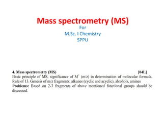
- 1. Mass spectrometry (MS) For M.Sc. I Chemistry SPPU
- 2. Introduction • Mass spectrometry is an analytical technique that measures the mass-to-charge ratio of ions. • Mass spectrometry is a powerful analytical technique used to quantify known materials, to identify unknown compounds within a sample, and to elucidate the structure and chemical properties of different molecules. • The complete process involves the conversion of the sample into gaseous ions, with or without fragmentation, which are then characterized by their mass to charge ratios (m/z) and relative abundance. • A mass spectrometer generates multiple ions from the sample under investigation, it then separates them according to their specific mass-to-charge ratio (m/z), and then records the relative abundance of each ion type. 2AISC/SBS/MSc-I
- 3. The formed ions are separated by Deflection in magnetic field according to their Mass to charge (m/z) Ions formed Further break up into smaller ion (Fragment ions or Daughter ions formed) Mass Spectroscopy Basic Principles 3 Molecules converted into highly energetic positively charged ions (Molecular ions or Parent ions) Organic Molecules are bombarded with electron Mass Spectrum m/z ratio Vs intensity (relative abundance of peak) AISC/SBS/MSc-I
- 4. Basic Principle • The first step in the mass spectrometric analysis of compounds is the production of gas phase ions of the compound, basically by electron ionization with bombarded of electron to form molecular ions or parent ion. • This molecular ion undergoes fragmentation to from positively charged ions and neutral ions or radicals. • Each primary product ion derived from the molecular ion, in turn, undergoes fragmentation, and so on. • The ions are separated in the mass spectrometer according to their mass-to- charge ratio, and are detected in proportion to their abundance. • A mass spectrum of the molecule is thus produced. • It displays the result in the form of a plot of ion abundance versus mass-to-charge ratio. • Ions provide information concerning the nature and the structure of their precursor molecule. • In the spectrum of a pure compound, the molecular ion, if present, appears at the highest value of m/z (followed by ions containing heavier isotopes) and gives the molecular mass of the compound. 4AISC/SBS/MSc-I
- 5. • In MS technique, molecules are bombarded with a beam of energetic electrons. • The molecules are ionized into ions and broken up into many fragments, some of which are positive ions. • Each kind of ion has a particular ratio of mass to charge, i.e. m/e ratio(value). • For most ions, the charge is one and thus, m/e ratio is simply the molecular mass of the ion. 5AISC/SBS/MSc-I
- 7. Working of MS • There are four key stages in the process for Mass Spectrometry. – Ionization – Acceleration – Deflection – Detection 7AISC/SBS/MSc-I
- 8. Stage 1: Ionisation 8 • The initial sample may be a solid, liquid, or gas. • The sample is vaporized into a gas and then ionized by the ion source, usually by losing an electron to become a cation • Electro Ionisation is the most common type of ionisation. • The sample is bombarded by electrons which come from a heated filament. • When the sample passes through the electron stream, the high energy electrons in the stream knock electrons out of the sample to form ions. • The ionization chamber is kept in a vacuum so the ions that are produced can progress through the instrument without running into molecules from air. Stage 2: Acceleration • Acceleration is a simple step where the ions are placed between a set of charges parallel plates. • The ions will then be repelled by one plate and attracted to the other. • There is a slit cut in the plate which the ions are attracted to. the force of attraction and repulsion forces the ions through the slit at an accelerated rate. • The speed of acceleration can be adjusted by changing the charge on the plates. • The purpose of acceleration is to give all species the same kinetic energy, like starting a race with all runners on the same line. AISC/SBS/MSc-I
- 9. Stage 3: Deflection • Ions are deflected by the magnetic field surrounding the instrument. • The amount of deflection depends on the mass and charge of the ions. • Lighter components or components with more ionic charge will deflect in the field more than heavier or less charged components. • The heavier ions, are deflected the least (Ion stream C) • The lightest ions are deflected the most (Ion Stream A) • The ions at the correct mass and charge travel to the detector. (Ion Stream B) • The mass to charge ratio (m/z) is determined from the ion that hits the detector. 9 Step 4: Detection • When the ion stream reached the detector the hit a wire. On hitting the wire they become neutralised by an electron jumping from the metal wire to the ion. • The amplifier picks up on this current being created between the wire and the ion and amplifies the signal being detected. • The computer picks up on this and converts it to mass/charge ratio and a spectrum is produced. AISC/SBS/MSc-I
- 10. •Ionizing technique and their source in MS There are many types of ionization methods are used in mass spectrometry methods. Gas Phase Sources. (Sources) • Electron Impact Ionization (EI). (By Energetic electrons) • Chemical Ionization (CI). (By reagent gaseous ions) • Field Ionizations (FI). (By high potential electrodes) Desorption Sources. • Field Desorption (FD). (By high potential electrodes) • Electrospray Ionization (ESI). ( by high electric field) • Matrix assisted desorption/Ionisation (MALDI). (By laser beam) • Plasma desorption (PD). (by fragments from 253Cf) • Fast Atom Bombardment (FAB). (by energetic atomic beam) • Thermospray Ionization (TS). (By high temperature) • Secondary Ion Mass Spectrometry (SIMS). (energetic atomic beam) 10AISC/SBS/MSc-I
- 12. Types of Peaks in MS • Molecular ion Peak • Base Peak • Metastable peak • Fragment ion peak • Rearrangement ion peak • Multicharged ion • Negative ion peak 12AISC/SBS/MSc-I
- 13. Molecular ion Peak • When a any sample is bombarded with electrons of 9 to 15 eV energy, the molecular ion is produced by loss of single electron. It is denoted by M (or M) • When the vaporized organic sample passes into the ionization chamber of a mass spectrometer, it is bombarded by a stream of electrons. These electrons have a high enough energy to knock an electron off an organic molecule to form a positive ion. This ion is called the molecular ion. (sometimes the parent ion.) • Molecular ion peak gives molecular weight of compounds M M 2e+ e molecular ion 13AISC/SBS/MSc-I
- 14. Molecular Ion peak in Ethanol 14AISC/SBS/MSc-I
- 15. Mass spectrum of Pentane (Molecular weight = 72) 15AISC/SBS/MSc-I
- 16. High Resolution Molecular ion peak • A unique molecular formula can often be derived accurate mass measurement alone in High Resolution Mass spectrometry (HRMS). So molecular ion in HRMS are called high resolution molecular ion peak. In HRMS masses obtained up to four or five decimal places. • The unique molecular formula possible because the nuclide masses are not integer. • Exact masses of some nuclides H 1.00783 C 12.0000 N 14.0031 O 15.9949 • AISC/SBS/MSc-I 16
- 17. Precise mass determination • if we have molecular formula C6H12O then its HRMS molecular ion mass is unique at 100.0888 • Mass = (number of Carbon X Mass of Carbon) + (number of hydrogen X Mass of Hydrogen )+ (number of Oxygen X mass of Oxygen) • Mass = 6 X Mass of Carbon + 12 X Mass of Hydrogen + 1 X mass of Oxygen = 6 x 12.0000 + 12 x 1.00783 + 1 x 15. 9949 = 72.0000 + 12.09396 + 15.9949 =100.08886 • For molecular formula C5H8O2 • Mass = 5 x Mass of Carbon + 8 x Mass of Hydrogen + 2 x mass of Oxygen = 5 x 12.0000 + 8 x 1.00783 + 2 x 15. 9949 = 60.0000 + 8.06264 + 31.9898 = 100.05244 AISC/SBS/MSc-I 17
- 18. Significance / Use of the Molecular Ion (M) • By using rule thirteen for molecular ion peak, one can determine the molecular formula. • From Molecular ion peak, it gives idea of isotopic element if present. • It gives idea of odd nitrogen present in compound (nitrogen rule). • The molecular ion along with other information from IR and NMR spectra can allow the identity of an unknown to be determined. • Now a days, Once the identity of the molecular ion has been determined much can be learned about the compound. One extremely valuable piece of information that can be determined from a high resolution mass spectrometer is the molecular formula of an unknown analyte. • If a molecular ion was identified to be at m/z 80 on an instrument with unit resolution little could be determined about the molecular formula. For example, some of the many possible molecular formulas include C4H4N2 (80.0375), C5H4O(80.0262), and C6H8 (80.0626). • A high resolution instrument measurement of this peak at 80.0372 ± 0.0005 would indicate that the empirical formula is C4H4N2. 18AISC/SBS/MSc-I
- 19. Base peak • The most intense ion peak or tallest peak in mass spectrum is assigned an abundance of 100, and it is referred to as the base peak. • The relative abundance of the tallest peak or base peak ion is assigned a value of 100, and the abundances of all other ions plotted in that mass spectr um are normalised to that value. • The base peak is not necessarily the same as the parent molecular ion peak. 19AISC/SBS/MSc-I
- 20. Base peak in pentane 20 In the mass spectrum for pentane, the positive ion with m/z = 43 produces the tallest spectrum. It is base of given spectrum. AISC/SBS/MSc-I
- 21. Mass Spectrum of ethyl Benzene (Base peak = 91) 21AISC/SBS/MSc-I
- 22. Metastable peaks 22 • Metastable Ions: Fragment of a parent ion will give rise to a new ion (daughter) plus either a neutral molecule or a radical. • Fragment ion produced in the analyzer follows abnormal flight path on its way to detector. This ions appear at an m/e ratio depend on its masses as well as the masses of original ions from which it formed. Such ions are called Metastable ions. • For example • M1 + M2 + + non charged fragment • An intermediate situation is possible; M1 + may decompose to M2 + while being accelerated. • The resultant daughter ion M2 + will not be recorded at either M1 or M2, but at a position M* as a rather broad, poorly focused peak. Such an ion is called a metastable ion. AISC/SBS/MSc-I
- 23. • Metastable ions have lower kinetic energy than normal ions and metastable peaks are smaller than the M1 and M2 peaks and also broader. These metastable ions arise from fragmentation that takes place during the flight down through ion rather than in the ionization chamber. Molecular ions formed in the ionization • Significance of Metastable ions: • Metastable ions are useful in helping to establish fragments routes. • Metastable ion peak can also be used to distinguish between fragmentation Processes, which occur in few microseconds. 23AISC/SBS/MSc-I
- 24. Fragment ion peak • Molecular ion peak in MS further undergo fragmentation gives new daughter ion peaks such peak produced in MS called as fragment ion peaks. 24AISC/SBS/MSc-I
- 25. Negative ion peak 25 • Negative ions are formed from electron bombardment of sample. These results due to the capture of electron by a molecule during collision of molecules . • Molecular ions observed in negative ion chemical ionization mass spectra are usually M or [M-H]-. • In the negative ion mode operation peaks corresponding to deprotonated analyte molecules are observed. • The formation of negative ions is very rare . AISC/SBS/MSc-I
- 26. *Nitrogen rule in Mass spectroscopy* • When m/z for M has an odd mass (odd number of amu), the corresponding molecular formula has an odd number of nitrogen atoms (1, 3, 5, etc.). • When m/z for M has an even mass (even number of amu), the corresponding molecular formula has no / zero nitrogen or an even number of nitrogen atoms (0, 2, 4, etc.). • This is called nitrogen rule. • Nitrogen rule is very useful for determining the nitrogen content of an unknown compound. 26AISC/SBS/MSc-I
- 27. Example of Nitrogen rule 27AISC/SBS/MSc-I
- 28. Calculate Mass and tally with Nitrogen rule 28 NH2 NO2 NH2 mass nitrogen odd / even odd / even odd / even odd / even odd / even odd / even H3C H3C H2N AISC/SBS/MSc-I
- 29. Calculate Mass and nitrogen 29 NH2 NO2 NH2 NO2 mass nitrogen odd / even odd / even odd / even odd / even odd / even odd / even AISC/SBS/MSc-I
- 30. *Rule of 13* • The Rule of 13 is a simple procedure for tabulating possible chemical formula for a given molecular mass. • The first step in applying the rule is to assume that only carbon and hydrogen are present in the molecule and that the molecule comprises some number of CH "units" each of which has a nominal mass of 13. • If the molecular weight of the molecule in question is M, the number of possible CH units is n and • where r is the remainder. • The base formula for the molecule is CnHn+r 30AISC/SBS/MSc-I
- 31. 31AISC/SBS/MSc-I Q2 ) for a m+ = 106, n=8 (106/13) with r of 2. A possible molecular formula for this ion C8H8+2 = C8H10
- 32. Other way Rule of 13 32AISC/SBS/MSc-I
- 33. The nominal mass of a substance is 140. What is its molecular formula? • By using The rule of 13: • Divide the nominal mass by 13: • A hydrocarbon with this molecular weight would have 10 C atoms • Multiply the remainder by 13: 0.769*13 = 9.997 or 10; • A hydrocarbon with this molecular weight would have (n+r) (10+10) or 20 hydrogens • ‘CH’Molecular formula will be C10H20 33AISC/SBS/MSc-I
- 34. By using rule of 13, molecular formula determination with other element. • From parent hydrocarbon which is derived from rule of 13, other elements (like O, N) can be added to that hydrocarbon. • For addition of oxygen / nitrogen we have to remove same mass of oxygen or nitrogen from hydrocarbon formula. • For addition of ‘O’ remove CH4 • For addition of ‘N’ remove CH2 • For addition of ‘C’ remove 12 H 34AISC/SBS/MSc-I
- 35. Q) A carboxylic acid of molecular ion peak (m/z) at 122. find its molecular formula with the help of rule of 13. • M = 122 • Therefore become :. CH Molecular formula CnHn+r = C9H14 Carboxylic acid contain at least 2oxygen to form –COOH group C9H14 +O -CH4 C8H10O +O -CH4 C7H6O2 35 :. Molecular formula for give mass 122 is C7H6O2 AISC/SBS/MSc-I
- 36. Q) A hydrocarbon compound with mass 128 and 8 hydrogen. find its molecular formula with the help of rule of 13 36 • M = 128 • Therefore become :. CH Molecular formula CnHn+r = C9H20 C9H20 remove12H add C C10H8 AISC/SBS/MSc-I
- 37. Q) A hydrocarbon compound with mass 109 and PMR shows 7 hydrogen. find its molecular formula with the help of rule of 13 37 • M = 109 • Therefore become :. CH Molecular formula CnHn+r = C8H13 mass is odd there it contain odd no. of Nitrogen (Nitrogen Rule) C8H13 +N -CH2 C8H11N +O -CH4 C7H7ON :. Molecular formula for give mass 109 is C7H7ON AISC/SBS/MSc-I
- 38. Genesis of m/z fragments: Alkanes (cyclic and acyclic), alcohols, amines 38AISC/SBS/MSc-I • Genesis means mode of formation of something. Here in MS, it is used for formation of fragments from molecular ions. Fragments which having positive charge (cations) which detected in MS and shows peak in mass spectrum. • In this we have to show how fragment for a given value of m/z is formed from parent ion or by daughter ions. • Some compounds follows fragmentation rule for particular function groups to gives new fragments.
- 39. Formation of molecular ion M+ and Fragmentation of the molecular ion AISC/SBS/MSc-I 39 • When a any molecule / sample is bombarded with electrons, the molecular ion is produced by loss of single electron with same mass of its molecular weight. Molecular ion is also denoted by a radical cation as below A B e A B M o le c u la r io n M 2 e • These Molecular ion or a radical cation undergo fragmentation to break a bond between A&B to form a radical and a cation as given below. • Only cations or fragment with positive charge is detected in MS. Molecular ion a radical cation A B A + B A + B radical radical cation cation
- 40. Fragmentation of Alkanes 40AISC/SBS/MSc-I • Fragmentation of straight chain alkane is observed by breaking of any C-C bond from molecular ion. • Alkane gives following m/z fragments due loose of group M-15 (due to loose of -CH3 methyl group) M-29 (due to loose of –CH2CH3 ethyl group) M-43 (due to loose of -CH2CH2CH3 propyl group) • Lose of largest substituent is more fevered due to stability . • Stability of cation : • methyl < primary < secondary < tertiary
- 41. Genesis of butane Mass (m/z): 15, 29, 43(100%), 58. AISC/SBS/MSc-I 41 CH3CH2CH2 CH3 m/z = 58 CH3CH2CH2 CH3+ m/z = 43 CH3CH2CH2 CH3 m/z = 58 CH3CH3CH2 CH3+ m/z = 43 CH3CH2 CH2CH3 CH3CH2 + CH2CH3 m/z = 58 m/z = 29 CH3CH2CH2CH3 molecular ion m/z = 58 CH3CH2CH2CH3 Molecular Weight = 58 i) ii) iii) iv) C4H10
- 42. Q.2) Give the genesis for pentane Mass (m/z): 15, 29, 43(100%), 57, 72 42AISC/SBS/MSc-I Molecular ion m/z = 72 CH3CH2CH2CH2CH3 e CH3CH2CH2CH2CH3 MW =72 Answer
- 43. Another way it is also written like this AISC/SBS/MSc-I 43
- 45. Fragmentation of Branched alkane • Genesis of 2-methyl pentane: • m/z = 15, 29, 43 (100%),71, 86 AISC/SBS/MSc-I 45
- 46. Q) Show the genesis for following branched alkanes. AISC/SBS/MSc-I 46
- 47. Fragmentation of Cycloalkanes • Loss of side chains if present, by cleavage • Loss of ethylene or double bond fragment from ring • Cleavage takes place by homolytic fasion (show single headed arrow mechanism) AISC/SBS/MSc-I 47
- 48. Fragmentation in cyclopentane AISC/SBS/MSc-I 48 e- + MW = M/Z = molecular ion 70 70 + M/Z = 70 + + CH2 CH2 m/z =42 28C3H6 C5H10 C5H10
- 49. Fragmentation in cyclohexane AISC/SBS/MSc-I 49 e- + MW = M/Z = molecular ion 84 84 + M/Z = 84 + + CH2 CH2 m/z =56 28C4H8 C6H12 + + m/z =42 C3H6 i) ii) iii) C6H12
- 50. Genesis of 1-metyl cyclopentane AISC/SBS/MSc-I 50 e- + MW = M/Z = molecular ion 84 84 + + + CH2 CH2 m/z =56 28C4H8 C6H12 CH3 CH3 + M/Z = 84 C6H12 + CH3 m/z = 69 M/Z = 84 C6H12 i) ii) iii)
- 51. Genesis for 1-methyl cyclohexane AISC/SBS/MSc-I 51 e- + MW = M/Z = molecular ion98 98 + M/Z = 98 + + CH2 CH2 m/z =70 28C5H10 C6H12 + +m/z =56 i) ii) iii) + M/Z = 98 + CH3 m/z = 83 iv)
- 52. Fragmentation of Alcohols 52AISC/SBS/MSc-I • The molecular ion of alcohols is usually small and sometimes undetectable especially in tertiary alcohols. • The identification of the molecular ion is complicated by the prevalence of a M-1 peak caused by the loss of a single hydrogen from the α carbon in primary and secondary alcohols. • Fragmentation pattern in alcohol – -Bond Cleavage to oxygen – loss of water molecule Dehydration – Rearrangement and loss of water molecule – -Bond Cleavage to oxygen giving m/z= 31
- 53. -cleavage in alcohol • The most important fragmentation reaction in alcohols is the loss of an alkyl group which are . • Example or AISC/SBS/MSc-I 53 e- molecular ion i) ii) OH + + OH + OH OHa-cleavage
- 54. • In tertiary alcohol, there are chances of braking each all three -bond to alcohol • Alcohols also undergo dehydration to loss water molecule either 1,2 elimination oe 1,4 elimination resulting (M-18) peak. AISC/SBS/MSc-I 54 1,2 elimination 1,4 elimination
- 55. • Alcohols also frequently undergo the rearrangement resulting in a M-18 peak from the loss of water. This peak is most easily visible in primary alcohols but can be found in secondary and tertiary alcohols as well. Primary alcohols also can lose both water and an alkene. • Alcohol containing four or more carbon may undergo such rearrangement simultaneous loss water and ethylene as AISC/SBS/MSc-I 55 Or
- 56. • Alcohols also frequently cleave to give resonance stabilized cations due to the breaking of the β bond. As a result of this cleavage, alcohols show a prominent peak at m/z 31 AISC/SBS/MSc-I 56
- 57. Genesis of 1-butanol mass (m/z) = 31, 43, 73, 74. AISC/SBS/MSc-I 57
- 58. AISC/SBS/MSc-I 58 • Q1. Give Genesis of 1-pentanol mass (m/z) = 31, 42 (100%), 55, 70, 87,88. • Q2. Give Genesis of 2-pentanol mass (m/z) = 42, 45 (100%), 55, 73, 87,88. • Q3. Give Genesis of 2methyl-2-butanol mass (m/z) = 31, 43, 55, 59 (100%),70, 73, 88.
- 59. Q4) Give the genesis for following base peak fragments for their molecular ion. AISC/SBS/MSc-I 59
- 60. Cyclic alcohol may undergo fragmentation by at least three different pathway 60AISC/SBS/MSc-I
- 61. Fragmentation of Amines 61AISC/SBS/MSc-I The fragmentation reaction in amines are similar to alcohol • -cleavage in amines:
- 62. • Primary amine that are not branched at the carbon next to nitrogen, the most intense peak occurs at m/e = 30. it arises from cleavage. AISC/SBS/MSc-I 62
- 63. Genesis of dipropyl amine m/z = 30, 58, 72, 100 ,101 63AISC/SBS/MSc-I
- 64. Questions to solve 64AISC/SBS/MSc-I • Q1. Give Genesis for Ethyl amine mass (m/z) = 30 (100%) , 44, 45. • Q2. Give Genesis for diethyl amine mass (m/z) = 30, 58 (100%) , 72, 73. • Q1. Give Genesis for triethyl amine mass (m/z) = 30, 58, 86 (100%) , 100, 101.
- 65. Applications of Mass Spectrometry • Mass Spectrometry as a technique can be coupled with other techniques such as HPLC and GC. • As it is used in the identification of compounds it is used in all areas of science. • Some of its uses are: • Trace Gas Analysis • Pharmaceutical Industry • Space Exploration • Forensic Toxicology • Archaeological Dating. 65AISC/SBS/MSc-I
- 66. Question from question papers 66AISC/SBS/MSc-I i) ii) iii) iv)
- 69. Calculation of intensity of isotopic (M+1) peak • M+1 peak possible due to isotopes of C, H, and N. M+1 peak intensity can be calculated by number of atoms present with their isotopic abundance • The formula for calculating the intensity of the M+1 peak is as follows • % (M+1) = 1.1 X number of C atoms + 0.016 X number of H atoms + 0.38 X number of N atoms +… AISC/SBS/MSc-I 69
- 70. What will be the M+1 intensity for propene C3H6 • Propene C3H6 • % (M+1) = 1.1 X number of C atoms + 0.016 X number of H atoms + 0.38 X number of N atoms +… % (M+1) = 1.1 x 3 + 0.016 x 6 = 3.3 + 0.096 = 3.396 % Calculate intensity of M+1 peak for Aniline Aniline C6H7N % (M+1) = 1.1 X number of C atoms + 0.016 X number of H atoms + 0.38 X number of N atoms +… % (M+1) = 1.1 x 6 + 0.016 x 7 + 0.38 x 1 = 6.6 + 0.112 + 0.38 = 7.1 % AISC/SBS/MSc-I 70