
Recommended
More Related Content
Similar to GC-MS.ppt
Similar to GC-MS.ppt (20)
Recently uploaded
Recently uploaded (20)
GC-MS.ppt
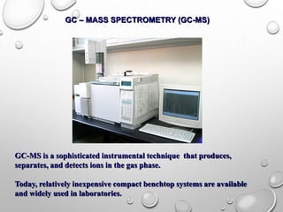
- 1. GC – MASS SPECTROMETRY (GC-MS) GC-MS is a sophisticated instrumental technique that produces, separates, and detects ions in the gas phase. Today, relatively inexpensive compact benchtop systems are available and widely used in laboratories.
- 2. • Mass spectroscopy is used to determine the molecular formula of the unknown compound. • Mass spectroscopy data that provides structural information tends to be unreliable and thus will only be used to verify a possible structure or in the event that the other spectral techniques are unsuccessful. GC – MASS SPECTROMETRY (GC-MS)
- 3. GAS CHROMATOGRAPHY – MASS SPECTROMETRY (GCMS) Process When a peak is detected in gas chromatography, some of the component is sent to a mass spectrometer A mass spectrometer has three main parts...
- 4. GAS CHROMATOGRAPHY – MASS SPECTROMETRY (GCMS) Process When a peak is detected in gas chromatography, some of the component is sent to a mass spectrometer A mass spectrometer has three main parts... Ionizer - the sample is bombarded with electrons and ionized - a positive molecular ion is formed - the molecular ion can break up into smaller ions - positive ions are accelerated toward the analyser
- 5. GAS CHROMATOGRAPHY – MASS SPECTROMETRY (GCMS) Process When a peak is detected in gas chromatography, some of the component is sent to a mass spectrometer A mass spectrometer has three main parts... Ioniser - the sample is bombarded with electrons and ionised - a positive molecular ion is formed - the molecular ion can break up into smaller ions - positive ions are accelerated towards the analyser Analyser - positive ions separate according to mass/charge ratio - higher mass/charge ratio = smaller deflection
- 6. GAS CHROMATOGRAPHY – MASS SPECTROMETRY (GCMS) Process When a peak is detected in gas chromatography, some of the component is sent to a mass spectrometer A mass spectrometer has three main parts... Ionizer - the sample is bombarded with electrons and ionized - a positive molecular ion is formed - the molecular ion can break up into smaller ions - positive ions are accelerated toward the analyser Analyzer - positive ions separate according to mass/charge ratio - higher mass/charge ratio = smaller deflection Detector - records the identity and abundance of each ion - compounds have a unique mass spectrum - the final peak (molecular ion) gives the molecular mass
- 7. GAS CHROMATOGRAPHY – MASS SPECTROMETRY (GCMS) • Process When a peak is detected in gas chromatography, some of the component is sent to a mass spectrometer A mass spectrometer has three main parts... Ionizer - the sample is bombarded with electrons and ionized - a positive molecular ion is formed - the molecular ion can break up into smaller ions - positive ions are accelerated toward the analyzer Analyzer - positive ions separate according to mass/charge ratio - higher mass/charge ratio = smaller deflection Detector - records the identity and abundance of each ion - compounds have a unique mass spectrum - the final peak (molecular ion) gives the molecular mass
- 8. A MASS SPECTROMETER ION SOURCE ANALYSER DETECTOR IONISATION • gaseous atoms are bombarded by electrons from an electron gun and are IONISED • sufficient energy is given to form ions of 1+ charge ACCELERATION • ions are charged so can be ACCELERATED by an electric field DEFLECTION • charged particles will be DEFLECTED by a magnetic or electric field DETECTION • by electric or photographic methods
- 9. PRINCIPLE OF GC-MS Block diagram of mass spectroscopy
- 10. • The inlet transfers the sample into the vacuum of the mass spectrometer. In the source region, neutral sample molecules are ionized and then accelerated into the mass analyzer. • The mass analyzer is the heart of the mass spectrometer. This section separates ions, either in space or in time, according to their mass-to-charge ratio. • After the ions are separated, they are detected and the signal is transferred to a data system for analysis. • All mass spectrometers also have a vacuum system to maintain the low pressure, which is also called a high vacuum, required for operation. • High vacuum minimizes ion-molecule reactions, scattering, and neutralization of the ions. • In some experiments, the pressure in the source region or a part of the mass spectrometer is intentionally increased to study these ion-molecule reactions. Under normal operation, however, any collisions will interfere with the analysis.
- 12. ELECTRON IMPACT (EI) • Electron Ionization (EI) is the most common ionization technique used for mass spectrometry. EI works well for many gas phase molecules, but it does have some limitations. • Although the mass spectra are very reproducible and are widely used for spectral libraries, EI causes extensive fragmentation so that the molecular ion is not observed for many compounds. Fragmentation is useful because it provides structural information for interpreting unknown spectra.
- 14. A) Ionizing electron approaches the electron cloud of a molecule B)Electron cloud distorted by ionizing electron C) Electron cloud further distorted by ionizing electron ELECTRON IONIZATION PROCESS
- 15. E) Electron cloud of molecule ejecting an electron F) Molecular ion and ejected electron. D) Ionizing electron passes by the molecule
- 16. CHEMICAL IONIZATION • Chemical Ionization (CI) is a “soft” ionization technique that produces ions with little excess energy. As a result, less fragmentation is observed in the mass spectrum. • Since this increases the abundance of the molecular ion, the technique is complementary to 70 eV EI. • CI is often used to verify the molecular mass of an unknown. Only slight modifications of an EI source region are required for CI experiments.
- 17. • In Chemical Ionization the source is enclosed in a small cell with openings for the electron beam, the reagent gas, and the sample. • The reagent gas is added to this cell at approximately 10 Pa (0.1 torrs) pressure. • This is higher than the 10-3 Pa (10-5 torr) pressure typical for a mass spectrometer source. At 10-3 Pa the mean free path between collisions is approximately 2 meters and ion-molecule reactions are unlikely. • In the CI source, however, the mean free path between collisions is only 10-4 meters and analyte molecules undergo many collisions with the reagent gas. • The reagent gas in the CI source is ionized with an electron beam to produce a cloud of ions. The reagent gas ions in this cloud react and produce adduct ions like CH5+, which are excellent proton donors.
- 18. • When analyte molecules (M) are introduced to a source region with this cloud of ions, the reagent gas ions donate a proton to the analyte molecule and produce MH+ ions. • The energy of the proton transfer is controlled by using different reagent gases. • The most common reagent gases are methane, isobutane, and ammonia. Methane is the strongest proton donor commonly used with a proton affinity (PA) of 5.7 eV. For softer ionization, isobutane (PA 8.5 eV) and ammonia (PA 9.0 eV) are frequently used. • Acid base chemistry is frequently used to describe chemical ionization reactions. The reagent gas must be a strong enough Brønsted acid to transfer a proton to the analyte. • Fragmentation is minimized in CI by reducing the amount of excess energy produced by the reaction. Because the adduct ions have little excess energy and are relatively stable, CI is very useful for molecular mass determination.
- 19. MASS ANALYZER • After ions are formed in the source region they are accelerated into the mass analyzer by an electric field. The mass analyzer separates these ions according to their m/z value. • The selection of a mass analyzer depends upon the resolution, mass range, scan rate, and detection limits required for an application. • Each analyzer has very different operating characteristics and the selection of an instrument involves important tradeoffs.
- 20. QUADRUPOLE MASS FILTER • The quadrupole mass spectrometer is the most common mass analyzer. • Its compact size, fast scan rate, high transmission efficiency, and modest vacuum requirements are ideal for small inexpensive instruments. • It ‘filter’ and only allow specific ions to pass. • Most quadrupole instruments are limited to unit m/z resolution and have a mass range of m/z 1000. • Many benchtop instruments have a mass range of m/z 500 but research instruments are available with a mass range of up to m/z 4000.
- 21. QUADRUPOLE MASS ANALYZER • Only compounds with a specific m/z ratio will resonate along the field (stable path) • Achieved by rapidly varying the voltage
- 22. TIME OF FLIGHT ANALYZER • The time-of-flight (TOF) mass analyzer separates ions in time as they travel down a flight tube. • This is a very simple mass spectrometer that uses fixed voltages and does not require a magnetic field. The greatest drawback is that TOF instruments have the poor mass resolution, usually less than 500. • These instruments have high transmission efficiency, no upper m/z limit, very low detection limits, and fast scan rates. • For some applications these advantages outweigh the low resolution. • Recent developments in pulsed ionization techniques and new instrument designs with improved resolution have renewed interest in TOF-MS.
- 23. • This picture shows the working principle of a linear time of flight mass spectrometer. • To allow the ions to fly through the flight path without hitting anything else, all the air molecules have been pumped out to create an ultra-high vacuum. • Ions of different m/z ratio travel at different velocities. • The difference in arrival times separates two (or more) ions.
- 24. Graph of ion intensity versus mass-to-charge ratio (m/z) (units daltons, Da)