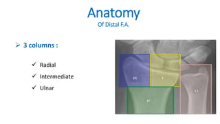
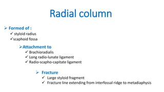
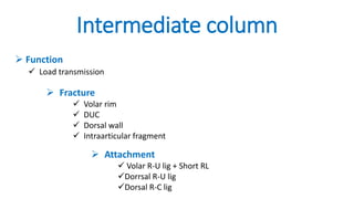
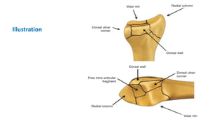
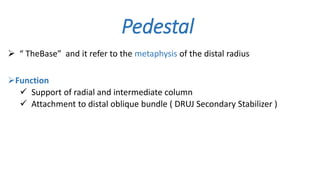
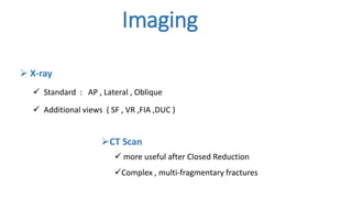
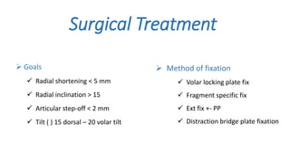
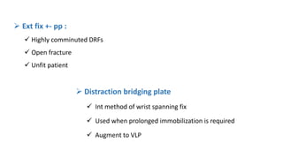
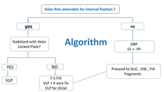
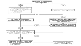
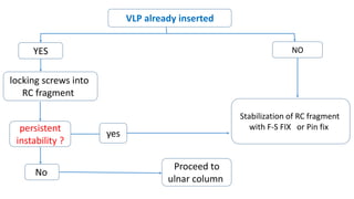
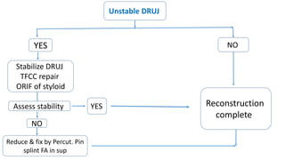
Journal Club
Complex Distal Radius
Fracture
Presented by :
Ibraheim El Wasif
Orthopedic Surgery Resident (R3)
Abdallah Jomaa El Azanki
Orthopedic Surgery As. Lecturer
Cedar Tree of
Lebanon
About the article
 Authors :
 Peter Charles Rhee, DO, MS
 Robert J. Medoff, MD
 Alexander Y. Shin, MD
Journal of the American Academy of Orthopedic Surgeons , 2017 .
Department of Orthopedic Surgery, San Antonio Military Medical Center
Department of Orthopedic Surgery, University of Hawaii, Kailua
Department of Orthopedic Surgery, Mayo Clinic
Anatomy
Of Distal F.A.
 3 columns :
 Radial
 Intermediate
 Ulnar
Radial column
 Formed of :
 styloid radius
scaphoid fossa
Attachment to
 Brachioradialis
 Long radio-lunate ligament
 Radio-scapho-capitate ligament
 Fracture
 Large styloid fragment
 Fracture line extending from interfossal ridge to metadiaphysis
Intermediate column
 Function
 Load transmission
 Attachment
 Volar R-U lig + Short RL
Dorrsal R-U lig
Dorsal R-C lig
 Fracture
 Volar rim
 DUC
 Dorsal wall
 Intraarticular fragment
Illustration
Ulnar column
Formed of :
 Distal ulna
 TFCC
 Attachments :
Dorsal RU lig
Volar RU lig
Function :
 DRUJ stability
Pedestal
 “ TheBase” and it refer to the metaphysis of the distal radius
Function
 Support of radial and intermediate column
 Attachment to distal oblique bundle ( DRUJ Secondary Stabilizer )
Imaging
 X-ray
 Standard : AP , Lateral , Oblique
 Additional views ( SF , VR ,FIA ,DUC )
CT Scan
 more useful after Closed Reduction
Complex , multi-fragmentary fractures
Surgical Treatment
 Goals
 Radial shortening < 5 mm
 Radial inclination > 15
 Articular step-off < 2 mm
 Tilt ( ) 15 dorsal – 20 volar tilt
 Method of fixation
 Volar locking plate fix
 Fragment specific fix
 Ext fix +- PP
 Distraction bridge plate fixation
 Fragment specific fix :
 App of individualized low profile implant
 Create multi-planar , load shearing construct
 Useful when # fragments too small or distal
 Volar locking plate :
Fixed angle construct
Mainstay for fix of DRFs
Additional fix may be required
 Distraction bridging plate
 Int method of wrist spanning fix
 Used when prolonged immobilization is required
 Augment to VLP
 Ext fix +- pp :
 Highly comminuted DRFs
 Open fracture
 Unfit patient
Reconstruction
 Sequence
1. Intermediate
2. Radial
3. Ulnar
Volar Rim amenable for internal fixation ?
noyes
DBP
EF +- PP
Stabilized with Volar
Locked Plate?
NOYYES
VLP
F-S FIX
VLP + K wire fix
VLP far distal
Proceed to DUC , DW , FIA
fragments
Algorithm
VLP already inserted
NOYES
locking screws into
RC fragment
persistent
instability ?
yes
Stabilization of RC fragment
with F-S FIX or Pin fix
No
Proceed to
ulnar column
Unstable DRUJ
NOYES
Stabilize DRUJ
TFCC repair
ORIF of styloid
Assess stability YES Reconstruction
complete
NO
Reduce & fix by Percut. Pin
splint FA in sup
Summary
 Surgical treatment of complex DRFs very challenging .
 Understanding anatomy & goals of reconstruction are beneficial in
preoperative planning and decision making .
 Successful surgical management require familiarity of various
surgical methods.
Complex Distal Radius Fracture  #dr_azanki

Complex Distal Radius Fracture #dr_azanki

  • 1.
    Journal Club Complex DistalRadius Fracture Presented by : Ibraheim El Wasif Orthopedic Surgery Resident (R3) Abdallah Jomaa El Azanki Orthopedic Surgery As. Lecturer Cedar Tree of Lebanon
  • 2.
    About the article Authors :  Peter Charles Rhee, DO, MS  Robert J. Medoff, MD  Alexander Y. Shin, MD Journal of the American Academy of Orthopedic Surgeons , 2017 . Department of Orthopedic Surgery, San Antonio Military Medical Center Department of Orthopedic Surgery, University of Hawaii, Kailua Department of Orthopedic Surgery, Mayo Clinic
  • 3.
    Anatomy Of Distal F.A. 3 columns :  Radial  Intermediate  Ulnar
  • 4.
    Radial column  Formedof :  styloid radius scaphoid fossa Attachment to  Brachioradialis  Long radio-lunate ligament  Radio-scapho-capitate ligament  Fracture  Large styloid fragment  Fracture line extending from interfossal ridge to metadiaphysis
  • 5.
    Intermediate column  Function Load transmission  Attachment  Volar R-U lig + Short RL Dorrsal R-U lig Dorsal R-C lig  Fracture  Volar rim  DUC  Dorsal wall  Intraarticular fragment
  • 6.
  • 7.
    Ulnar column Formed of:  Distal ulna  TFCC  Attachments : Dorsal RU lig Volar RU lig Function :  DRUJ stability
  • 8.
    Pedestal  “ TheBase”and it refer to the metaphysis of the distal radius Function  Support of radial and intermediate column  Attachment to distal oblique bundle ( DRUJ Secondary Stabilizer )
  • 9.
    Imaging  X-ray  Standard: AP , Lateral , Oblique  Additional views ( SF , VR ,FIA ,DUC ) CT Scan  more useful after Closed Reduction Complex , multi-fragmentary fractures
  • 10.
    Surgical Treatment  Goals Radial shortening < 5 mm  Radial inclination > 15  Articular step-off < 2 mm  Tilt ( ) 15 dorsal – 20 volar tilt  Method of fixation  Volar locking plate fix  Fragment specific fix  Ext fix +- PP  Distraction bridge plate fixation
  • 11.
     Fragment specificfix :  App of individualized low profile implant  Create multi-planar , load shearing construct  Useful when # fragments too small or distal  Volar locking plate : Fixed angle construct Mainstay for fix of DRFs Additional fix may be required
  • 12.
     Distraction bridgingplate  Int method of wrist spanning fix  Used when prolonged immobilization is required  Augment to VLP  Ext fix +- pp :  Highly comminuted DRFs  Open fracture  Unfit patient
  • 13.
  • 14.
    Volar Rim amenablefor internal fixation ? noyes DBP EF +- PP Stabilized with Volar Locked Plate? NOYYES VLP F-S FIX VLP + K wire fix VLP far distal Proceed to DUC , DW , FIA fragments Algorithm
  • 16.
    VLP already inserted NOYES lockingscrews into RC fragment persistent instability ? yes Stabilization of RC fragment with F-S FIX or Pin fix No Proceed to ulnar column
  • 17.
    Unstable DRUJ NOYES Stabilize DRUJ TFCCrepair ORIF of styloid Assess stability YES Reconstruction complete NO Reduce & fix by Percut. Pin splint FA in sup
  • 18.
    Summary  Surgical treatmentof complex DRFs very challenging .  Understanding anatomy & goals of reconstruction are beneficial in preoperative planning and decision making .  Successful surgical management require familiarity of various surgical methods.