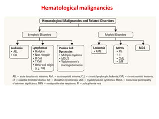
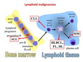
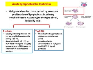
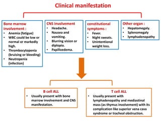
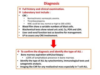
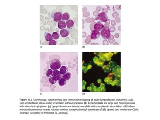
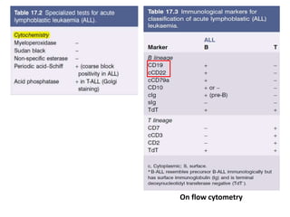
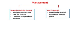
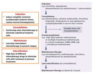
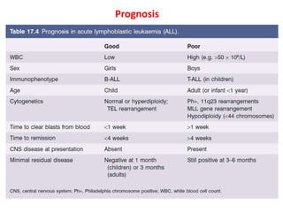
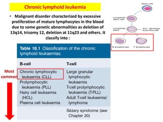
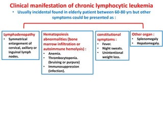
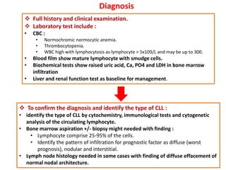
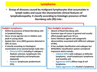
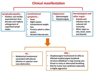
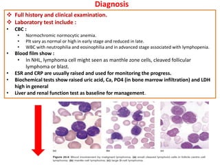
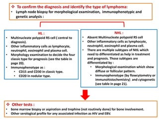
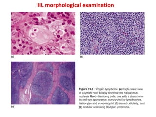
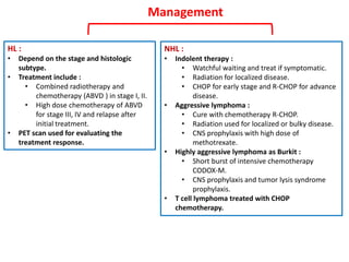
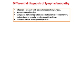
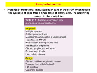
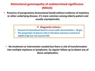
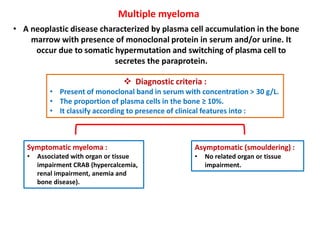
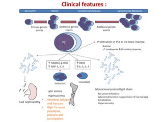
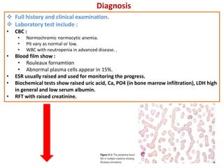
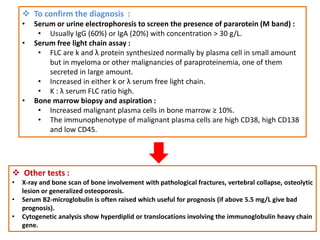
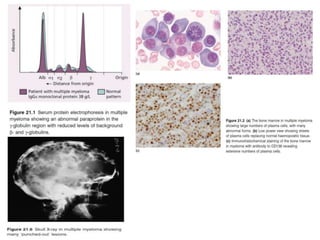
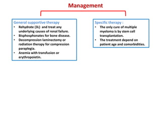
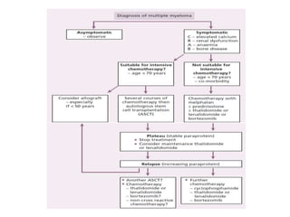
Lymphoid disorders
Mohammed AlHinai
Hematological malignancies
stem
cell
lymphoid
progenitor
progenitor-B
pre-B
immature
B-cell
memory
B-cell
plasma cell
DLBCL,
FL, HL
ALL
CLL
MM
germinal
center
B-cell
mature
naive
B-cell
Lymphoid malignancies
Lymphoid disorders
Acute lymphoblastic leukemia
• Malignant disorder characterized by excessive
proliferation of lymphoblast in primary
lymphoid tissue. According to the type of cell,
it classify into :
T cell ALL
• Usually affecting childhood,
adolescence and young
adult.
• Associated with
abnormalities in TCR gene
and NOTCH1 signal
pathway.
B cell ALL
• Usually affecting children < 6
yrs but could also present in
eldery > 60 yrs.
• Associated with t(9 ; 22) in
BCR-ABL1 oncogene, t(12;21),
rearrangment of MLL gene or
alteration in chromosome
number.
Clinical manifestation
Bone marrow
involvement :
• Anemia (fatigue)
• WBC could be low or
normal or markedly
high.
• Thrombocytopenia
(bruising or bleeding)
• Neutropenia
(infection)
constitutional
symptoms :
• Fever.
• Night sweats.
• Unintentional
weight loss.
Other organ :
• Hepatomegaly.
• Splenomegaly
• lymphadenopathy
CNS involvement
• Headache.
• Nausea and
vomiting.
• Blurring vision or
diplopia.
• Papilloedema.
T cell ALL
• Usually present with
lymphadenopathy and mediastinal
mass (as thymus involvement) with its
complication like superior vena cava
syndrome or tracheal obstruction.
B cell ALL
• Usually present with bone
marrow involvement and CNS
manifestation.
Diagnosis
 Full history and clinical examination.
 Laboratory test include :
• CBC :
• Normochromic normocytic anemia.
• Thrombocytopenia.
• WBC could be low, normal or high to 200 x109/L
• Blood film show a variable numbers of blast cells.
• Biochemical tests show raised uric acid, Ca, PO4 and LDH.
• Liver and renal function test as baseline for management.
• LP to assess any CNS involvement.
 To confirm the diagnosis and identify the type of ALL :
• Bone marrow aspiration with/out biopsy :
• ≥20% of lymphoblast presence in bone marrow.
• identify the type of ALL by cytochemistry, immunological tests and
cytogenetic analysis.
• Imaging like CXR for any mediastinal mass especially in T cell-ALL.
On flow cytometry
Management
Specific therapy :
• Chemotherapy with/out
radiotherapy in several
phases.
General supportive therapy
• Blood product transfusion.
• Treat any infection.
• Correction of any metabolic
imbalance.
Induction
• Induce complete remission
(undetectable leukemic blasts,
restore normal hematopoiesis).
Consolidation
• Continuing same chemotherapy to
eliminate subclinical leukemic
cells.
Maintainace
• Low dose intermittent
chemotherapy to prevent relapse.
Intensification
• High dose of different
chemotherapy drugs to eliminate
cells with resistance to primary
treatment.
Prognosis
Chronic lymphoid leukemia
• Malignant disorder characterized by excessive
proliferation of mature lymphocytes in the blood
due to some genetic abnormalities as deletion of
13q14, trisomy 12, deletion at 11q23 and others. It
classify into :
Most
common
Clinical manifestation of chronic lymphocytic leukemia
• Usually incidental found in elderly patient between 60-80 yrs but other
symptoms could be presented as :
Lymphadenopathy
• Symmetrical
enlargement of
cervical, axillary or
inguinal lymph
nodes.
constitutional
symptoms :
• Fever.
• Night sweats.
• Unintentional
weight loss.
Other organ :
• Splenomegaly
• Hepatomegaly.
Hematopoiesis
abnormalities (bone
marrow infiltration or
autoimmune hemolysis) :
• Anemia.
• Thrombocytopenia.
(bruising or purpura)
• Immunosuppression
(infection).
Diagnosis
 Full history and clinical examination.
 Laboratory test include :
• CBC :
• Normochromic normocytic anemia.
• Thrombocytopenia.
• WBC high with lymphocytosis as lymphocyte > 5x10⁹/L and may be up to 300.
• Blood film show mature lymphocyte with smudge cells.
• Biochemical tests show raised uric acid, Ca, PO4 and LDH in bone marrow
infiltration
• Liver and renal function test as baseline for management.
 To confirm the diagnosis and identify the type of CLL :
• identify the type of CLL by cytochemistry, immunological tests and cytogenetic
analysis of the circulating lymphocyte.
• Bone marrow aspiration +/- biopsy might needed with finding :
• Lymphocyte comprise 25-95% of the cells.
• Identify the pattern of infiltration for prognostic factor as diffuse (worst
prognosis), nodular and interstitial.
• Lymph node histology needed in some cases with finding of diffuse effacement of
normal nodal architecture.
Treatment
• The aiming of treatment is conservative to control the symptoms as the cure
is rare.
• If the patient present with autoimmune hemolytic or bone marrow failure,
treated initially with prednisolone only until recovery of blood parameters.
• Then combination therapy of R-FC (retuximab, fludarabine and
cyclophosphamide) given every 4 weeks with 4-6 courses until the
symptoms are controlled. As side effect like pneumocytoss carinii and
herpes infection, co-trimoxazole and aciclovir are given for 6 months.
• Other treatment may include :
• Chlorambucil for elderly patients.
• Radiotherapy to reducing the size of bulky lymph node groups.
• R-CHOP chemotherapy used in late stage cases.
Lymphoma
• Group of diseases caused by malignant lymphocytes that accumulate in
lymph nodes and cause the characteristic clinical features of
lymphadenopathy. It classify according to histologic presence of Red-
Sternberg cells (RS) into :
Non-Hodgkin lymphoma :
• Absent of Reed-Sternberg cells.
• Common type of cancer in general and usually
affect people aged over 65 yrs.
• Some infectious agents and cytogenetic
abnormalities associated with different subtypes
of NHL.
• It has multiple classification and subtypes but
WHO/REAL classification system considered
important which include :
• Indolent (40%) with subtypes as follicular
lymphoma, small lymphocytic lymphoma
and manthle cell.
• Aggressive (50%) diffuse large B cell
lymphoma.
• Highly aggressive (5%) Burkitt’s lymphoma.
Hodgkin lymphoma :
• Define by presence of Reed-Sternberg cells
in lymphoid biopsy.
• Occur at any age but rare in children and
peak in young adult.
• Epstein-Barr virus (EBV) genome detected in
50% of cases.
• It classify according to histological
examination of an excised lymph node into :
• Classical (95%) including 4 subtypes (
Nodular sclerosis, lymphocyte rich,
mixed cellularity and lymphocyte
depleted).
• Nodular lymphocyte predominant
(5%).
Histological classification of HL :
Infectious agents and cytogenetic abnormalities in
NHL :
Clinical manifestation
Lymphadenopathy :
• Painless, non-tender,
asymmetrical, firm,
discrete and rubbery
enlargement of
superficial lymph
nodes.
constitutional
symptoms :
• Fever.
• Night sweats.
• Unintentional weight
loss.
• Pruritus which is often
severe.
• Alcohol induced pain.
Abdominal :
• Splenomegaly
• Hepatomegaly.
NHL :
• Oropharyngeal involvement (5-10%) as
affected oropharyngeal lymphoid
structure (Waldeyer’s ring) causing sore
throat or noisy or obstructed breathing.
• Risk for tumor lysis syndrome especially
in highly aggressive
HL :
• Mediastinal involvement
associated with pleural
effusion or superior vena
cava obstruction.
Hematological and
other organ:
• Anemia and
infection due to
reduced cell
immunity.
• Other organ as
skin, brain, testis
and thyroid.
Diagnosis
 Full history and clinical examination.
 Laboratory test include :
• CBC :
• Normochromic normocytic anemia.
• Plt vary as normal or high in early stage and reduced in late.
• WBC with neutrophilia and eosinophilia and in advanced stage associated with lymphopenia.
• Blood film show :
• In NHL, lymphoma cell might seen as manthle zone cells, cleaved follicular
lymphoma or blast.
• ESR and CRP are usually raised and used for monitoring the progress.
• Biochemical tests show raised uric acid, Ca, PO4 (in bone marrow infiltration) and LDH
high in general
• Liver and renal function test as baseline for management.
 To confirm the diagnosis and identify the type of lymphoma:
• Lymph node biopsy for morphological examination, immunophenotypic and
genetic analysis :
HL :
• Multinucleate polypoid RS cell ( central to
diagnosis).
• Other inflammatory cells as lymphocyte,
neutrophil, eosinophil and plasma cell.
• Morphology examination to decide the four
classic type for prognosis (see the table in
page 20).
• Immunophenotype as :
• CD15 and CD30 in classic type.
• CD20 in nodular type.
NHL :
• Absent Multinucleate polypoid RS cell
• Other inflammatory cells as lymphocyte,
neutrophil, eosinophil and plasma cell.
• There are multiple subtypes of NHL which
need to differentiated as help in treatment
and prognosis. Those subtypes are
differentiated by :
• Morphological examination which show
diffuse or follicular pattern.
• Immunophenotype (by flowcytometry or
immunohistochemistry) and cytogenetic
(see table in page 21).
 Other tests :
• bone marrow biopsy or aspiration and trephine (not routinely done) for bone involvement.
• Other serological profile for any associated infection as HIV and EBV.
HL morphological examination
NHL morphological examination
Starry sky appearance in Burkit
lymphoma
 Staging :
• Important for management and prognosis which use combined clinical examination and
imaging to detect superficial and deep involved lymph nodes with other tissue
involvement. The imaging include :
• CXR for any hilar lymph node or mediastinal mass.
• Chest, abdomen and pelvic CT.
• PET to detect any tiny foci.
• MRI and bone scan needed in some patient.
Management
NHL :
• Indolent therapy :
• Watchful waiting and treat if symptomatic.
• Radiation for localized disease.
• CHOP for early stage and R-CHOP for advance
disease.
• Aggressive lymphoma :
• Cure with chemotherapy R-CHOP.
• Radiation used for localized or bulky disease.
• CNS prophylaxis with high dose of
methotrexate.
• Highly aggressive lymphoma as Burkit :
• Short burst of intensive chemotherapy
CODOX-M.
• CNS prophylaxis and tumor lysis syndrome
prophylaxis.
• T cell lymphoma treated with CHOP
chemotherapy.
HL :
• Depend on the stage and histologic
subtype.
• Treatment include :
• Combined radiotherapy and
chemotherapy (ABVD ) in stage I, II.
• High dose chemotherapy of ABVD
for stage III, IV and relapse after
initial treatment.
• PET scan used for evaluating the
treatment response.
Differential diagnosis of lymphadenopathy
• Infection : present with painful smooth lymph node.
• Autoimmune disorders
• Malignant hematological disease as leukemia : bone marrow
and peripheral vascular predominant involving.
• Metastasis from other primary tumor.
Para-proteinaemia
• Presence of monoclonal immunoglobulin band in the serum which reflects
the synthesis of band from a single clone of plasma cells. The underlying
causes of this classify into :
Monoclonal gammopathy of undetermined significance
(MGUS)
• Presence of paraproteins (monoclonal band) without evidence of myeloma
or other underlying disease. It’s more common among elderly patient and
usually asymptomatic.
 Diagnostic criteria :
• Present of monoclonal band in serum with concentration < 30 g/L.
• The proportion of plasma cells in the bone marrow is normal or
slightly high but not exceed 10%.
• No treatment or intervention needed but there is risk of transformation
into multiple myeloma or lymphoma. So regular follow up to detect any of
these complication.
Multiple myeloma
• A neoplastic disease characterized by plasma cell accumulation in the bone
marrow with presence of monoclonal protein in serum and/or urine. It
occur due to somatic hypermutation and switching of plasma cell to
secretes the paraprotein.
 Diagnostic criteria :
• Present of monoclonal band in serum with concentration > 30 g/L.
• The proportion of plasma cells in the bone ≥ 10%.
• It classify according to presence of clinical features into :
Asymptomatic (smouldering) :
• No related organ or tissue
impairment.
Symptomatic myeloma :
• Associated with organ or tissue
impairment CRAB (hypercalcemia,
renal impairment, anemia and
bone disease).
• Vertebral collapse
and fracture.
• High Ca cause
polydipsia,
polyuria and
constipation.
Clinical features :
Diagnosis
 Full history and clinical examination.
 Laboratory test include :
• CBC :
• Normochromic normocytic anemia.
• Plt vary as normal or low.
• WBC with neutropenia in advanced disease. .
• Blood film show :
• Rouleaux fornamtion
• Abnormal plasma cells appear in 15%.
• ESR usually raised and used for monitoring the progress.
• Biochemical tests show raised uric acid, Ca, PO4 (in bone marrow infiltration), LDH high
in general and low serum albumin.
• RFT with raised creatinine.
 To confirm the diagnosis :
• Serum or urine electrophoresis to screen the presence of pararotein (M band) :
• Usually IgG (60%) or IgA (20%) with concentration > 30 g/L.
• Serum free light chain assay :
• FLC are k and λ protein synthesized normally by plasma cell in small amount
but in myeloma or other malignancies of paraproteinemia, one of them
secreted in large amount.
• Increased in either k or λ serum free light chain.
• K : λ serum FLC ratio high.
• Bone marrow biopsy and aspiration :
• Increased malignant plasma cells in bone marrow ≥ 10%.
• The immunophenotype of malignant plasma cells are high CD38, high CD138
and low CD45.
 Other tests :
• X-ray and bone scan of bone involvement with pathological fractures, vertebral collapse, osteolytic
lesion or generalized osteoporosis.
• Serum B2-microglobulin is often raised which useful for prognosis (if above 5.5 mg/L give bad
prognosis).
• Cytogenetic analysis show hyperdiplid or translocations involving the immunoglobulin heavy chain
gene.
Management
Specific therapy :
• The only cure of multiple
myeloma is by stem cell
transplantation.
• The treatment depend on
patient age and comorbidities.
General supportive therapy
• Rehydrate (3L) and treat any
underlying causes of renal failure.
• Bisphosphonates for bone disease.
• Decompression laminectomy or
radiation therapy for compression
paraplegia.
• Anemia with transfusion or
erythropoietin.
Lymphoid malignancies

Lymphoid malignancies

  • 1.
  • 2.
  • 3.
  • 4.
  • 5.
    Acute lymphoblastic leukemia •Malignant disorder characterized by excessive proliferation of lymphoblast in primary lymphoid tissue. According to the type of cell, it classify into : T cell ALL • Usually affecting childhood, adolescence and young adult. • Associated with abnormalities in TCR gene and NOTCH1 signal pathway. B cell ALL • Usually affecting children < 6 yrs but could also present in eldery > 60 yrs. • Associated with t(9 ; 22) in BCR-ABL1 oncogene, t(12;21), rearrangment of MLL gene or alteration in chromosome number.
  • 6.
    Clinical manifestation Bone marrow involvement: • Anemia (fatigue) • WBC could be low or normal or markedly high. • Thrombocytopenia (bruising or bleeding) • Neutropenia (infection) constitutional symptoms : • Fever. • Night sweats. • Unintentional weight loss. Other organ : • Hepatomegaly. • Splenomegaly • lymphadenopathy CNS involvement • Headache. • Nausea and vomiting. • Blurring vision or diplopia. • Papilloedema. T cell ALL • Usually present with lymphadenopathy and mediastinal mass (as thymus involvement) with its complication like superior vena cava syndrome or tracheal obstruction. B cell ALL • Usually present with bone marrow involvement and CNS manifestation.
  • 7.
    Diagnosis  Full historyand clinical examination.  Laboratory test include : • CBC : • Normochromic normocytic anemia. • Thrombocytopenia. • WBC could be low, normal or high to 200 x109/L • Blood film show a variable numbers of blast cells. • Biochemical tests show raised uric acid, Ca, PO4 and LDH. • Liver and renal function test as baseline for management. • LP to assess any CNS involvement.  To confirm the diagnosis and identify the type of ALL : • Bone marrow aspiration with/out biopsy : • ≥20% of lymphoblast presence in bone marrow. • identify the type of ALL by cytochemistry, immunological tests and cytogenetic analysis. • Imaging like CXR for any mediastinal mass especially in T cell-ALL.
  • 9.
  • 10.
    Management Specific therapy : •Chemotherapy with/out radiotherapy in several phases. General supportive therapy • Blood product transfusion. • Treat any infection. • Correction of any metabolic imbalance.
  • 11.
    Induction • Induce completeremission (undetectable leukemic blasts, restore normal hematopoiesis). Consolidation • Continuing same chemotherapy to eliminate subclinical leukemic cells. Maintainace • Low dose intermittent chemotherapy to prevent relapse. Intensification • High dose of different chemotherapy drugs to eliminate cells with resistance to primary treatment.
  • 12.
  • 13.
    Chronic lymphoid leukemia •Malignant disorder characterized by excessive proliferation of mature lymphocytes in the blood due to some genetic abnormalities as deletion of 13q14, trisomy 12, deletion at 11q23 and others. It classify into : Most common
  • 14.
    Clinical manifestation ofchronic lymphocytic leukemia • Usually incidental found in elderly patient between 60-80 yrs but other symptoms could be presented as : Lymphadenopathy • Symmetrical enlargement of cervical, axillary or inguinal lymph nodes. constitutional symptoms : • Fever. • Night sweats. • Unintentional weight loss. Other organ : • Splenomegaly • Hepatomegaly. Hematopoiesis abnormalities (bone marrow infiltration or autoimmune hemolysis) : • Anemia. • Thrombocytopenia. (bruising or purpura) • Immunosuppression (infection).
  • 15.
    Diagnosis  Full historyand clinical examination.  Laboratory test include : • CBC : • Normochromic normocytic anemia. • Thrombocytopenia. • WBC high with lymphocytosis as lymphocyte > 5x10⁹/L and may be up to 300. • Blood film show mature lymphocyte with smudge cells. • Biochemical tests show raised uric acid, Ca, PO4 and LDH in bone marrow infiltration • Liver and renal function test as baseline for management.  To confirm the diagnosis and identify the type of CLL : • identify the type of CLL by cytochemistry, immunological tests and cytogenetic analysis of the circulating lymphocyte. • Bone marrow aspiration +/- biopsy might needed with finding : • Lymphocyte comprise 25-95% of the cells. • Identify the pattern of infiltration for prognostic factor as diffuse (worst prognosis), nodular and interstitial. • Lymph node histology needed in some cases with finding of diffuse effacement of normal nodal architecture.
  • 18.
    Treatment • The aimingof treatment is conservative to control the symptoms as the cure is rare. • If the patient present with autoimmune hemolytic or bone marrow failure, treated initially with prednisolone only until recovery of blood parameters. • Then combination therapy of R-FC (retuximab, fludarabine and cyclophosphamide) given every 4 weeks with 4-6 courses until the symptoms are controlled. As side effect like pneumocytoss carinii and herpes infection, co-trimoxazole and aciclovir are given for 6 months. • Other treatment may include : • Chlorambucil for elderly patients. • Radiotherapy to reducing the size of bulky lymph node groups. • R-CHOP chemotherapy used in late stage cases.
  • 20.
    Lymphoma • Group ofdiseases caused by malignant lymphocytes that accumulate in lymph nodes and cause the characteristic clinical features of lymphadenopathy. It classify according to histologic presence of Red- Sternberg cells (RS) into : Non-Hodgkin lymphoma : • Absent of Reed-Sternberg cells. • Common type of cancer in general and usually affect people aged over 65 yrs. • Some infectious agents and cytogenetic abnormalities associated with different subtypes of NHL. • It has multiple classification and subtypes but WHO/REAL classification system considered important which include : • Indolent (40%) with subtypes as follicular lymphoma, small lymphocytic lymphoma and manthle cell. • Aggressive (50%) diffuse large B cell lymphoma. • Highly aggressive (5%) Burkitt’s lymphoma. Hodgkin lymphoma : • Define by presence of Reed-Sternberg cells in lymphoid biopsy. • Occur at any age but rare in children and peak in young adult. • Epstein-Barr virus (EBV) genome detected in 50% of cases. • It classify according to histological examination of an excised lymph node into : • Classical (95%) including 4 subtypes ( Nodular sclerosis, lymphocyte rich, mixed cellularity and lymphocyte depleted). • Nodular lymphocyte predominant (5%).
  • 21.
  • 22.
    Infectious agents andcytogenetic abnormalities in NHL :
  • 24.
    Clinical manifestation Lymphadenopathy : •Painless, non-tender, asymmetrical, firm, discrete and rubbery enlargement of superficial lymph nodes. constitutional symptoms : • Fever. • Night sweats. • Unintentional weight loss. • Pruritus which is often severe. • Alcohol induced pain. Abdominal : • Splenomegaly • Hepatomegaly. NHL : • Oropharyngeal involvement (5-10%) as affected oropharyngeal lymphoid structure (Waldeyer’s ring) causing sore throat or noisy or obstructed breathing. • Risk for tumor lysis syndrome especially in highly aggressive HL : • Mediastinal involvement associated with pleural effusion or superior vena cava obstruction. Hematological and other organ: • Anemia and infection due to reduced cell immunity. • Other organ as skin, brain, testis and thyroid.
  • 25.
    Diagnosis  Full historyand clinical examination.  Laboratory test include : • CBC : • Normochromic normocytic anemia. • Plt vary as normal or high in early stage and reduced in late. • WBC with neutrophilia and eosinophilia and in advanced stage associated with lymphopenia. • Blood film show : • In NHL, lymphoma cell might seen as manthle zone cells, cleaved follicular lymphoma or blast. • ESR and CRP are usually raised and used for monitoring the progress. • Biochemical tests show raised uric acid, Ca, PO4 (in bone marrow infiltration) and LDH high in general • Liver and renal function test as baseline for management.
  • 26.
     To confirmthe diagnosis and identify the type of lymphoma: • Lymph node biopsy for morphological examination, immunophenotypic and genetic analysis : HL : • Multinucleate polypoid RS cell ( central to diagnosis). • Other inflammatory cells as lymphocyte, neutrophil, eosinophil and plasma cell. • Morphology examination to decide the four classic type for prognosis (see the table in page 20). • Immunophenotype as : • CD15 and CD30 in classic type. • CD20 in nodular type. NHL : • Absent Multinucleate polypoid RS cell • Other inflammatory cells as lymphocyte, neutrophil, eosinophil and plasma cell. • There are multiple subtypes of NHL which need to differentiated as help in treatment and prognosis. Those subtypes are differentiated by : • Morphological examination which show diffuse or follicular pattern. • Immunophenotype (by flowcytometry or immunohistochemistry) and cytogenetic (see table in page 21).  Other tests : • bone marrow biopsy or aspiration and trephine (not routinely done) for bone involvement. • Other serological profile for any associated infection as HIV and EBV.
  • 27.
  • 28.
  • 29.
    Starry sky appearancein Burkit lymphoma
  • 30.
     Staging : •Important for management and prognosis which use combined clinical examination and imaging to detect superficial and deep involved lymph nodes with other tissue involvement. The imaging include : • CXR for any hilar lymph node or mediastinal mass. • Chest, abdomen and pelvic CT. • PET to detect any tiny foci. • MRI and bone scan needed in some patient.
  • 32.
    Management NHL : • Indolenttherapy : • Watchful waiting and treat if symptomatic. • Radiation for localized disease. • CHOP for early stage and R-CHOP for advance disease. • Aggressive lymphoma : • Cure with chemotherapy R-CHOP. • Radiation used for localized or bulky disease. • CNS prophylaxis with high dose of methotrexate. • Highly aggressive lymphoma as Burkit : • Short burst of intensive chemotherapy CODOX-M. • CNS prophylaxis and tumor lysis syndrome prophylaxis. • T cell lymphoma treated with CHOP chemotherapy. HL : • Depend on the stage and histologic subtype. • Treatment include : • Combined radiotherapy and chemotherapy (ABVD ) in stage I, II. • High dose chemotherapy of ABVD for stage III, IV and relapse after initial treatment. • PET scan used for evaluating the treatment response.
  • 33.
    Management NHL : • Indolenttherapy : • Watchful waiting and treat if symptomatic. • Radiation for localized disease. • CHOP for early stage and R-CHOP for advance disease. • Aggressive lymphoma : • Cure with chemotherapy R-CHOP. • Radiation used for localized or bulky disease. • CNS prophylaxis with high dose of methotrexate. • Highly aggressive lymphoma as Burkit : • Short burst of intensive chemotherapy CODOX-M. • CNS prophylaxis and tumor lysis syndrome prophylaxis. • T cell lymphoma treated with CHOP chemotherapy. HL : • Depend on the stage and histologic subtype. • Treatment include : • Combined radiotherapy and chemotherapy (ABVD ) in stage I, II. • High dose chemotherapy of ABVD for stage III, IV and relapse after initial treatment. • PET scan used for evaluating the treatment response.
  • 34.
    Differential diagnosis oflymphadenopathy • Infection : present with painful smooth lymph node. • Autoimmune disorders • Malignant hematological disease as leukemia : bone marrow and peripheral vascular predominant involving. • Metastasis from other primary tumor.
  • 35.
    Para-proteinaemia • Presence ofmonoclonal immunoglobulin band in the serum which reflects the synthesis of band from a single clone of plasma cells. The underlying causes of this classify into :
  • 36.
    Monoclonal gammopathy ofundetermined significance (MGUS) • Presence of paraproteins (monoclonal band) without evidence of myeloma or other underlying disease. It’s more common among elderly patient and usually asymptomatic.  Diagnostic criteria : • Present of monoclonal band in serum with concentration < 30 g/L. • The proportion of plasma cells in the bone marrow is normal or slightly high but not exceed 10%. • No treatment or intervention needed but there is risk of transformation into multiple myeloma or lymphoma. So regular follow up to detect any of these complication.
  • 37.
    Multiple myeloma • Aneoplastic disease characterized by plasma cell accumulation in the bone marrow with presence of monoclonal protein in serum and/or urine. It occur due to somatic hypermutation and switching of plasma cell to secretes the paraprotein.  Diagnostic criteria : • Present of monoclonal band in serum with concentration > 30 g/L. • The proportion of plasma cells in the bone ≥ 10%. • It classify according to presence of clinical features into : Asymptomatic (smouldering) : • No related organ or tissue impairment. Symptomatic myeloma : • Associated with organ or tissue impairment CRAB (hypercalcemia, renal impairment, anemia and bone disease).
  • 38.
    • Vertebral collapse andfracture. • High Ca cause polydipsia, polyuria and constipation. Clinical features :
  • 39.
    Diagnosis  Full historyand clinical examination.  Laboratory test include : • CBC : • Normochromic normocytic anemia. • Plt vary as normal or low. • WBC with neutropenia in advanced disease. . • Blood film show : • Rouleaux fornamtion • Abnormal plasma cells appear in 15%. • ESR usually raised and used for monitoring the progress. • Biochemical tests show raised uric acid, Ca, PO4 (in bone marrow infiltration), LDH high in general and low serum albumin. • RFT with raised creatinine.
  • 40.
     To confirmthe diagnosis : • Serum or urine electrophoresis to screen the presence of pararotein (M band) : • Usually IgG (60%) or IgA (20%) with concentration > 30 g/L. • Serum free light chain assay : • FLC are k and λ protein synthesized normally by plasma cell in small amount but in myeloma or other malignancies of paraproteinemia, one of them secreted in large amount. • Increased in either k or λ serum free light chain. • K : λ serum FLC ratio high. • Bone marrow biopsy and aspiration : • Increased malignant plasma cells in bone marrow ≥ 10%. • The immunophenotype of malignant plasma cells are high CD38, high CD138 and low CD45.  Other tests : • X-ray and bone scan of bone involvement with pathological fractures, vertebral collapse, osteolytic lesion or generalized osteoporosis. • Serum B2-microglobulin is often raised which useful for prognosis (if above 5.5 mg/L give bad prognosis). • Cytogenetic analysis show hyperdiplid or translocations involving the immunoglobulin heavy chain gene.
  • 42.
    Management Specific therapy : •The only cure of multiple myeloma is by stem cell transplantation. • The treatment depend on patient age and comorbidities. General supportive therapy • Rehydrate (3L) and treat any underlying causes of renal failure. • Bisphosphonates for bone disease. • Decompression laminectomy or radiation therapy for compression paraplegia. • Anemia with transfusion or erythropoietin.