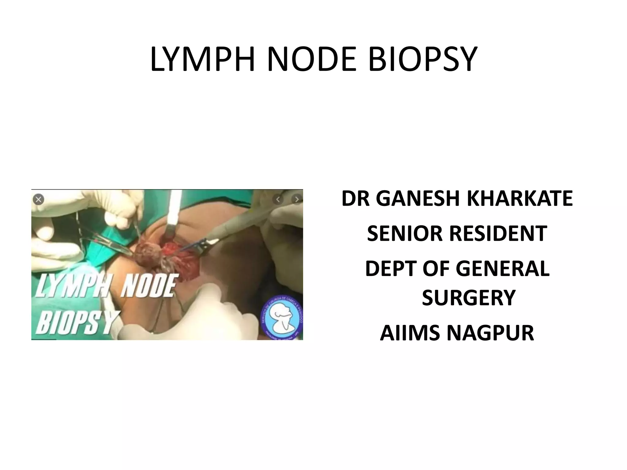
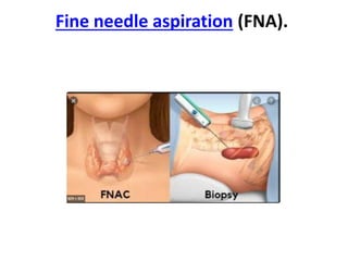
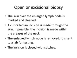
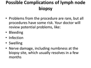
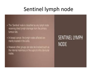
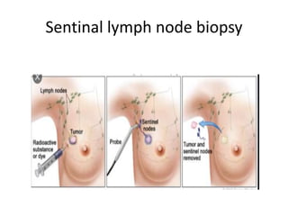
LYMPH NODE BIOPSY
DR GANESH KHARKATE
SENIOR RESIDENT
DEPT OF GENERAL
SURGERY
AIIMS NAGPUR
WHAT ARE Lymph Nodes
• Lymph nodes are small glandular structures in
the course of lymphatic vessels.
1]Dense connective tissue is the outermost
cover.
2]Cortex is composed of Primary and secondary
lymph follicles.
Functions of Lymph nodes
• Filtration of Lymph –Absorption of water and
electrolytes.
• Bacteria and other toxic substances are
destroyed by Macrophages of Lymph nodes
• Lymph nodes are found throughout the body.
• Part of the body’s immune system.
• These nodes help fight infection by producing
special white blood cells.
• They also work by trapping bacteria, viruses,
and cancer cells.
• Normally, lymph nodes cannot be felt unless
they are swollen.
Enlargement of lymph nodes:
• INFLAMMATORY : Acute and chronic lymphadinitis.
• Chronic is again subdivided into specific i.e TB, Syphlis,Filarial,
lymphpgranuloma inguinale. & Non specific or pyogenic.
• NEOPLASTIC: Almost always malignant. Primary and Secondary
Primary[reticulosis]: Hodgkin’s disease, Lymphosarcoma,
Reticulosarcoma, lymphtic leukaemia.
Secondary: Carcinoma, Melanoma, Sarcoma
DIFFERENT TYPES OF LYMPH NODE
BIOPSY
• Sentinel lymph node biopsy
• Fine needle aspiration (FNA).
• Core needle biopsy.
• Open biopsy.
Fine needle aspiration (FNA).
FNAC
• Needle into the lymph nodes to remove
fluid and cells, which get examined .
• local anesthesia is needed sometimes.
• 23 GAUGE NEEDLE IS REQUIRED
• Can be USG OR CT GUIDED(radiologist help
may be required )
Open or excisional biopsy
• The skin over the enlarged lymph node is
marked and cleaned.
• A cut called an incision is made through the
skin. If possible, the incision is made within
the creases of the neck.
• The enlarged lymph node is removed. It is sent
to a lab for testing.
• The incision is closed with stitches.
excisional biopsy
Core needle biopsy
Core needle biopsy devices
Types of core needles
• Regions for Procedure
• Common areas for biopsy include:
• Groin
• Armpit
• Neck
• Under the jaw and chin
• Behind the ears
Possible Complications of lymph node
biopsy
• Problems from the procedure are rare, but all
procedures have some risk. Your doctor will
review potential problems, like:
• Bleeding
• Infection
• Swelling
• Nerve damage, including numbness at the
biopsy site, which usually resolves in a few
months
Sentinel lymph node
Sentinal lymph node biopsy
Sentinal lymph node
Sentinal lymph node
• ISOPHANE BLUE
• METHYLENE BLUE
• 99mTc
• 99mTc-labelled colloidal
rhenium sulphide),
gamma probe and
gamma camera for
radiolocalisation and
monitoring
Sentinal lymph node
• MINIMUM 2 LN TO BE
TAKEN FOR BREAST
• MINIMUM 4 LN TO BE
TAKEN FOR AXILLARY
SAMPLING
Lymph node biopsy
Lymph node biopsy
Lymph node biopsy
Lymph node biopsy

Lymph node biopsy

  • 1.
    LYMPH NODE BIOPSY DRGANESH KHARKATE SENIOR RESIDENT DEPT OF GENERAL SURGERY AIIMS NAGPUR
  • 2.
    WHAT ARE LymphNodes • Lymph nodes are small glandular structures in the course of lymphatic vessels. 1]Dense connective tissue is the outermost cover. 2]Cortex is composed of Primary and secondary lymph follicles.
  • 3.
    Functions of Lymphnodes • Filtration of Lymph –Absorption of water and electrolytes. • Bacteria and other toxic substances are destroyed by Macrophages of Lymph nodes
  • 4.
    • Lymph nodesare found throughout the body. • Part of the body’s immune system. • These nodes help fight infection by producing special white blood cells. • They also work by trapping bacteria, viruses, and cancer cells. • Normally, lymph nodes cannot be felt unless they are swollen.
  • 5.
    Enlargement of lymphnodes: • INFLAMMATORY : Acute and chronic lymphadinitis. • Chronic is again subdivided into specific i.e TB, Syphlis,Filarial, lymphpgranuloma inguinale. & Non specific or pyogenic. • NEOPLASTIC: Almost always malignant. Primary and Secondary Primary[reticulosis]: Hodgkin’s disease, Lymphosarcoma, Reticulosarcoma, lymphtic leukaemia. Secondary: Carcinoma, Melanoma, Sarcoma
  • 6.
    DIFFERENT TYPES OFLYMPH NODE BIOPSY • Sentinel lymph node biopsy • Fine needle aspiration (FNA). • Core needle biopsy. • Open biopsy.
  • 7.
  • 8.
    FNAC • Needle intothe lymph nodes to remove fluid and cells, which get examined . • local anesthesia is needed sometimes. • 23 GAUGE NEEDLE IS REQUIRED • Can be USG OR CT GUIDED(radiologist help may be required )
  • 9.
    Open or excisionalbiopsy • The skin over the enlarged lymph node is marked and cleaned. • A cut called an incision is made through the skin. If possible, the incision is made within the creases of the neck. • The enlarged lymph node is removed. It is sent to a lab for testing. • The incision is closed with stitches.
  • 10.
  • 11.
  • 12.
  • 13.
  • 15.
    • Regions forProcedure • Common areas for biopsy include: • Groin • Armpit • Neck • Under the jaw and chin • Behind the ears
  • 16.
    Possible Complications oflymph node biopsy • Problems from the procedure are rare, but all procedures have some risk. Your doctor will review potential problems, like: • Bleeding • Infection • Swelling • Nerve damage, including numbness at the biopsy site, which usually resolves in a few months
  • 17.
  • 18.
  • 19.
  • 20.
    Sentinal lymph node •ISOPHANE BLUE • METHYLENE BLUE • 99mTc • 99mTc-labelled colloidal rhenium sulphide), gamma probe and gamma camera for radiolocalisation and monitoring
  • 21.
    Sentinal lymph node •MINIMUM 2 LN TO BE TAKEN FOR BREAST • MINIMUM 4 LN TO BE TAKEN FOR AXILLARY SAMPLING