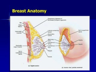
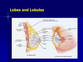
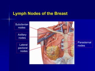
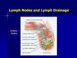
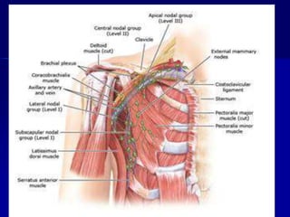
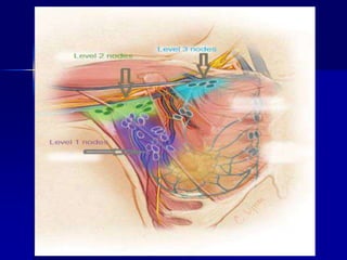
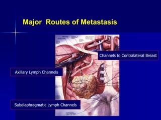
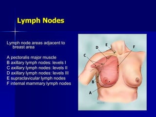
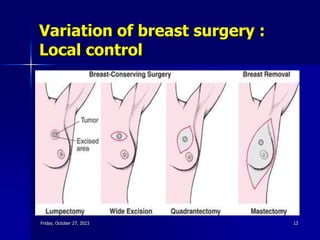
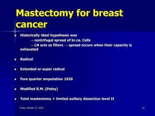
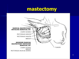
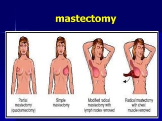
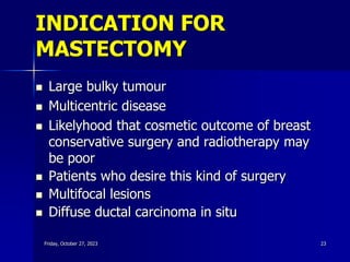
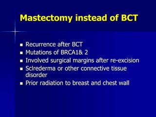
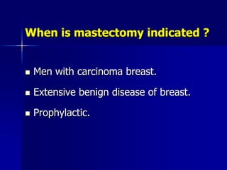
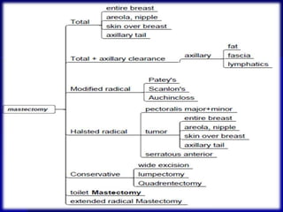
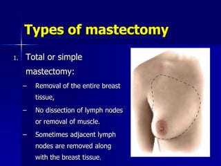
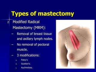
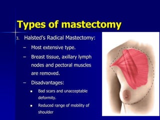
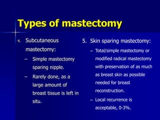
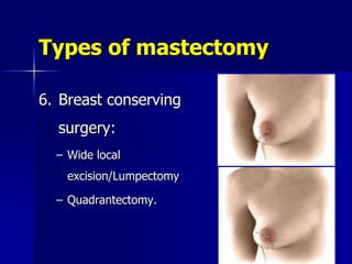
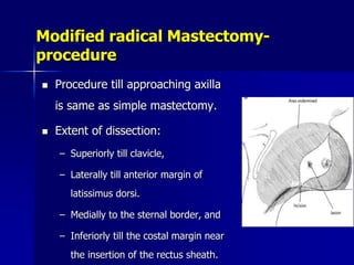
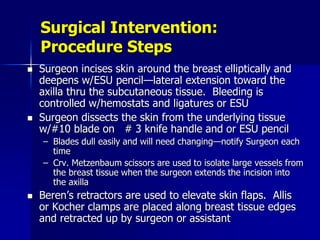
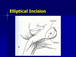
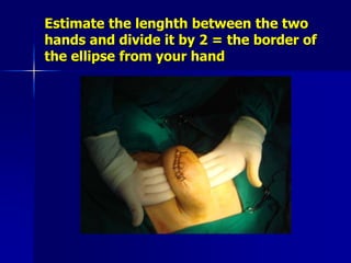
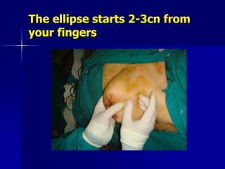
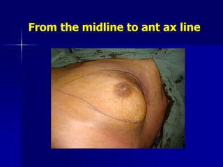
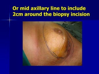
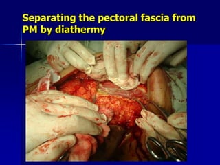
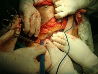
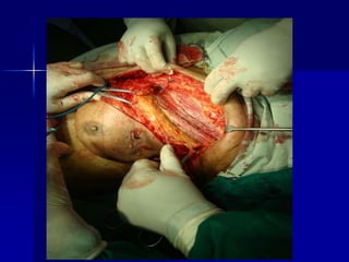
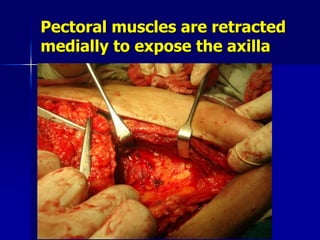
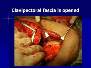
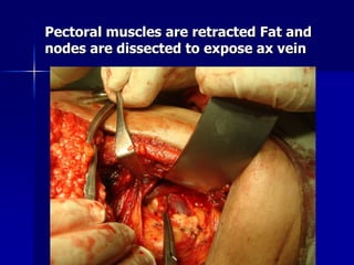
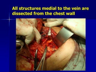
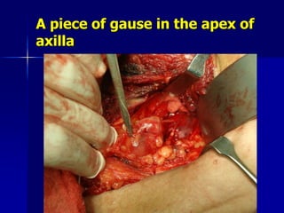
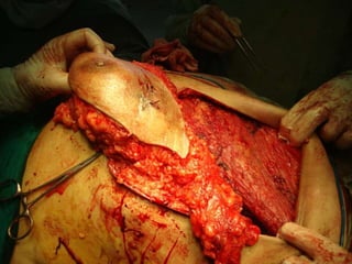
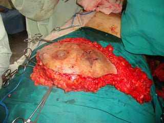
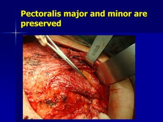
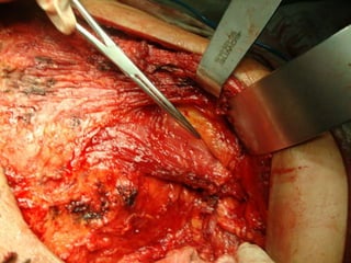
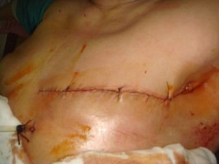
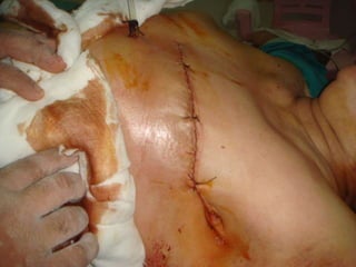
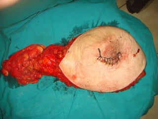
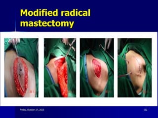
Breast Cancer
Modified Radical Mastectomy
Hamed Rashad
Professor of surgery Banha faculty of
medicine - Egypt
‫الرحيم‬ ‫الرحمن‬ ‫هللا‬ ‫بسم‬
Breast Anatomy
Lobes and Lobules
Subclavian
nodes
Axillary
nodes
Lateral
pectoral
nodes
Parasternal
nodes
Lymph Nodes of the Breast
Lymph Nodes and Lymph Drainage
Axillary
Nodes
Subdiaphragmatic Lymph Channels
Channels to Contralateral Breast
Axillary Lymph Channels
Major Routes of Metastasis
Lymph Nodes
Lymph node areas adjacent to
breast area
A pectoralis major muscle
B axillary lymph nodes: levels I
C axillary lymph nodes: levels II
D axillary lymph nodes: levels III
E supraclavicular lymph nodes
F internal mammary lymph nodes
Biopsy of palpable lesions
 Excision biopsy
- tumours less than 2cm with safety
margin
- excision for frozen section
 Incisional biopsy
- bigger tumours
- for diagnostic purposes
Friday, October 27, 2023 11
Recommended incision lines for
excisional biopsy
Not recommended Recommended
Friday, October 27, 2023 12
Variation of breast surgery :
Local control
Friday, October 27, 2023 13
Mastectomy
Mastectomy for breast
cancer
 Historically ideal hypothesis was
→ centrifugal spread of br.ca. Cells
→ LN acts as filters → spread occurs when their capacity is
exhausted
 Radical
 Extended or super radical
 Fore quarter amputation 1920
 Modified R.M. (Patey)
 Total mastectomy + limited axillary dissection level II
Friday, October 27, 2023 15
mastectomy
Friday, October 27, 2023 16
mastectomy
Friday, October 27, 2023 17
Mastectomy
‘Surgical Removal of the
Breast’
 Radical mastectomy – removal of the
breast, pectoralis major and minor m., and
Level I-III axillary nodes
 Modified radical mastectomy – removal of
the breast and Level I or Level I and II
nodes
 Simple mastectomy – removal of breast
 Skin-sparing mastectomy – mastectomy
with removal of the nipple-areolar complex,
but with preservation of the rest of the
Modified Radical
Mastectomy (MRM)
Patey:
 removal of pectoralis minor muscle to allow
Level III node dissection
Madden and Auchiinclloss:
 preservation of both pectoralis major and
minor; only level I-II dissection Higher
chance of medial pectoral nerve
preservation Reduce arm swelling
Absolute Indications for
Mastectomy
= Absolute contraindications for BCT
 Multicentricity or diffuse malignant appearing
microcalcifications
 Persistent positive margins after reasonable
surgical attempts
 History of prior therapeutic irradiation to the breast
region
 Pregnancy (first or second trimester)
Relative Indications for
Mastectomy
 Large tumor in a small breast
 Tumor size (> 4-5 cm)
 Breast size (large or pendulous breasts)
 History of collagen vascular disease
 (scleroderma or active SLE)
 Multifocality
INDICATION FOR
MASTECTOMY
 Large bulky tumour
 Multicentric disease
 Likelyhood that cosmetic outcome of breast
conservative surgery and radiotherapy may
be poor
 Patients who desire this kind of surgery
 Multifocal lesions
 Diffuse ductal carcinoma in situ
Friday, October 27, 2023 23
 Recurrence after BCT
 Mutations of BRCA1& 2
 Involved surgical margins after re-excision
 Sclrederma or other connective tissue
disorder
 Prior radiation to breast and chest wall
Mastectomy instead of BCT
When is mastectomy indicated ?
 Men with carcinoma breast.
 Extensive benign disease of breast.
 Prophylactic.
? Indications for
Mastectomy
 Prophylactic mastectomy for familial or
 high-risk women
 Cost and inconvenience of irradiation
 Attitude of patient/relatives/Friends
 Because the doctors say so
Types of mastectomy
1. Total or simple
mastectomy:
– Removal of the entire breast
tissue,
– No dissection of lymph nodes
or removal of muscle.
– Sometimes adjacent lymph
nodes are removed along
with the breast tissue.
Types of mastectomy
2. Modified Radical
Mastectomy (MRM):
– Removal of breast tissue
and axillary lymph nodes.
– No removal of pectoral
muscle.
– 3 modifications:
a. Patey’s
b. Scanlon’s.
c. Auchincloss.
Types of mastectomy
3. Halsted’s Radical Mastectomy:
– Most extensive type.
– Breast tissue, axillary lymph
nodes and pectoral muscles
are removed.
– Disadvantages:
 Bad scars and unacceptable
deformity.
 Reduced range of mobility of
shoulder
Types of mastectomy
4. Subcutaneous
mastectomy:
– Simple mastectomy
sparing nipple.
– Rarely done, as a
large amount of
breast tissue is left in
situ.
5. Skin sparing mastectomy:
– Total/simple mastectomy or
modified radical mastectomy
with preservation of as much
as breast skin as possible
needed for breast
reconstruction.
– Local recurrence is
acceptable, 0-3%.
Types of mastectomy
6. Breast conserving
surgery:
– Wide local
excision/Lumpectomy
– Quadrantectomy.
Types of mastectomy
7. Extended radical
mastectomy:
– Radical mastectomy +
enbloc resection of
internal mammary lymph
nodes + supraclavicular
lymph nodes.
– Obsolete.
8. Toilet mastectomy:
– Done in fungating
or ulcerative
growths.
– Palliative simple
mastectomy.
Which procedure is suitable for the
given patient ?
 Age
 Size of the tumor
 Axillary lymph node status.
 Stage of the malignancy
 Biologic aggressiveness of
the tumor
 Receptor status of the tumor.
 Multicentricity or multifocality
 Menstrual status.
 Size of the breast
 Availability of
radiotherapy.
 Patients choice.
 Prophylactic/therapeutic/
palliative.
Which procedure is best ?
 When the tumor size is ≥ 1cm, becomes
systemic.
 No single method is considered better in terms
of disease free survival or mortality.
 Suitable local therapy + systemic therapy is the
most appropriate approach.
Which procedure is best ?
 Loco-Regional therapy include:
a. Surgery
b. Radiotherapy
 Systemic therapy:
a. Chemotherapy
b. Hormonal therapy
c. Monoclonal antibodies.
However surgery is important to get rid of gross cancer
Pre-operative management
 Triple assessment.
 Metastatic workup.
 Routine blood investigations.
 Pre-anesthetic evaluation.
 Control of medical conditions like diabetes and hypertension.
 Counseling and written informed consent.
 Parts preparation- neck to mid thigh including pelvic region,
axilla and arm.
Operative procedures-Mastectomy
1. Simple mastectomy.
2. Modified radical mastectomy.
3. Breast conserving surgery.
Operative procedure
 Anesthesia
– General anesthesia.
 Position
– The patient is placed in supine position with the
arm abducted < 90 degree.
– Sandbag or folded sheet is placed under the
thorax and shoulder of affected side.
Introduction
 Definition: surgical removal of breast
tissue partially or completely.
 In a study conducted in 2004,
– Highest mastectomies were done in Europe 60-70%.
– USA- 56%.
– Australia and New Zealand: 34%.
Surgical Intervention: Skin Prep
 Method of hair removal
 Anatomic perimeters
– Shoulder, upper arm extending down to
the elbow (circumferentially), the axilla,
& chest to table line and to the shoulder
opposite from affected side—access to
underarm for AND and possible extend
to fingertips of operative side
– Arm on operative side should be draped
free using stockinette & drapes that
allow free movement of the arm to
facilitate access to the axilla
 Solution options
– Betadine or Hibiclens
Surgical Intervention:
Procedure Steps Overview
 Breast incised elliptically
 Incision deepened to encompass entire
breast
 Breast removed en bloc w/ALNs
 Axillary lymph nodes are removed
 Wound is closed
Operative procedures-
Simple Mastectomy
 Indications:
– Stage I and stage IIa carcinoma
– Large cancers that persist after adjuvant therapy
– Multifocal or multicentric CIS.
 Incision:
– Horizontal elliptical incision is marked so as to include the
entire areolar complex.
– Should be 1-2cm away from the tumor margins.
– Skin sparing incision- if breast reconstruction is planned
– Two skin edges should be of equivalent length
Simple Mastectomy-procedure
 Skin incision is deepened with
electro-cautery.
 A plane between breast fat and
the subcutaneous fat, seen as
white fibrous plane.
 Dissection is carried in this plane
and flaps are raised inferiorly and
superiorly.
 Ideally thickness of the flap
should be 7-10mm.
Lines of incision
Lines of incision
Reach the plane between the skin and
subcutaneous tissues and the breast tissues
Simple Mastectomy-procedure
 Extent of dissection:
– Superiorly till clavicle,
– Laterally till P.major lateral border
– Medially to the sternal border, and
– Inferiorly till infra-mammary fold
 Breast tissue along with the pectoral fascia
(controversial) is dissected from the P.major.
Simple Mastectomy-procedure
 Usually started superiorly and the proceeded clock-wise
ending in the axillary region.
 Care must be taken to ligate perforating branches of
lateral thoracic and anterior intercostal vessels.
 Lateral branches of the medial pectoral neurovascular
bundle is carefully dissected while removing axillary tail.
 Wound irrigated with sterile water to crenate (shrivel or
shrink) cancerous cells.
 Subcutaneous tissue is closed using 00 absorbable
interrupted sutures.
 Skin closed using 00 absorbable sucuticular sutures or
using staples.
Operative procedures-
Modified radical Mastectomy
 Indications:
– LABC
– Residual large cancers that persist after
adjuvant therapy
– Multifocal or multicentric disease.
Anatomic Boundaries of
MRM
 Lateral - anterior margin of latissimus
dorsi muscle
 Medial - midline of the sternum
 Superior - subclavius muscle
 Inferior - caudal extension of the
breast 2 to 3 cm inferior to the
inframammary fold
Modified Radical
Mastectomy (MRM)
 ‘‘Total mastectomy with en bloc
removal of breast tissue,, pectoralis
fascia,, nipple/areolar complex,,
axillary lymphatics,, and overlying skin
near the tumor with a 2-cm margin’’
 Preservation of pectoralis major muscle
Operative procedures-
Modified radical Mastectomy
 Incision:
– Oblique elliptical incision angled towards axilla.
– Should include the entire areolar complex and
previous scars, if present.
– Should be 1-2cm away from the tumor margins.
– Two skin edges should be of equivalent length
Modified radical Mastectomy-
procedure
 Procedure till approaching axilla
is same as simple mastectomy.
 Extent of dissection:
– Superiorly till clavicle,
– Laterally till anterior margin of
latissimus dorsi.
– Medially to the sternal border, and
– Inferiorly till the costal margin near
the insertion of the rectus sheath.
Surgical Intervention:
Procedure Steps
 Surgeon incises skin around the breast elliptically and
deepens w/ESU pencil—lateral extension toward the
axilla thru the subcutaneous tissue. Bleeding is
controlled w/hemostats and ligatures or ESU
 Surgeon dissects the skin from the underlying tissue
w/#10 blade on # 3 knife handle and or ESU pencil
– Blades dull easily and will need changing—notify Surgeon each
time
– Crv. Metzenbaum scissors are used to isolate large vessels from
the breast tissue when the surgeon extends the incision into
the axilla
 Beren’s retractors are used to elevate skin flaps. Allis
or Kocher clamps are placed along breast tissue edges
and retracted up by surgeon or assistant
Elliptical Incision
Surgical Intervention:
Procedure Steps
 The margins of skin flaps are covered w/warm moist
lap pads and held away w/retractors.
 The intercostal arteries and veins are clamped and
ligated.
 The axillary flap is retracted for complete dissection
of the axilla.
 Careful attention is directed to preventing injury to
the axillary vein & medial and lateral nerves of the
pectoralis major muscle
 The fascia is dissected from the lateral edge of the
pectoralis muscle. Ligation of vessels is performed in
the axilla & adjacent to sternum. The fascia is then
dissected to the serratus anterior muscle. The
thoracic & thoracodorsal nerves are preserved
Modified radical Mastectomy-
procedure
 The specimen is retracted upwards and laterally to
expose P.minor.
 The dissection is continued to axillary lymph node
clearance.
 Care must be taken not to injure medial pectoral
nerve and vessels.
 The axillary investing fascia is incised to expose
the axillary group of lymph nodes.
Modified radical Mastectomy-
procedure
1. Patey’s procedure:
– The P.minor is removed for better visualization and
easy dissection of level III lymph nodes.
2. Scanlon’s procedure:
– P.minor is retracted to expose level III nodes and
dissected out.
3. Auchincloss procedure:
– Level I and II lymph nodes are cleared, level III
nodes are left behind.
Modified radical Mastectomy-
procedure
 The inter-pectoral (Rotter) group of lymph nodes are
removed.
 Then dissection can be done either from medial to lateral or
vise-versa.
 The loose lateral areolar tissue in axillary space is dissected
to expose the axillary vein.
 The investing layer of axillary vessels is cut, the tributaries
are transfixed and cut.
 Dissection is carried out laterally including lateral grp (level
I) of lymph nodes.
Modified radical Mastectomy-
procedure
 Thoracodorsal neurovascular bundle lies over
the lat.dorsi, with nerve more laterally placed,
subscapular (level I) nodes are removed.
 The level II lymph nodes between superior
trunk of intercostobranchial bundle and axillary
vein are removed.
Modified radical Mastectomy-
procedure
 The central grp of lymph nodes are removed carefully
separating from axillary vein and its tributaries.
 While dissecting medially, long thoracic nerve is
encountered, which lies anterior to the subscapular
muscle. The dissection carried out anterior and medial
to long thoracic nerve and the specimen delivered.
Modified radical Mastectomy-
procedure
 Care must be taken while dissecting in
axillary area to preserve,
– Medial and lateral pectoral nerve.
– Long thoracic vessels and nerve
– Nerve to latissimus dorsi.
– Axillary vein.
Modified radical Mastectomy-
procedure
 Wound irrigated with sterile water to
shrink/crenate cancerous cells.
 2 drains, 1 below and other above P.major
are secured.
 Subcutaneous tissue is closed using 00
absorbable interrupted sutures.
 Skin closed using 00 absorbable subcuticular
sutures or using staples.
Surgical Intervention:
Procedure Steps
 Be sure to keep exposed tissue moist with lap
packs for protection
 Surgeon dissects the breast and axillary
fascia away from the latissimus dorsi muscle
and suspensory ligaments—from near the
clavicle to midportion of the sternum. The
pectoralis major muscle is left intact.
 The specimen is passed to STSR
 Wound is inspected for bleeding sites, which
are ligated & electrocoagulated, then irrigated
(NS).
Surgical Intervention:
Procedure Steps
 Surgeon places closed-wound suction drainage
tube(s) thru stab wounds and secured to skin
w/nonabsorbable suture on a cutting needle
 A few absorbable suture may be used in the
subcutaneous tissue to approximate the skin edges.
 Surgeon closes w/interrupted nonabsorbable
sutures or staple, anchors drains, and connects to
closed suction reservoir.
 The dressing may be one of several: simple gauze,
bulky held in place w/Surgi-Bra, or gauze and
elastic wrap.
Post-operative care
 Wound examined on post-op day 3.
 Drain can be removed when it is < 50ml.
 Any collection is to be aspirated under aseptic
precautions.
 Stitches if done can be removed after 10days.
 Arm movements started in the 1st week..
 Active shoulder and upper limb exercises are
started from 2 weeks
Technique of mastectomy - summary
 Incision : - elliptical with equal lengths to avoid
dog ear
- including nipple ,areola & biopsy
site with 4cm on either side
- extends from lateral border of
sternum to mid-axillary line
 Skin flap : - up to clavicle
- medially to sternum
- down to rectus sheath
- lateral to edge of latissmus dorsi
Friday, October 27, 2023 71
 Dissection :
- the breast and pectoral fascia are dissected from
medial to lateral till edge of pectoralis major
- clavipectoral fascia is incised and reflected for
exposure of pectoralis minor
- pectoralis minor is - left & retracted medially
- cut at insertion
- removed completely to
expose apex of axilla
NB : care of dissection of lateral pectoral nerve (medial
nerve)
NB : inter-pectoral ( Roter’s ) nodes are in close
relation to this nerve and vessel ( dissected with care )
Friday, October 27, 2023 72
 All fat & nodes medial and down to axillary vein are
dissected → it requires ligation and division of several
axillary vein branches
 Inter costo brachial nerve has to be divided to facilitate
dissection
 Nerve to latissmus dorsi & serratus has to be preserved
 Inter-nerve nodes → subscapular ?
Friday, October 27, 2023 73
 Closure
- suction drain
- closure should not be under tension
- subcutaneous & subcuticular absorbable
sutures
- dressing
- drain removed when drainage is less
than 50ml/24h
- arm exercise next day
Friday, October 27, 2023 74
Biopsy site closed
The breast is hold between the two hands
stretching the skin on the chest wall
Estimate the lenghth between the two
hands and divide it by 2 = the border of
the ellipse from your hand
The ellipse starts 2-3cn from
your fingers
From the midline to ant ax line
Or mid axillary line to include
2cm around the biopsy incision
There is an avascular plane of dissection
between the skin and breast (white line)
Incising the pectoral
fascia
Separating the pectoral fascia from
PM by diathermy
Pectoral muscles are retracted
medially to expose the axilla
Clavipectoral fascia is opened
Pectoral muscles are retracted Fat and
nodes are dissected to expose ax vein
All structures medial to the vein are
dissected from the chest wall
A piece of gause in the apex of
axilla
Intercostobrachial nerve
Nerve to serratus anterior
Breast dissection is completed
Breast and axillary nodes are
removed en-mass
Pectoralis major and minor are
preserved
The two nerves are preserved
Skin is closed with tube drain
Modified radical
mastectomy
Friday, October 27, 2023 112
After removal of the breast
and axilla
Friday, October 27, 2023 113
Friday, October 27, 2023 114
Drain or No Drain
 Following MRM, standard practice involves
insertion of suction drains deep to
mastectomy flaps and in the axilla
 Drains are left in situ until fluid drainage is
less than 40-50 ml/day usually 6-14 days
after operation
 Despite the use of suction drains, seromas
requiring aspiration still occur in 10-52
percent of patients
Complication after
mastectomy
 Seroma → low pressure suction drains
 Infection → good surgical technique
 Flap necrosis
- thin
- devascularized flaps
- tension
 Phantom breast → 50% of patients
 Rare complications
- pneumothorax
- injury of neurovascular bundle
 Lymphoedema
 More frequent when surgery is associated with
radiotherapy
Friday, October 27, 2023 116
Preservation of the
Intercostobrachial Nerve
 Oncological safe
 Alteration in sensation or presence of pain cannot
be solely attributed to the preservation or sacrifice
of the ICBN
 Some patients who had nerve sacrifice had normal
sensation and yet many with nerve preservation did
not
 No clear difference in pain
Freeman et al. EJSO 2003
Drain or No Drain
 Following MRM, the standard practice involves the
insertion of suction drains deep into mastectomy
flaps and in the axilla
 Drains are left in situ until fluid drainage is less
than 40-50 ml/day usually 6-14 days after the
operation
 Despite the use of suction drains, seromas
requiring aspiration still occur in 10-52 percent of
patients
Mastectomy without Drain at Pectoral
Area
: a Randomized Controlled Trial
Sixty patients underwent MRM Randomized to
 Group I: only 1 drain was inserted at the axilla area
 Group II: 2 drains were inserted into the pectoral
area and axilla area
No differences in total drainage contents and
complications
Puttawibul et al. J Med Assoc Thai. 2003
Methods to Reduce
Seroma Formation
 Fibrin glue -Prospective randomized trials
significantly decrease the duration and
quantity of serosanguinous drainage
Berger et al. Breast Cancer Res Treat 2001
no significant benefit on axillary lymphatic drainage,
drain removal time, or seroma formation
Ulusoy et al. Breast J. 2003
Methods to Reduce Seroma
Formation
 External compression dressing (circumferential
chest wrap of two 6-inch Ace bandages, held in
place by circumferential Elastoplast bandage)
 vs. Standard front-fastening Surgibra Fails to
decrease postoperative drainage and may
increase the incidence of seroma formation
after drain removal
O’Hea et al. Am J Surg 1999
Methods to Reduce Seroma
Formation
Octreotide
prospective randomized controlled trial
261 consecutive patients
Treatment group: 0.1 mg octreotide s.c.
3 times a day for 5 days
Control group: no treatment
Decreased amount and duration of seroma
Carcoforo et al. J Am Coll Surg 2003
Radiotherapy after mastectomy
 Radiotherapy after mastectomy has :
- no effect on survival
- marked reduction of local recurrence
 Indications of post mastectomy radiotherapy
1- large tumour >4cm
2- high grade tumour grade III
3- node positive >4 especially if extra-
nodal extension is seen
4- node negative tumour with wide
spread vascular / lymphatic invasion
Friday, October 27, 2023 123
A Middle radiation beam
B Side radiation beam
C Bright yellow: place
where radiation is given to
the breast
D Rib cage/chest wall
E Heart
F Lungs
G Backbone
H Sternum/breast bone
Cross-sectional view of beam
RT
www.breastcancer.org
The axilla in cancer breast
 The axilla is a heated debate
 Management ranges from →
- nothing
- through sampling
- limited axillary dissection ±
radiotherapy
To → formal clearance
 Axillary nodes are on the average 20 nodes
- 13.5 at level I
- 4.5 at level II
- 2.3 at level III
Friday, October 27, 2023 125
 Skip metastasis occur only in 1.3% of patients
 Axillary node status
- remains the single best prognostic
factor
- treatment decision is based upon
 Clinical examination of axilla correlates BADLY with true
nodal status so some form of sampling is needed
 Still there is no good imaging technique
Friday, October 27, 2023 126
Role of axillary surgery
 To stage the axilla
 To treat axillary disease
Friday, October 27, 2023 127
Ability to stage the axilla
1- axillary node sampling (4 nodes) to
inter-costo brachial nerve as a land mark
2- level I dissection (10 nodes)
3- level II clearance
4- level III clearance
 Any of them will adequately stage the axilla
Friday, October 27, 2023 128
Treatment of axillary disease
 Radical radiotherapy
 Full axillary clearance (level III)
NB : level I dissection can never represent level II & III
 In patients who have even one node positive at level I,
there is a significant chance of positive nodes at level
II or III
 Axillary recurrence is generally less with axillary
clearance than radical radiotherapy
Friday, October 27, 2023 129
CORRELATION OF +VE NODE NUMBER AT LEVEL
I WITH % OF NODE +VE AT LEVEL II & III
Node +ve at level I % +ve node at level II & III
1
2
3
4
5
12
19
37
40
84
Friday, October 27, 2023 130
Predicting which patient will have
involved axillary nodes
 There is direct relation between tumour size and
presence of node metastasis
 So in :
- patients with impalpable breast cancer, axillary node
sampling or level I dissection is enough
- patients with palpable or invasive breast cancer, level
III clearance should be done
Friday, October 27, 2023 131
Definitions of levels I–III of
the axilla
Friday, October 27, 2023 132
Axillary vein
II
Latissmus dorsi muscle
Serratus anterior muscle
I
III
Techniques of axillary surgery
A- sentinel node biopsy
1- injection of radio labelled (technetium) sulpher colloid
or blue dye into the tumour or skin overlying it
2- identification of the sentinel node either by :
- visual injection (blue)
- or radiation monitor using gamma
detecting probe
3- incision small axillary → remove sentinel node
for biopsy or frozen section
Friday, October 27, 2023 133
sentinel node biopsy
Friday, October 27, 2023 134
B- axillary node sampling
- through separate axillary incision in skin crease
- immediately prior to the wide local excision
- edges of pectoralis major and latissmus dorsi are identified
- finger passed around behind the lower axillary fat , situated
between two muscles , this make nodes in this fat easier to feel
- at least 4 palpable nodes are excised & sent for histology
- if 4 nodes not felt in lower axilla (level I) then palpable nodes
from level II or III are excised
- haemostasis
- no need for drain
- closure in layer
Friday, October 27, 2023 135
C- axillary node clearance
- a lazy s incision is made along the skin crease of axilla
- skin flaps dissected to edges of pectoralis major &
latissmus dorsi muscles
- pectoralis minor is cleared of tissues
- pect. Minor is divided to allow thorough axillary clearance
if there is evidence of extensive nodal disease
- the front of axillary vein is identified & contents of axilla
below the vein are cleared to the apex of axilla
Friday, October 27, 2023 136
Axillary Lymph Node
Dissection
Axillary dissection (different
incision lines)
Friday, October 27, 2023 137
Axillary incision
Friday, October 27, 2023 138
Axillary Lymph Node
Dissection
Friday, October 27, 2023 139
Level III axillary evacuation
Friday, October 27, 2023 140
Treatment of internal mammary
& supraclavicular nodes
 The value of irradiation is unproven
 Supraclavicular can be included when axillary
radiotherapy is given with no added morbidity
 90% of women with +ve int. Mammary nodes have
axillary node involvement so treated accordingly
 5-10% may have int.mammary alone
→ medial cancer alone
→ ?? Irradiated ?? Complex field that touches heart
so they are not now irradiated
Friday, October 27, 2023 141
Conclusion
 Patients with breast cancer should be
informed of options available during
treatment planning
 MRM remains an important tool for
locoregional control of breast cancer
 Various methods to reduce postoperative
complications remain inconclusive
THAK YOU

Br Ca- MRM the lect.ppt

  • 1.
    Breast Cancer Modified RadicalMastectomy Hamed Rashad Professor of surgery Banha faculty of medicine - Egypt
  • 2.
  • 3.
  • 4.
  • 5.
  • 6.
    Lymph Nodes andLymph Drainage Axillary Nodes
  • 9.
    Subdiaphragmatic Lymph Channels Channelsto Contralateral Breast Axillary Lymph Channels Major Routes of Metastasis
  • 10.
    Lymph Nodes Lymph nodeareas adjacent to breast area A pectoralis major muscle B axillary lymph nodes: levels I C axillary lymph nodes: levels II D axillary lymph nodes: levels III E supraclavicular lymph nodes F internal mammary lymph nodes
  • 11.
    Biopsy of palpablelesions  Excision biopsy - tumours less than 2cm with safety margin - excision for frozen section  Incisional biopsy - bigger tumours - for diagnostic purposes Friday, October 27, 2023 11
  • 12.
    Recommended incision linesfor excisional biopsy Not recommended Recommended Friday, October 27, 2023 12
  • 13.
    Variation of breastsurgery : Local control Friday, October 27, 2023 13
  • 14.
  • 15.
    Mastectomy for breast cancer Historically ideal hypothesis was → centrifugal spread of br.ca. Cells → LN acts as filters → spread occurs when their capacity is exhausted  Radical  Extended or super radical  Fore quarter amputation 1920  Modified R.M. (Patey)  Total mastectomy + limited axillary dissection level II Friday, October 27, 2023 15
  • 16.
  • 17.
  • 18.
    Mastectomy ‘Surgical Removal ofthe Breast’  Radical mastectomy – removal of the breast, pectoralis major and minor m., and Level I-III axillary nodes  Modified radical mastectomy – removal of the breast and Level I or Level I and II nodes  Simple mastectomy – removal of breast  Skin-sparing mastectomy – mastectomy with removal of the nipple-areolar complex, but with preservation of the rest of the
  • 19.
    Modified Radical Mastectomy (MRM) Patey: removal of pectoralis minor muscle to allow Level III node dissection Madden and Auchiinclloss:  preservation of both pectoralis major and minor; only level I-II dissection Higher chance of medial pectoral nerve preservation Reduce arm swelling
  • 20.
    Absolute Indications for Mastectomy =Absolute contraindications for BCT  Multicentricity or diffuse malignant appearing microcalcifications  Persistent positive margins after reasonable surgical attempts  History of prior therapeutic irradiation to the breast region  Pregnancy (first or second trimester)
  • 21.
    Relative Indications for Mastectomy Large tumor in a small breast  Tumor size (> 4-5 cm)  Breast size (large or pendulous breasts)  History of collagen vascular disease  (scleroderma or active SLE)  Multifocality
  • 23.
    INDICATION FOR MASTECTOMY  Largebulky tumour  Multicentric disease  Likelyhood that cosmetic outcome of breast conservative surgery and radiotherapy may be poor  Patients who desire this kind of surgery  Multifocal lesions  Diffuse ductal carcinoma in situ Friday, October 27, 2023 23
  • 24.
     Recurrence afterBCT  Mutations of BRCA1& 2  Involved surgical margins after re-excision  Sclrederma or other connective tissue disorder  Prior radiation to breast and chest wall Mastectomy instead of BCT
  • 25.
    When is mastectomyindicated ?  Men with carcinoma breast.  Extensive benign disease of breast.  Prophylactic.
  • 26.
    ? Indications for Mastectomy Prophylactic mastectomy for familial or  high-risk women  Cost and inconvenience of irradiation  Attitude of patient/relatives/Friends  Because the doctors say so
  • 28.
    Types of mastectomy 1.Total or simple mastectomy: – Removal of the entire breast tissue, – No dissection of lymph nodes or removal of muscle. – Sometimes adjacent lymph nodes are removed along with the breast tissue.
  • 29.
    Types of mastectomy 2.Modified Radical Mastectomy (MRM): – Removal of breast tissue and axillary lymph nodes. – No removal of pectoral muscle. – 3 modifications: a. Patey’s b. Scanlon’s. c. Auchincloss.
  • 30.
    Types of mastectomy 3.Halsted’s Radical Mastectomy: – Most extensive type. – Breast tissue, axillary lymph nodes and pectoral muscles are removed. – Disadvantages:  Bad scars and unacceptable deformity.  Reduced range of mobility of shoulder
  • 31.
    Types of mastectomy 4.Subcutaneous mastectomy: – Simple mastectomy sparing nipple. – Rarely done, as a large amount of breast tissue is left in situ. 5. Skin sparing mastectomy: – Total/simple mastectomy or modified radical mastectomy with preservation of as much as breast skin as possible needed for breast reconstruction. – Local recurrence is acceptable, 0-3%.
  • 32.
    Types of mastectomy 6.Breast conserving surgery: – Wide local excision/Lumpectomy – Quadrantectomy.
  • 33.
    Types of mastectomy 7.Extended radical mastectomy: – Radical mastectomy + enbloc resection of internal mammary lymph nodes + supraclavicular lymph nodes. – Obsolete. 8. Toilet mastectomy: – Done in fungating or ulcerative growths. – Palliative simple mastectomy.
  • 34.
    Which procedure issuitable for the given patient ?  Age  Size of the tumor  Axillary lymph node status.  Stage of the malignancy  Biologic aggressiveness of the tumor  Receptor status of the tumor.  Multicentricity or multifocality  Menstrual status.  Size of the breast  Availability of radiotherapy.  Patients choice.  Prophylactic/therapeutic/ palliative.
  • 35.
    Which procedure isbest ?  When the tumor size is ≥ 1cm, becomes systemic.  No single method is considered better in terms of disease free survival or mortality.  Suitable local therapy + systemic therapy is the most appropriate approach.
  • 36.
    Which procedure isbest ?  Loco-Regional therapy include: a. Surgery b. Radiotherapy  Systemic therapy: a. Chemotherapy b. Hormonal therapy c. Monoclonal antibodies. However surgery is important to get rid of gross cancer
  • 37.
    Pre-operative management  Tripleassessment.  Metastatic workup.  Routine blood investigations.  Pre-anesthetic evaluation.  Control of medical conditions like diabetes and hypertension.  Counseling and written informed consent.  Parts preparation- neck to mid thigh including pelvic region, axilla and arm.
  • 38.
    Operative procedures-Mastectomy 1. Simplemastectomy. 2. Modified radical mastectomy. 3. Breast conserving surgery.
  • 39.
    Operative procedure  Anesthesia –General anesthesia.  Position – The patient is placed in supine position with the arm abducted < 90 degree. – Sandbag or folded sheet is placed under the thorax and shoulder of affected side.
  • 40.
    Introduction  Definition: surgicalremoval of breast tissue partially or completely.  In a study conducted in 2004, – Highest mastectomies were done in Europe 60-70%. – USA- 56%. – Australia and New Zealand: 34%.
  • 41.
    Surgical Intervention: SkinPrep  Method of hair removal  Anatomic perimeters – Shoulder, upper arm extending down to the elbow (circumferentially), the axilla, & chest to table line and to the shoulder opposite from affected side—access to underarm for AND and possible extend to fingertips of operative side – Arm on operative side should be draped free using stockinette & drapes that allow free movement of the arm to facilitate access to the axilla  Solution options – Betadine or Hibiclens
  • 42.
    Surgical Intervention: Procedure StepsOverview  Breast incised elliptically  Incision deepened to encompass entire breast  Breast removed en bloc w/ALNs  Axillary lymph nodes are removed  Wound is closed
  • 43.
    Operative procedures- Simple Mastectomy Indications: – Stage I and stage IIa carcinoma – Large cancers that persist after adjuvant therapy – Multifocal or multicentric CIS.  Incision: – Horizontal elliptical incision is marked so as to include the entire areolar complex. – Should be 1-2cm away from the tumor margins. – Skin sparing incision- if breast reconstruction is planned – Two skin edges should be of equivalent length
  • 45.
    Simple Mastectomy-procedure  Skinincision is deepened with electro-cautery.  A plane between breast fat and the subcutaneous fat, seen as white fibrous plane.  Dissection is carried in this plane and flaps are raised inferiorly and superiorly.  Ideally thickness of the flap should be 7-10mm.
  • 46.
  • 47.
  • 48.
    Reach the planebetween the skin and subcutaneous tissues and the breast tissues
  • 50.
    Simple Mastectomy-procedure  Extentof dissection: – Superiorly till clavicle, – Laterally till P.major lateral border – Medially to the sternal border, and – Inferiorly till infra-mammary fold  Breast tissue along with the pectoral fascia (controversial) is dissected from the P.major.
  • 51.
    Simple Mastectomy-procedure  Usuallystarted superiorly and the proceeded clock-wise ending in the axillary region.  Care must be taken to ligate perforating branches of lateral thoracic and anterior intercostal vessels.  Lateral branches of the medial pectoral neurovascular bundle is carefully dissected while removing axillary tail.  Wound irrigated with sterile water to crenate (shrivel or shrink) cancerous cells.  Subcutaneous tissue is closed using 00 absorbable interrupted sutures.  Skin closed using 00 absorbable sucuticular sutures or using staples.
  • 52.
    Operative procedures- Modified radicalMastectomy  Indications: – LABC – Residual large cancers that persist after adjuvant therapy – Multifocal or multicentric disease.
  • 53.
    Anatomic Boundaries of MRM Lateral - anterior margin of latissimus dorsi muscle  Medial - midline of the sternum  Superior - subclavius muscle  Inferior - caudal extension of the breast 2 to 3 cm inferior to the inframammary fold
  • 54.
    Modified Radical Mastectomy (MRM) ‘‘Total mastectomy with en bloc removal of breast tissue,, pectoralis fascia,, nipple/areolar complex,, axillary lymphatics,, and overlying skin near the tumor with a 2-cm margin’’  Preservation of pectoralis major muscle
  • 55.
    Operative procedures- Modified radicalMastectomy  Incision: – Oblique elliptical incision angled towards axilla. – Should include the entire areolar complex and previous scars, if present. – Should be 1-2cm away from the tumor margins. – Two skin edges should be of equivalent length
  • 56.
    Modified radical Mastectomy- procedure Procedure till approaching axilla is same as simple mastectomy.  Extent of dissection: – Superiorly till clavicle, – Laterally till anterior margin of latissimus dorsi. – Medially to the sternal border, and – Inferiorly till the costal margin near the insertion of the rectus sheath.
  • 57.
    Surgical Intervention: Procedure Steps Surgeon incises skin around the breast elliptically and deepens w/ESU pencil—lateral extension toward the axilla thru the subcutaneous tissue. Bleeding is controlled w/hemostats and ligatures or ESU  Surgeon dissects the skin from the underlying tissue w/#10 blade on # 3 knife handle and or ESU pencil – Blades dull easily and will need changing—notify Surgeon each time – Crv. Metzenbaum scissors are used to isolate large vessels from the breast tissue when the surgeon extends the incision into the axilla  Beren’s retractors are used to elevate skin flaps. Allis or Kocher clamps are placed along breast tissue edges and retracted up by surgeon or assistant
  • 58.
  • 59.
    Surgical Intervention: Procedure Steps The margins of skin flaps are covered w/warm moist lap pads and held away w/retractors.  The intercostal arteries and veins are clamped and ligated.  The axillary flap is retracted for complete dissection of the axilla.  Careful attention is directed to preventing injury to the axillary vein & medial and lateral nerves of the pectoralis major muscle  The fascia is dissected from the lateral edge of the pectoralis muscle. Ligation of vessels is performed in the axilla & adjacent to sternum. The fascia is then dissected to the serratus anterior muscle. The thoracic & thoracodorsal nerves are preserved
  • 60.
    Modified radical Mastectomy- procedure The specimen is retracted upwards and laterally to expose P.minor.  The dissection is continued to axillary lymph node clearance.  Care must be taken not to injure medial pectoral nerve and vessels.  The axillary investing fascia is incised to expose the axillary group of lymph nodes.
  • 61.
    Modified radical Mastectomy- procedure 1.Patey’s procedure: – The P.minor is removed for better visualization and easy dissection of level III lymph nodes. 2. Scanlon’s procedure: – P.minor is retracted to expose level III nodes and dissected out. 3. Auchincloss procedure: – Level I and II lymph nodes are cleared, level III nodes are left behind.
  • 62.
    Modified radical Mastectomy- procedure The inter-pectoral (Rotter) group of lymph nodes are removed.  Then dissection can be done either from medial to lateral or vise-versa.  The loose lateral areolar tissue in axillary space is dissected to expose the axillary vein.  The investing layer of axillary vessels is cut, the tributaries are transfixed and cut.  Dissection is carried out laterally including lateral grp (level I) of lymph nodes.
  • 63.
    Modified radical Mastectomy- procedure Thoracodorsal neurovascular bundle lies over the lat.dorsi, with nerve more laterally placed, subscapular (level I) nodes are removed.  The level II lymph nodes between superior trunk of intercostobranchial bundle and axillary vein are removed.
  • 64.
    Modified radical Mastectomy- procedure The central grp of lymph nodes are removed carefully separating from axillary vein and its tributaries.  While dissecting medially, long thoracic nerve is encountered, which lies anterior to the subscapular muscle. The dissection carried out anterior and medial to long thoracic nerve and the specimen delivered.
  • 65.
    Modified radical Mastectomy- procedure Care must be taken while dissecting in axillary area to preserve, – Medial and lateral pectoral nerve. – Long thoracic vessels and nerve – Nerve to latissimus dorsi. – Axillary vein.
  • 66.
    Modified radical Mastectomy- procedure Wound irrigated with sterile water to shrink/crenate cancerous cells.  2 drains, 1 below and other above P.major are secured.  Subcutaneous tissue is closed using 00 absorbable interrupted sutures.  Skin closed using 00 absorbable subcuticular sutures or using staples.
  • 68.
    Surgical Intervention: Procedure Steps Be sure to keep exposed tissue moist with lap packs for protection  Surgeon dissects the breast and axillary fascia away from the latissimus dorsi muscle and suspensory ligaments—from near the clavicle to midportion of the sternum. The pectoralis major muscle is left intact.  The specimen is passed to STSR  Wound is inspected for bleeding sites, which are ligated & electrocoagulated, then irrigated (NS).
  • 69.
    Surgical Intervention: Procedure Steps Surgeon places closed-wound suction drainage tube(s) thru stab wounds and secured to skin w/nonabsorbable suture on a cutting needle  A few absorbable suture may be used in the subcutaneous tissue to approximate the skin edges.  Surgeon closes w/interrupted nonabsorbable sutures or staple, anchors drains, and connects to closed suction reservoir.  The dressing may be one of several: simple gauze, bulky held in place w/Surgi-Bra, or gauze and elastic wrap.
  • 70.
    Post-operative care  Woundexamined on post-op day 3.  Drain can be removed when it is < 50ml.  Any collection is to be aspirated under aseptic precautions.  Stitches if done can be removed after 10days.  Arm movements started in the 1st week..  Active shoulder and upper limb exercises are started from 2 weeks
  • 71.
    Technique of mastectomy- summary  Incision : - elliptical with equal lengths to avoid dog ear - including nipple ,areola & biopsy site with 4cm on either side - extends from lateral border of sternum to mid-axillary line  Skin flap : - up to clavicle - medially to sternum - down to rectus sheath - lateral to edge of latissmus dorsi Friday, October 27, 2023 71
  • 72.
     Dissection : -the breast and pectoral fascia are dissected from medial to lateral till edge of pectoralis major - clavipectoral fascia is incised and reflected for exposure of pectoralis minor - pectoralis minor is - left & retracted medially - cut at insertion - removed completely to expose apex of axilla NB : care of dissection of lateral pectoral nerve (medial nerve) NB : inter-pectoral ( Roter’s ) nodes are in close relation to this nerve and vessel ( dissected with care ) Friday, October 27, 2023 72
  • 73.
     All fat& nodes medial and down to axillary vein are dissected → it requires ligation and division of several axillary vein branches  Inter costo brachial nerve has to be divided to facilitate dissection  Nerve to latissmus dorsi & serratus has to be preserved  Inter-nerve nodes → subscapular ? Friday, October 27, 2023 73
  • 74.
     Closure - suctiondrain - closure should not be under tension - subcutaneous & subcuticular absorbable sutures - dressing - drain removed when drainage is less than 50ml/24h - arm exercise next day Friday, October 27, 2023 74
  • 75.
  • 76.
    The breast ishold between the two hands stretching the skin on the chest wall
  • 77.
    Estimate the lenghthbetween the two hands and divide it by 2 = the border of the ellipse from your hand
  • 78.
    The ellipse starts2-3cn from your fingers
  • 79.
    From the midlineto ant ax line
  • 80.
    Or mid axillaryline to include 2cm around the biopsy incision
  • 81.
    There is anavascular plane of dissection between the skin and breast (white line)
  • 84.
  • 85.
    Separating the pectoralfascia from PM by diathermy
  • 88.
    Pectoral muscles areretracted medially to expose the axilla
  • 89.
  • 90.
    Pectoral muscles areretracted Fat and nodes are dissected to expose ax vein
  • 91.
    All structures medialto the vein are dissected from the chest wall
  • 92.
    A piece ofgause in the apex of axilla
  • 93.
  • 94.
  • 95.
  • 96.
    Breast and axillarynodes are removed en-mass
  • 99.
    Pectoralis major andminor are preserved
  • 101.
    The two nervesare preserved
  • 103.
    Skin is closedwith tube drain
  • 112.
  • 113.
    After removal ofthe breast and axilla Friday, October 27, 2023 113
  • 114.
  • 115.
    Drain or NoDrain  Following MRM, standard practice involves insertion of suction drains deep to mastectomy flaps and in the axilla  Drains are left in situ until fluid drainage is less than 40-50 ml/day usually 6-14 days after operation  Despite the use of suction drains, seromas requiring aspiration still occur in 10-52 percent of patients
  • 116.
    Complication after mastectomy  Seroma→ low pressure suction drains  Infection → good surgical technique  Flap necrosis - thin - devascularized flaps - tension  Phantom breast → 50% of patients  Rare complications - pneumothorax - injury of neurovascular bundle  Lymphoedema  More frequent when surgery is associated with radiotherapy Friday, October 27, 2023 116
  • 117.
    Preservation of the IntercostobrachialNerve  Oncological safe  Alteration in sensation or presence of pain cannot be solely attributed to the preservation or sacrifice of the ICBN  Some patients who had nerve sacrifice had normal sensation and yet many with nerve preservation did not  No clear difference in pain Freeman et al. EJSO 2003
  • 118.
    Drain or NoDrain  Following MRM, the standard practice involves the insertion of suction drains deep into mastectomy flaps and in the axilla  Drains are left in situ until fluid drainage is less than 40-50 ml/day usually 6-14 days after the operation  Despite the use of suction drains, seromas requiring aspiration still occur in 10-52 percent of patients
  • 119.
    Mastectomy without Drainat Pectoral Area : a Randomized Controlled Trial Sixty patients underwent MRM Randomized to  Group I: only 1 drain was inserted at the axilla area  Group II: 2 drains were inserted into the pectoral area and axilla area No differences in total drainage contents and complications Puttawibul et al. J Med Assoc Thai. 2003
  • 120.
    Methods to Reduce SeromaFormation  Fibrin glue -Prospective randomized trials significantly decrease the duration and quantity of serosanguinous drainage Berger et al. Breast Cancer Res Treat 2001 no significant benefit on axillary lymphatic drainage, drain removal time, or seroma formation Ulusoy et al. Breast J. 2003
  • 121.
    Methods to ReduceSeroma Formation  External compression dressing (circumferential chest wrap of two 6-inch Ace bandages, held in place by circumferential Elastoplast bandage)  vs. Standard front-fastening Surgibra Fails to decrease postoperative drainage and may increase the incidence of seroma formation after drain removal O’Hea et al. Am J Surg 1999
  • 122.
    Methods to ReduceSeroma Formation Octreotide prospective randomized controlled trial 261 consecutive patients Treatment group: 0.1 mg octreotide s.c. 3 times a day for 5 days Control group: no treatment Decreased amount and duration of seroma Carcoforo et al. J Am Coll Surg 2003
  • 123.
    Radiotherapy after mastectomy Radiotherapy after mastectomy has : - no effect on survival - marked reduction of local recurrence  Indications of post mastectomy radiotherapy 1- large tumour >4cm 2- high grade tumour grade III 3- node positive >4 especially if extra- nodal extension is seen 4- node negative tumour with wide spread vascular / lymphatic invasion Friday, October 27, 2023 123
  • 124.
    A Middle radiationbeam B Side radiation beam C Bright yellow: place where radiation is given to the breast D Rib cage/chest wall E Heart F Lungs G Backbone H Sternum/breast bone Cross-sectional view of beam RT www.breastcancer.org
  • 125.
    The axilla incancer breast  The axilla is a heated debate  Management ranges from → - nothing - through sampling - limited axillary dissection ± radiotherapy To → formal clearance  Axillary nodes are on the average 20 nodes - 13.5 at level I - 4.5 at level II - 2.3 at level III Friday, October 27, 2023 125
  • 126.
     Skip metastasisoccur only in 1.3% of patients  Axillary node status - remains the single best prognostic factor - treatment decision is based upon  Clinical examination of axilla correlates BADLY with true nodal status so some form of sampling is needed  Still there is no good imaging technique Friday, October 27, 2023 126
  • 127.
    Role of axillarysurgery  To stage the axilla  To treat axillary disease Friday, October 27, 2023 127
  • 128.
    Ability to stagethe axilla 1- axillary node sampling (4 nodes) to inter-costo brachial nerve as a land mark 2- level I dissection (10 nodes) 3- level II clearance 4- level III clearance  Any of them will adequately stage the axilla Friday, October 27, 2023 128
  • 129.
    Treatment of axillarydisease  Radical radiotherapy  Full axillary clearance (level III) NB : level I dissection can never represent level II & III  In patients who have even one node positive at level I, there is a significant chance of positive nodes at level II or III  Axillary recurrence is generally less with axillary clearance than radical radiotherapy Friday, October 27, 2023 129
  • 130.
    CORRELATION OF +VENODE NUMBER AT LEVEL I WITH % OF NODE +VE AT LEVEL II & III Node +ve at level I % +ve node at level II & III 1 2 3 4 5 12 19 37 40 84 Friday, October 27, 2023 130
  • 131.
    Predicting which patientwill have involved axillary nodes  There is direct relation between tumour size and presence of node metastasis  So in : - patients with impalpable breast cancer, axillary node sampling or level I dissection is enough - patients with palpable or invasive breast cancer, level III clearance should be done Friday, October 27, 2023 131
  • 132.
    Definitions of levelsI–III of the axilla Friday, October 27, 2023 132 Axillary vein II Latissmus dorsi muscle Serratus anterior muscle I III
  • 133.
    Techniques of axillarysurgery A- sentinel node biopsy 1- injection of radio labelled (technetium) sulpher colloid or blue dye into the tumour or skin overlying it 2- identification of the sentinel node either by : - visual injection (blue) - or radiation monitor using gamma detecting probe 3- incision small axillary → remove sentinel node for biopsy or frozen section Friday, October 27, 2023 133
  • 134.
    sentinel node biopsy Friday,October 27, 2023 134
  • 135.
    B- axillary nodesampling - through separate axillary incision in skin crease - immediately prior to the wide local excision - edges of pectoralis major and latissmus dorsi are identified - finger passed around behind the lower axillary fat , situated between two muscles , this make nodes in this fat easier to feel - at least 4 palpable nodes are excised & sent for histology - if 4 nodes not felt in lower axilla (level I) then palpable nodes from level II or III are excised - haemostasis - no need for drain - closure in layer Friday, October 27, 2023 135
  • 136.
    C- axillary nodeclearance - a lazy s incision is made along the skin crease of axilla - skin flaps dissected to edges of pectoralis major & latissmus dorsi muscles - pectoralis minor is cleared of tissues - pect. Minor is divided to allow thorough axillary clearance if there is evidence of extensive nodal disease - the front of axillary vein is identified & contents of axilla below the vein are cleared to the apex of axilla Friday, October 27, 2023 136
  • 137.
    Axillary Lymph Node Dissection Axillarydissection (different incision lines) Friday, October 27, 2023 137
  • 138.
  • 139.
  • 140.
    Level III axillaryevacuation Friday, October 27, 2023 140
  • 141.
    Treatment of internalmammary & supraclavicular nodes  The value of irradiation is unproven  Supraclavicular can be included when axillary radiotherapy is given with no added morbidity  90% of women with +ve int. Mammary nodes have axillary node involvement so treated accordingly  5-10% may have int.mammary alone → medial cancer alone → ?? Irradiated ?? Complex field that touches heart so they are not now irradiated Friday, October 27, 2023 141
  • 142.
    Conclusion  Patients withbreast cancer should be informed of options available during treatment planning  MRM remains an important tool for locoregional control of breast cancer  Various methods to reduce postoperative complications remain inconclusive
  • 143.