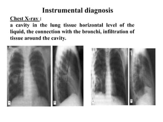
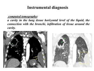
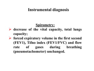
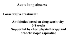
Acute lung abscess
Arivalagan Vetrivel
Group No;-21
DEFINITION:
Lung abscess: A localized cavity with pus, resulting from
necrosis of lung tissue, with surrounding pneumonitis. A lung
abscess may be putrid (due to anaerobic bacteria) or
nonputrid (due to anaerobes or aerobes). "Gangrene of the
lung" denotes a similar though morediffuse and extensive
process in which necrosis predominates.
In acute lung abscess the duration is less than weeks and pus
has thin wall
Acute lung abscess
CLINICAL SIGNS :
• Hectic fever,
• chills,
• cough,
• expectoration,
• chest pain,
• weight loss,
• dyspnoea
• hemoptysis
Acute lung abscess
Clinical feature of acute lung abscess
1st period – before opening an abscess
(it lasts from 3-4 days to 2-4 weeks)
2nd period – after opening (when
abscess perforates a bronchus)
General inspection: diffuse cyanosis, clubbed
fingernails, forced posture lying on the affected side.
Inspection of chest: lagging of the affected side of chest;
dyspnea, tachypnoe.
Palpation of the chest: chest pain on the affected side,
intensified vocal fremitus.
Percussion of the lung - dull sound.
Auscultation: bronchial or diminished vesicular
breathing.
Bronchophony: intensified.
Egophony - heard a phonetic "ā" ('ay').
1st period clinial signs
2nd period clinical signs
Complaints:
- cough with purulent sputum in large amounts «full
mouth» - till 2 liters per day (purulent sputum, three
layers, hemoptysis or streaked with blood, may be
expectorate over a few hours or several days);
- putrid odor;
- inspiratory dyspnea;
- chest pain on the affected side (in involving the
pleura);
- general complaint: weakness.
CLASSIFIATION ACCORDING TO ETIOLOGY:
 Aerobic bacteria infection (streptococcus
pneumoniae, streptococcus pyogenes, staphylococus aereus,
klebsiella pneumonie, pseodomonas aeruginosa, proteus
vulgaris, escherichia coli, neisseria meningitidis, haemophillus
influenzae)
 Anaerobic bacteria infection
(clostridium hystoliticum, bacteroides fragilis).
 Mixed bacterial flora ( common)
 NON-BACTERIAL
Fungal infections
Protozoa infections (entamoeba histolytica)
Acute lung abscess
The most
common
CLASSIFIATION ACCORDING TO PATHOGENISIS;
• suction abscesses (including obstructive);
• pneumonic abscesses;
• embolic hematogenous abscesses;
• Post-traumatic abscesses;
• lymph node abscesses.
CLASSIFIATION ACCORDING TO LOCALIZATION:
• abscess central (basal);
• abscess peripheral (cortical, subpleural)
dAcute lung abscess
Classification:
PRIMARY ABSCESS:
which develops as a result of primary infection of the lung. They most
commonly arise from aspiration, necrotising pneumonia or chronic
pneumonia (pulmonary TB).
SECONDARY ABSCESS:
In lung tissue affected by: existing lung disease, metastatic tumors, lung
carcinoma, foreign body, infarction,emphysema.
Acute lung abscess
Diagnosis:
 clinical signs,
 laboratory diagnosis
 instrumental diagnosis
Acute lung abscess
Total blood count: neutrophylic hyperleukocytosis, shift
to the left, to the myelocytes, significantly ↑ ESR,
possible anemia.
Sputum examination
Macroscopically: large amounts, three layers, unpleasant
odor, purulent sputum, may be mixed with blood.
Microscopically: contains a large number of leucocytes,
neutrophils, erythrocytes, can be elastic fibers.
Dietrich's plugs (composed of cellular decomposition
various bacteria, droplets of fat, fatty acid crystals)
Laboratory diagnosis
Instrumental diagnosis
Chest X-ray :
a cavity in the lung tissue horizontal level of the
liquid, the connection with the bronchi, infiltration of
tissue around the cavity.
Instrumental diagnosis
computed tomography:
a cavity in the lung tissue horizontal level of the liquid, the
connection with the bronchi, infiltration of tissue around the
cavity.
Instrumental diagnosis
Spirometry:
 decrease of the vital capacity, total lungs
capacity;
 forced expiratory volume in the first second
(FEV1), Tifno index (FEV1/FVC) and flow
rate of gases during breathing
(pneumotachometer) unchanged.
Conservative treatment :
Antibiotics based on drug sensitivity-
6-8 weeks
Supported by chest physiotherapy and
bronchoscopic aspiration
Acute lung abscess
THERAPY:
It is thought that lung abscess in its early stage can regress
spontaneously in approximately 20-30% of cases.
If conservative treatment is ineffective or complications are observed
adequate invasive or surgical treatment should be initiated.
It is estimated that invasive treatment by intercostal tube drainage or
surgery is indispensable in 11-12% of patients in whom antibiotic
therapy was ineffective.
Acute lung abscess
INVASIVE THERAPY:
INJECTION OF THERAPEUTIC AGENTS DIRECTLY INTO AN ABSCESS
CAVITY:
The abscess is punctured through the chest wall and antibiotics are injected directly
into its cavity. This method is suitable for large peripheral abscesses where the
visceral and parietal pleura are accreted. The method is burdened with a risk of
pneumothorax or empyema formation.
ENDOSCOPIC ABSCESS EVACUATION:
If lung abscess is a result of pathology of the bronchial tree with its obstruction and
retention of septic debris within bronchi distally to the level of obstruction
bronchoscopy is an effective technique for abscess evacuation. The abscess
evacuation can be completed by the injection of antibiotic into the abscess cavity.
Acute lung abscess
INVASIVE MANAGEMENT:
CAVERNOSTOMY:
A method used in selected cases of lung abscess (Monaldi procedure). Obliteration
of the pleural cavity is an indispensable condition to perform this procedure. Short
fragments of two or three ribs are resected over a place where the distance between
an abscess capsule and the chest wall is the shortest. The surface of the lung is
visualized and the abscess cavity is opened. Then the abscess cavity is packed with
gauze saturated with an antiseptic solution. The gauze is changed regularly for
several weeks until the process of healing and contraction of a lung defect is
obtained.
PERCUTANEOUS CATHETER DRAINAGE
Percutaneous catheter drainage of lung abscess under fluoroscopic or computed
tomography guidance is a technique introduced into medical practice during last
three decades and brings relatively good results in selected cases. The limitation of
this method is a localization of an abscess near important anatomical structures that
makes an introduction of a catheter hazardous. The technique is burdened with some
serious complications such as pneumothorax accompanied by a brocho-pleural
fistula, pleural empyema, hemorrhage and cardiac arrest.
Acute lung abscess
Lung abscess drainage
Surgical treatment:
Method: RESECTION.
Indications:
• Massive hemoptysis,
• Pleural empyema,
• Bronchopleural fistula,
• Conservative treatment failure,
• Foreign body,
• Big abscess (big cavity, >6cm after 8 weeks of treatment),
• Cancer suspicion.
Mortality rate: 11%-28%.
Acute lung abscess
Surgical treatment:
METHODS OF SURGICAL TREATMENT:
A type of resection of pulmonary parenchyma is dependent on
localization and size of abscess and a patient’s general state
• Non-anatomical resection (wedge or marginal)
• Anatomical resections: (segmentectomy, lobectomy,
pneumonectomy)
Acute lung abscess
NON-ANATOMICAL LUNG RESECTION
Marginal or wedge pulmonary parenchyma resection for abscesses
using linear staplers: TA and GIA type is effective for small lesions
localized peripherally.
ANATOMICAL LUNG RESECTION
Large lung abscesses, abscesses localized deeply in pulmonary
parenchyma and multiple lung abscesses are all indications for
anatomical resections. The most frequent procedures are resections of
a pulmonary lobe (lobectomy) or resection of two pulmonary lobes
of the right lung (bilobectomy). In the cases of solitary abscess or
multiple abscesses with vast destruction of the lung the resection of
the whole lung (pneumonectomy) is sometimes necessary. Local
conditions rarely enable less extensive surgical procedures such as
semisegmentectomy or bisegmentectomy.
Acute lung abscess
METHODS OF SURGICAL TREATMENT :
VATS (Video Assisted Thoracic Surgery)
A method that is a combination of minithoracotomy and videothoracoscopic
technique (video telescope and endoscopic instruments). It enables resection of
some small, peripherally localized abscesses.
DECORTICATION + LUNG RESECTION
A method used for the treatment of lung abscesses accompanied by pleural
empyema. Decortication consists in the removal of a thick fibrinopurulent coat
from the lung surface. Besides it the part of a pleural empyema capsule that covers
the internal surface of the chest wall is also resected along with the parietal pleura.
The procedure is completed by resection of pulmonary parenchyma with abscess.
LUNG RESECTION + IRRIGATING DRAINAGE
A procedure used in the cases of lung abscess coexisting with acute pleural
empyema. When pulmonary parenchyma with lung abscess is resected two or three
tubes are introduced into the pleural cavity and continuous irrigating drainage with
antibiotic or antiseptic solution is carried out.
Acute lung abscess

lung-abscess

  • 1.
    Acute lung abscess ArivalaganVetrivel Group No;-21
  • 2.
    DEFINITION: Lung abscess: Alocalized cavity with pus, resulting from necrosis of lung tissue, with surrounding pneumonitis. A lung abscess may be putrid (due to anaerobic bacteria) or nonputrid (due to anaerobes or aerobes). "Gangrene of the lung" denotes a similar though morediffuse and extensive process in which necrosis predominates. In acute lung abscess the duration is less than weeks and pus has thin wall Acute lung abscess
  • 3.
    CLINICAL SIGNS : •Hectic fever, • chills, • cough, • expectoration, • chest pain, • weight loss, • dyspnoea • hemoptysis Acute lung abscess
  • 4.
    Clinical feature ofacute lung abscess 1st period – before opening an abscess (it lasts from 3-4 days to 2-4 weeks) 2nd period – after opening (when abscess perforates a bronchus)
  • 5.
    General inspection: diffusecyanosis, clubbed fingernails, forced posture lying on the affected side. Inspection of chest: lagging of the affected side of chest; dyspnea, tachypnoe. Palpation of the chest: chest pain on the affected side, intensified vocal fremitus. Percussion of the lung - dull sound. Auscultation: bronchial or diminished vesicular breathing. Bronchophony: intensified. Egophony - heard a phonetic "ā" ('ay'). 1st period clinial signs
  • 6.
    2nd period clinicalsigns Complaints: - cough with purulent sputum in large amounts «full mouth» - till 2 liters per day (purulent sputum, three layers, hemoptysis or streaked with blood, may be expectorate over a few hours or several days); - putrid odor; - inspiratory dyspnea; - chest pain on the affected side (in involving the pleura); - general complaint: weakness.
  • 7.
    CLASSIFIATION ACCORDING TOETIOLOGY:  Aerobic bacteria infection (streptococcus pneumoniae, streptococcus pyogenes, staphylococus aereus, klebsiella pneumonie, pseodomonas aeruginosa, proteus vulgaris, escherichia coli, neisseria meningitidis, haemophillus influenzae)  Anaerobic bacteria infection (clostridium hystoliticum, bacteroides fragilis).  Mixed bacterial flora ( common)  NON-BACTERIAL Fungal infections Protozoa infections (entamoeba histolytica) Acute lung abscess The most common
  • 8.
    CLASSIFIATION ACCORDING TOPATHOGENISIS; • suction abscesses (including obstructive); • pneumonic abscesses; • embolic hematogenous abscesses; • Post-traumatic abscesses; • lymph node abscesses. CLASSIFIATION ACCORDING TO LOCALIZATION: • abscess central (basal); • abscess peripheral (cortical, subpleural) dAcute lung abscess
  • 9.
    Classification: PRIMARY ABSCESS: which developsas a result of primary infection of the lung. They most commonly arise from aspiration, necrotising pneumonia or chronic pneumonia (pulmonary TB). SECONDARY ABSCESS: In lung tissue affected by: existing lung disease, metastatic tumors, lung carcinoma, foreign body, infarction,emphysema. Acute lung abscess
  • 10.
    Diagnosis:  clinical signs, laboratory diagnosis  instrumental diagnosis Acute lung abscess
  • 11.
    Total blood count:neutrophylic hyperleukocytosis, shift to the left, to the myelocytes, significantly ↑ ESR, possible anemia. Sputum examination Macroscopically: large amounts, three layers, unpleasant odor, purulent sputum, may be mixed with blood. Microscopically: contains a large number of leucocytes, neutrophils, erythrocytes, can be elastic fibers. Dietrich's plugs (composed of cellular decomposition various bacteria, droplets of fat, fatty acid crystals) Laboratory diagnosis
  • 12.
    Instrumental diagnosis Chest X-ray: a cavity in the lung tissue horizontal level of the liquid, the connection with the bronchi, infiltration of tissue around the cavity.
  • 13.
    Instrumental diagnosis computed tomography: acavity in the lung tissue horizontal level of the liquid, the connection with the bronchi, infiltration of tissue around the cavity.
  • 14.
    Instrumental diagnosis Spirometry:  decreaseof the vital capacity, total lungs capacity;  forced expiratory volume in the first second (FEV1), Tifno index (FEV1/FVC) and flow rate of gases during breathing (pneumotachometer) unchanged.
  • 15.
    Conservative treatment : Antibioticsbased on drug sensitivity- 6-8 weeks Supported by chest physiotherapy and bronchoscopic aspiration Acute lung abscess
  • 16.
    THERAPY: It is thoughtthat lung abscess in its early stage can regress spontaneously in approximately 20-30% of cases. If conservative treatment is ineffective or complications are observed adequate invasive or surgical treatment should be initiated. It is estimated that invasive treatment by intercostal tube drainage or surgery is indispensable in 11-12% of patients in whom antibiotic therapy was ineffective. Acute lung abscess
  • 17.
    INVASIVE THERAPY: INJECTION OFTHERAPEUTIC AGENTS DIRECTLY INTO AN ABSCESS CAVITY: The abscess is punctured through the chest wall and antibiotics are injected directly into its cavity. This method is suitable for large peripheral abscesses where the visceral and parietal pleura are accreted. The method is burdened with a risk of pneumothorax or empyema formation. ENDOSCOPIC ABSCESS EVACUATION: If lung abscess is a result of pathology of the bronchial tree with its obstruction and retention of septic debris within bronchi distally to the level of obstruction bronchoscopy is an effective technique for abscess evacuation. The abscess evacuation can be completed by the injection of antibiotic into the abscess cavity. Acute lung abscess
  • 18.
    INVASIVE MANAGEMENT: CAVERNOSTOMY: A methodused in selected cases of lung abscess (Monaldi procedure). Obliteration of the pleural cavity is an indispensable condition to perform this procedure. Short fragments of two or three ribs are resected over a place where the distance between an abscess capsule and the chest wall is the shortest. The surface of the lung is visualized and the abscess cavity is opened. Then the abscess cavity is packed with gauze saturated with an antiseptic solution. The gauze is changed regularly for several weeks until the process of healing and contraction of a lung defect is obtained. PERCUTANEOUS CATHETER DRAINAGE Percutaneous catheter drainage of lung abscess under fluoroscopic or computed tomography guidance is a technique introduced into medical practice during last three decades and brings relatively good results in selected cases. The limitation of this method is a localization of an abscess near important anatomical structures that makes an introduction of a catheter hazardous. The technique is burdened with some serious complications such as pneumothorax accompanied by a brocho-pleural fistula, pleural empyema, hemorrhage and cardiac arrest. Acute lung abscess
  • 19.
  • 20.
    Surgical treatment: Method: RESECTION. Indications: •Massive hemoptysis, • Pleural empyema, • Bronchopleural fistula, • Conservative treatment failure, • Foreign body, • Big abscess (big cavity, >6cm after 8 weeks of treatment), • Cancer suspicion. Mortality rate: 11%-28%. Acute lung abscess
  • 21.
    Surgical treatment: METHODS OFSURGICAL TREATMENT: A type of resection of pulmonary parenchyma is dependent on localization and size of abscess and a patient’s general state • Non-anatomical resection (wedge or marginal) • Anatomical resections: (segmentectomy, lobectomy, pneumonectomy) Acute lung abscess
  • 22.
    NON-ANATOMICAL LUNG RESECTION Marginalor wedge pulmonary parenchyma resection for abscesses using linear staplers: TA and GIA type is effective for small lesions localized peripherally. ANATOMICAL LUNG RESECTION Large lung abscesses, abscesses localized deeply in pulmonary parenchyma and multiple lung abscesses are all indications for anatomical resections. The most frequent procedures are resections of a pulmonary lobe (lobectomy) or resection of two pulmonary lobes of the right lung (bilobectomy). In the cases of solitary abscess or multiple abscesses with vast destruction of the lung the resection of the whole lung (pneumonectomy) is sometimes necessary. Local conditions rarely enable less extensive surgical procedures such as semisegmentectomy or bisegmentectomy. Acute lung abscess
  • 23.
    METHODS OF SURGICALTREATMENT : VATS (Video Assisted Thoracic Surgery) A method that is a combination of minithoracotomy and videothoracoscopic technique (video telescope and endoscopic instruments). It enables resection of some small, peripherally localized abscesses. DECORTICATION + LUNG RESECTION A method used for the treatment of lung abscesses accompanied by pleural empyema. Decortication consists in the removal of a thick fibrinopurulent coat from the lung surface. Besides it the part of a pleural empyema capsule that covers the internal surface of the chest wall is also resected along with the parietal pleura. The procedure is completed by resection of pulmonary parenchyma with abscess. LUNG RESECTION + IRRIGATING DRAINAGE A procedure used in the cases of lung abscess coexisting with acute pleural empyema. When pulmonary parenchyma with lung abscess is resected two or three tubes are introduced into the pleural cavity and continuous irrigating drainage with antibiotic or antiseptic solution is carried out. Acute lung abscess