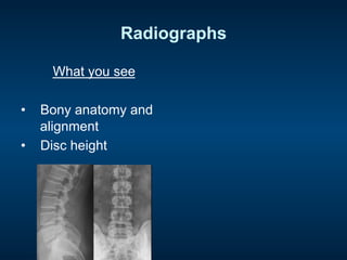
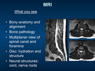
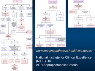
1) The document discusses appropriate imaging for back pain, describing different imaging modalities like X-rays, CT scans, bone scans, and MRI.
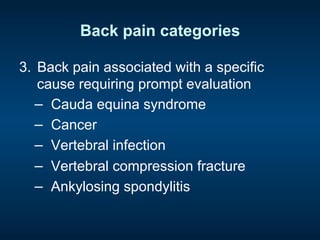
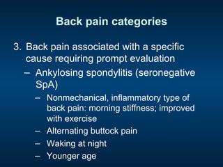
2) It categorizes back pain into 3 groups: nonspecific low back pain, back pain associated with radiculopathy, and back pain associated with a specific cause needing prompt evaluation.
3) Guidelines recommend triaging patients into these 3 categories and only imaging those with red flags, severe/progressive neurological symptoms, or if considering surgery/injections. Imaging is not recommended for nonspecific back pain.
Description of different ultrasound features of carpal tunnel syndrome before and after carpal tunnel release including Doppler imaging and elastography
This introductory presentation was directed to family physicians at the 2015 Family Medicine Forum. Following the presentation, there was a hands on demonstration for attendees.
Musculoskeletal Health Concerns of the Aging PopulationAllan Corpuz
A lecture on low back pain, osteoarthritis and soft tissue rheumatisms delivered to nurses, nursing attendants and institutional workers at the the Philippine General Hospital
This presentation is a comprehensive summary about all aspects of back pain. Back pain is one of the most common orthopaedic morbidity or orthopedic disability. Sciatica and lumbar disc diseases are common cause of spinal disability. Back pain are divided into Red flags, green flags and yellow flags for quick clinical screening. both treatment, prevention aspects are covered. Spinal anatomy and Biomechanics are covered. Epidemiology and role of various types of spine surgery, microdiscectomy, endoscopic spine surgery are also described.
Diffuse idiopathic skeletal hyperostosis (DISH) is a common skeletal process of uncertain etiology found in 12 to 18% of Indian populations above 50 years. The primary manifestations of DISH are calcification and ossification of the spinal ligaments, as well as entheseal ossification within extraspinal sites
Evaluation of Lumbar Spine Disease starts with understanding the clinical back grounds. It starts with good history and physical examination. This is a teaching lecture given twice by Prof. Dr. Mohamed Mohi Eldin, professor of neurosurgery, in the Basic Spine Course, Egyptian Medical Syndicate, Cairo, March 2009 and in 2010.
HOT NEW PRODUCT! BIG SALES FAST SHIPPING NOW FROM CHINA!! EU KU DB BK substit...GL Anaacs
Contact us if you are interested:
Email / Skype : kefaya1771@gmail.com
Threema: PXHY5PDH
New BATCH Ku !!! MUCH IN DEMAND FAST SALE EVERY BATCH HAPPY GOOD EFFECT BIG BATCH !
Contact me on Threema or skype to start big business!!
Hot-sale products:
NEW HOT EUTYLONE WHITE CRYSTAL!!
5cl-adba precursor (semi finished )
5cl-adba raw materials
ADBB precursor (semi finished )
ADBB raw materials
APVP powder
5fadb/4f-adb
Jwh018 / Jwh210
Eutylone crystal
Protonitazene (hydrochloride) CAS: 119276-01-6
Flubrotizolam CAS: 57801-95-3
Metonitazene CAS: 14680-51-4
Payment terms: Western Union,MoneyGram,Bitcoin or USDT.
Deliver Time: Usually 7-15days
Shipping method: FedEx, TNT, DHL,UPS etc.Our deliveries are 100% safe, fast, reliable and discreet.
Samples will be sent for your evaluation!If you are interested in, please contact me, let's talk details.
We specializes in exporting high quality Research chemical, medical intermediate, Pharmaceutical chemicals and so on. Products are exported to USA, Canada, France, Korea, Japan,Russia, Southeast Asia and other countries.
MANAGEMENT OF ATRIOVENTRICULAR CONDUCTION BLOCK.pdfJim Jacob Roy
Cardiac conduction defects can occur due to various causes.
Atrioventricular conduction blocks ( AV blocks ) are classified into 3 types.
This document describes the acute management of AV block.
Flu Vaccine Alert in Bangalore Karnatakaaddon Scans
As flu season approaches, health officials in Bangalore, Karnataka, are urging residents to get their flu vaccinations. The seasonal flu, while common, can lead to severe health complications, particularly for vulnerable populations such as young children, the elderly, and those with underlying health conditions.
Dr. Vidisha Kumari, a leading epidemiologist in Bangalore, emphasizes the importance of getting vaccinated. "The flu vaccine is our best defense against the influenza virus. It not only protects individuals but also helps prevent the spread of the virus in our communities," he says.
This year, the flu season is expected to coincide with a potential increase in other respiratory illnesses. The Karnataka Health Department has launched an awareness campaign highlighting the significance of flu vaccinations. They have set up multiple vaccination centers across Bangalore, making it convenient for residents to receive their shots.
To encourage widespread vaccination, the government is also collaborating with local schools, workplaces, and community centers to facilitate vaccination drives. Special attention is being given to ensuring that the vaccine is accessible to all, including marginalized communities who may have limited access to healthcare.
Residents are reminded that the flu vaccine is safe and effective. Common side effects are mild and may include soreness at the injection site, mild fever, or muscle aches. These side effects are generally short-lived and far less severe than the flu itself.
Healthcare providers are also stressing the importance of continuing COVID-19 precautions. Wearing masks, practicing good hand hygiene, and maintaining social distancing are still crucial, especially in crowded places.
Protect yourself and your loved ones by getting vaccinated. Together, we can help keep Bangalore healthy and safe this flu season. For more information on vaccination centers and schedules, residents can visit the Karnataka Health Department’s official website or follow their social media pages.
Stay informed, stay safe, and get your flu shot today!
micro teaching on communication m.sc nursing.pdfAnurag Sharma
Microteaching is a unique model of practice teaching. It is a viable instrument for the. desired change in the teaching behavior or the behavior potential which, in specified types of real. classroom situations, tends to facilitate the achievement of specified types of objectives.
These lecture slides, by Dr Sidra Arshad, offer a quick overview of physiological basis of a normal electrocardiogram.
Learning objectives:
1. Define an electrocardiogram (ECG) and electrocardiography
2. Describe how dipoles generated by the heart produce the waveforms of the ECG
3. Describe the components of a normal electrocardiogram of a typical bipolar leads (limb II)
4. Differentiate between intervals and segments
5. Enlist some common indications for obtaining an ECG
Study Resources:
1. Chapter 11, Guyton and Hall Textbook of Medical Physiology, 14th edition
2. Chapter 9, Human Physiology - From Cells to Systems, Lauralee Sherwood, 9th edition
3. Chapter 29, Ganong’s Review of Medical Physiology, 26th edition
4. Electrocardiogram, StatPearls - https://www.ncbi.nlm.nih.gov/books/NBK549803/
5. ECG in Medical Practice by ABM Abdullah, 4th edition
6. ECG Basics, http://www.nataliescasebook.com/tag/e-c-g-basics
Tom Selleck Health: A Comprehensive Look at the Iconic Actor’s Wellness Journeygreendigital
Tom Selleck, an enduring figure in Hollywood. has captivated audiences for decades with his rugged charm, iconic moustache. and memorable roles in television and film. From his breakout role as Thomas Magnum in Magnum P.I. to his current portrayal of Frank Reagan in Blue Bloods. Selleck's career has spanned over 50 years. But beyond his professional achievements. fans have often been curious about Tom Selleck Health. especially as he has aged in the public eye.
Follow us on: Pinterest
Introduction
Many have been interested in Tom Selleck health. not only because of his enduring presence on screen but also because of the challenges. and lifestyle choices he has faced and made over the years. This article delves into the various aspects of Tom Selleck health. exploring his fitness regimen, diet, mental health. and the challenges he has encountered as he ages. We'll look at how he maintains his well-being. the health issues he has faced, and his approach to ageing .
Early Life and Career
Childhood and Athletic Beginnings
Tom Selleck was born on January 29, 1945, in Detroit, Michigan, and grew up in Sherman Oaks, California. From an early age, he was involved in sports, particularly basketball. which played a significant role in his physical development. His athletic pursuits continued into college. where he attended the University of Southern California (USC) on a basketball scholarship. This early involvement in sports laid a strong foundation for his physical health and disciplined lifestyle.
Transition to Acting
Selleck's transition from an athlete to an actor came with its physical demands. His first significant role in "Magnum P.I." required him to perform various stunts and maintain a fit appearance. This role, which he played from 1980 to 1988. necessitated a rigorous fitness routine to meet the show's demands. setting the stage for his long-term commitment to health and wellness.
Fitness Regimen
Workout Routine
Tom Selleck health and fitness regimen has evolved. adapting to his changing roles and age. During his "Magnum, P.I." days. Selleck's workouts were intense and focused on building and maintaining muscle mass. His routine included weightlifting, cardiovascular exercises. and specific training for the stunts he performed on the show.
Selleck adjusted his fitness routine as he aged to suit his body's needs. Today, his workouts focus on maintaining flexibility, strength, and cardiovascular health. He incorporates low-impact exercises such as swimming, walking, and light weightlifting. This balanced approach helps him stay fit without putting undue strain on his joints and muscles.
Importance of Flexibility and Mobility
In recent years, Selleck has emphasized the importance of flexibility and mobility in his fitness regimen. Understanding the natural decline in muscle mass and joint flexibility with age. he includes stretching and yoga in his routine. These practices help prevent injuries, improve posture, and maintain mobilit
Ethanol (CH3CH2OH), or beverage alcohol, is a two-carbon alcohol
that is rapidly distributed in the body and brain. Ethanol alters many
neurochemical systems and has rewarding and addictive properties. It
is the oldest recreational drug and likely contributes to more morbidity,
mortality, and public health costs than all illicit drugs combined. The
5th edition of the Diagnostic and Statistical Manual of Mental Disorders
(DSM-5) integrates alcohol abuse and alcohol dependence into a single
disorder called alcohol use disorder (AUD), with mild, moderate,
and severe subclassifications (American Psychiatric Association, 2013).
In the DSM-5, all types of substance abuse and dependence have been
combined into a single substance use disorder (SUD) on a continuum
from mild to severe. A diagnosis of AUD requires that at least two of
the 11 DSM-5 behaviors be present within a 12-month period (mild
AUD: 2–3 criteria; moderate AUD: 4–5 criteria; severe AUD: 6–11 criteria).
The four main behavioral effects of AUD are impaired control over
drinking, negative social consequences, risky use, and altered physiological
effects (tolerance, withdrawal). This chapter presents an overview
of the prevalence and harmful consequences of AUD in the U.S.,
the systemic nature of the disease, neurocircuitry and stages of AUD,
comorbidities, fetal alcohol spectrum disorders, genetic risk factors, and
pharmacotherapies for AUD.
5. Radiographs
What you see
• Bony anatomy and
alignment
• Disc height
Disadvantages
• Radiation
• Nonspecific
– OA changes in most
adults
• Insensitive
– No direct visualisation
of neural and other
nonbony structures
7. Bone scan
What you see
• Bone pathology
– Osteoblastic activity
Disadvantages
• Radiation
• Very nonspecific
• Relatively poor
anatomical resolution
– (Improved with
SPECT; SPECT/CT)
– No direct visualisation
of neural and other
nonbony structures
8. CT
What you see
• Bony anatomy and
alignment
• Cross sectional view
of spinal canal and
foramina
• Disc, thecal sac,
nerve roots
9. CT
What you see
• Bony anatomy and
alignment
• Cross sectional view
of spinal canal and
foramina
• Disc, thecal sac,
nerve roots
Disadvantages
• Radiation
• Nonspecific
– Most adults have
‘findings’
• Poor visualisation of
individual neural
structures and disc
anatomy
10. Radiation doses
Imaging test Effective
dose (mSv)
CXRs Background
exposure
Flying hours
CXR 0.02 1 3 days 4
Lumbar X-ray 1.5 75 6/12 300
Lumbar CT 2-10 100-500 8/12 - 3 years 400 - 1800
Bone scan 6 300 2 years 1200
11. MRI
What you see
• Bony anatomy and
alignment
• Bone pathology
• Multiplanar view of
spinal canal and
foramina
• Disc: hydration and
structure
• Neural structures:
cord, nerve roots
12. MRI
What you see
• Bony anatomy and
alignment
• Bone pathology
• Multiplanar view of
spinal canal and
foramina
• Disc: hydration and
structure
• Neural structures:
cord, nerve roots
Disadvantages
• Availability, cost
• Pacemakers,
claustrophobia
• Nonspecific (too
sensitive)
– Most adults have
‘findings’
13. Appropriate imaging for back pain
• Imaging modalities
• Clinical presentations: classification into 3
broad categories
1. Nonspecific low back pain
2. Back pain associated with radiculopathy
3. Back pain associated with a specific
cause requiring prompt evaluation
• Guidelines
15. Back pain categories
2. Back pain associated with radiculopathy
a) Unilateral acute nerve root compression
(sciatica)
– Leg pain >> back pain
– Disc herniation
b) Unilateral chronic nerve root compression
– Disc herniation or spinal stenosis
c) Bilateral chronic nerve root compression
– Spinal stenosis
– DD vascular claudication
d) Bilateral acute nerve root compression =
‘cauda equina syndrome’
16. Cauda equina syndrome
• Bilateral acute nerve root compression
– Massive disc protrusion/ sequestration
• Sudden onset bilateral leg pain
• Saddle anaesthesia
• Rapidly progressive or severe neurological
deficits
– Motor deficits at >1 level
– Faecal incontinence
– Urinary retention
17. Back pain categories
3. Back pain associated with a specific
cause requiring prompt evaluation
− Cauda equina syndrome
− Cancer
− Vertebral infection
− Vertebral compression fracture
− Ankylosing spondylitis
18. Back pain categories
3. Back pain associated with a specific
cause requiring prompt evaluation
− Cauda equina syndrome
− Clinical scenario
− Cancer
− Vertebral infection
− Vertebral compression fracture
− Ankylosing spondylitis
19. Back pain categories
3. Back pain associated with a specific
cause requiring prompt evaluation
− Cancer
− Hx of Ca + new onset LBP
− Unexplained weight loss +/-
persistent symptoms +/- age > 50
− Vertebral infection
− Vertebral compression fracture
− Ankylosing spondylitis
20. Back pain categories
3. Back pain associated with a specific
cause requiring prompt evaluation
− Vertebral infection
− Fever
− iv drug use
− Recent infection
− Vertebral compression fracture
− Ankylosing spondylitis
21. Back pain categories
3. Back pain associated with a specific
cause requiring prompt evaluation
− Vertebral compression fracture
− Hx of osteoporosis
− Steroid use
− Old age +/- minor trauma
− Ankylosing spondylitis
22. Back pain categories
3. Back pain associated with a specific
cause requiring prompt evaluation
− Ankylosing spondylitis (seronegative
SpA)
− Nonmechanical, inflammatory type of
back pain: morning stiffness; improved
with exercise
− Alternating buttock pain
− Waking at night
− Younger age
23. Appropriate imaging for back pain
• Imaging modalities
• Clinical presentations
• Guidelines
– Multiple: different countries and
associations
– Common theme:
• Triage into 3 broad categories as
described
24. LOW BACK PAIN GUIDELINES
Diagnostic triage
1. Non-specific LBP
2. Radiculopathy
3. Specific LBP
• ‘Red flags’
‘Red Flags’
• Cauda equina syndrome
• Known 10 tumour
• Weight loss
• Severe symptoms, not
settling
• Fever
• Recent infection or Sx
• Osteoporosis
• Steroid use
• Non-mechanical pain
• Child*
25. Back pain in children and adolescents
Presentation Associated Sx DD Ix
Night pain Fever, malaise Tumour, infection X-ray
MRI
Acute pain Radiculopathy
+ve SLR
Disc herniation
Spondylosis
X-ray
MRI
Chronic pain Rigid kyphosis
Morning stiffness
“Scheuermann’s”
Inflammatory arthropathy
X-ray
Pain with extension
Sport: eg rowing
Hamstring tightness Spondylolysis
‘Stress reaction’
X-ray
MRI
Pain + recent onset
scoliosis
Fever, malaise,
+ve SLR
Idiopathic scoliosis
Tumour, infection, syrinx,
disc herniation
X-ray
MRI
Am Fam Phys 2007;76:1669-76
26. LOW BACK PAIN GUIDELINES
• American College of Physicians & American
Pain Society Recommendations
1. Focused Hx and examination to place patients
into 1 of 3 categories
2. No imaging for nonspecific LBP
3. Imaging for LBP + severe or progressive
neurological deficits OR risk factors for specific
cause
4. Imaging for LBP and radiculopathy if candidates
for surgery or epidural injection
Ann Intern Med 2007;147:478-491
27. Diagnostic work-up
Possible cause Imaging Additional studies
Nonspecific LBP None None
Radiculopathy MRI (CT)
Cauda equina MRI
Cancer MRI for known 10; X-ray
for other eg wt loss
ESR
Vertebral infection MRI ESR, CRP
Vertebral compression # X-ray
Ankylosing spondylitis X-ray, incl pelvis (MRI) HLA-B27; ESR, CRP
Ann Intern Med 2007;147:478-491
29. Ineffectiveness of imaging for
nonspecific LBP
• Favourable natural Hx
– Most improve by 4 weeks; unaffected by imaging
• Nonspecificity: loose association between findings
and symptoms
– ‘Abnormalities’ or normal aging?
• Potential harms:
– Radiation
– ‘Labelling’
– Incidental findings
Ann Intern Med 2011;154:181-190
30.
31. • 85 year old female
• Severe acute on chronic mechanical
back pain
– Can’t sleep
– Limited walking to only a few steps
• Spontaneous onset
• No known trauma
Radiograph (X-ray)
44. • 62 year old male
• Severe low back pain of rapid onset
• Febrile and unwell
• 4 weeks ago underwent abdominal
surgery for perforated diverticulitis
MRI