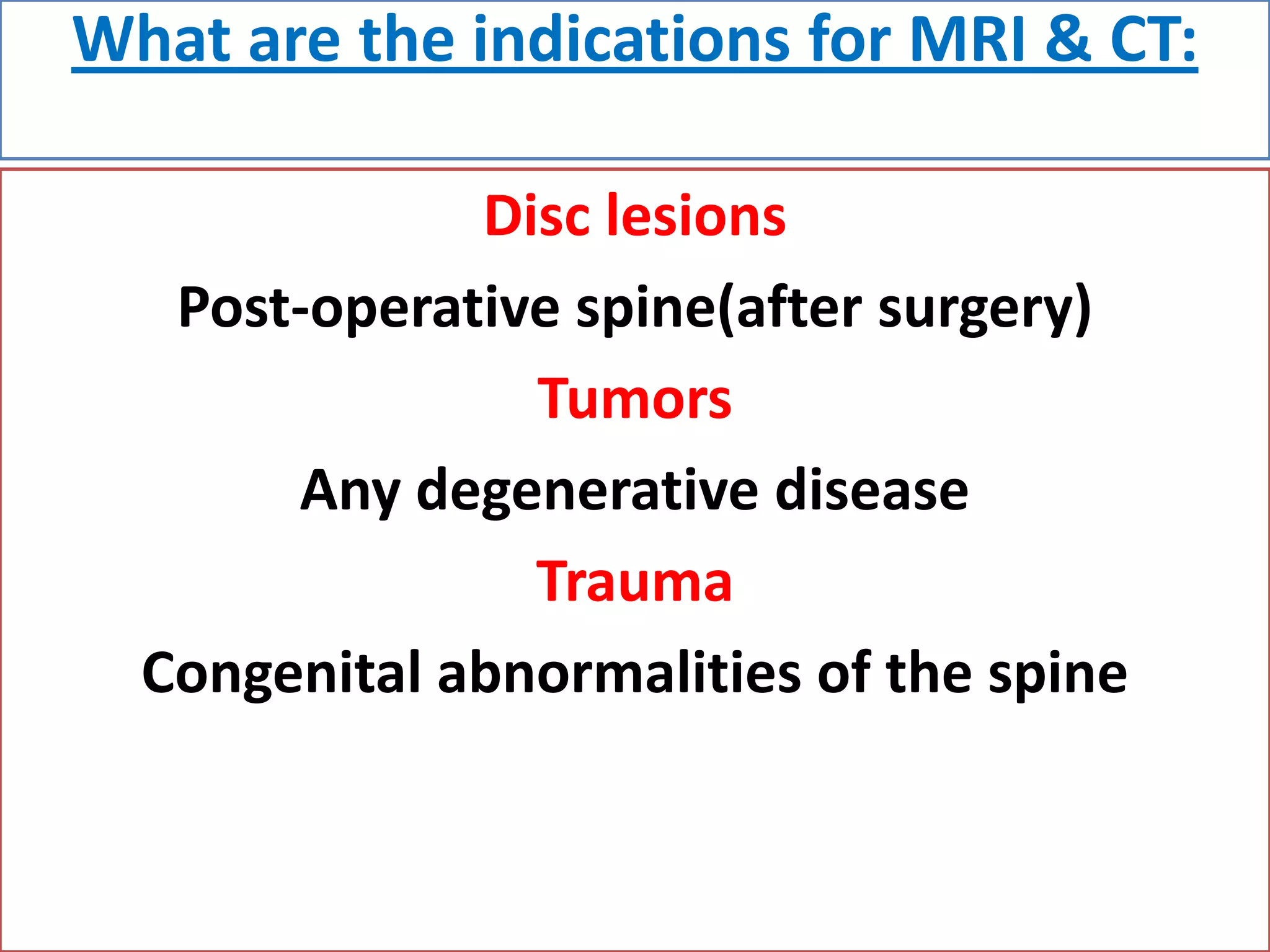
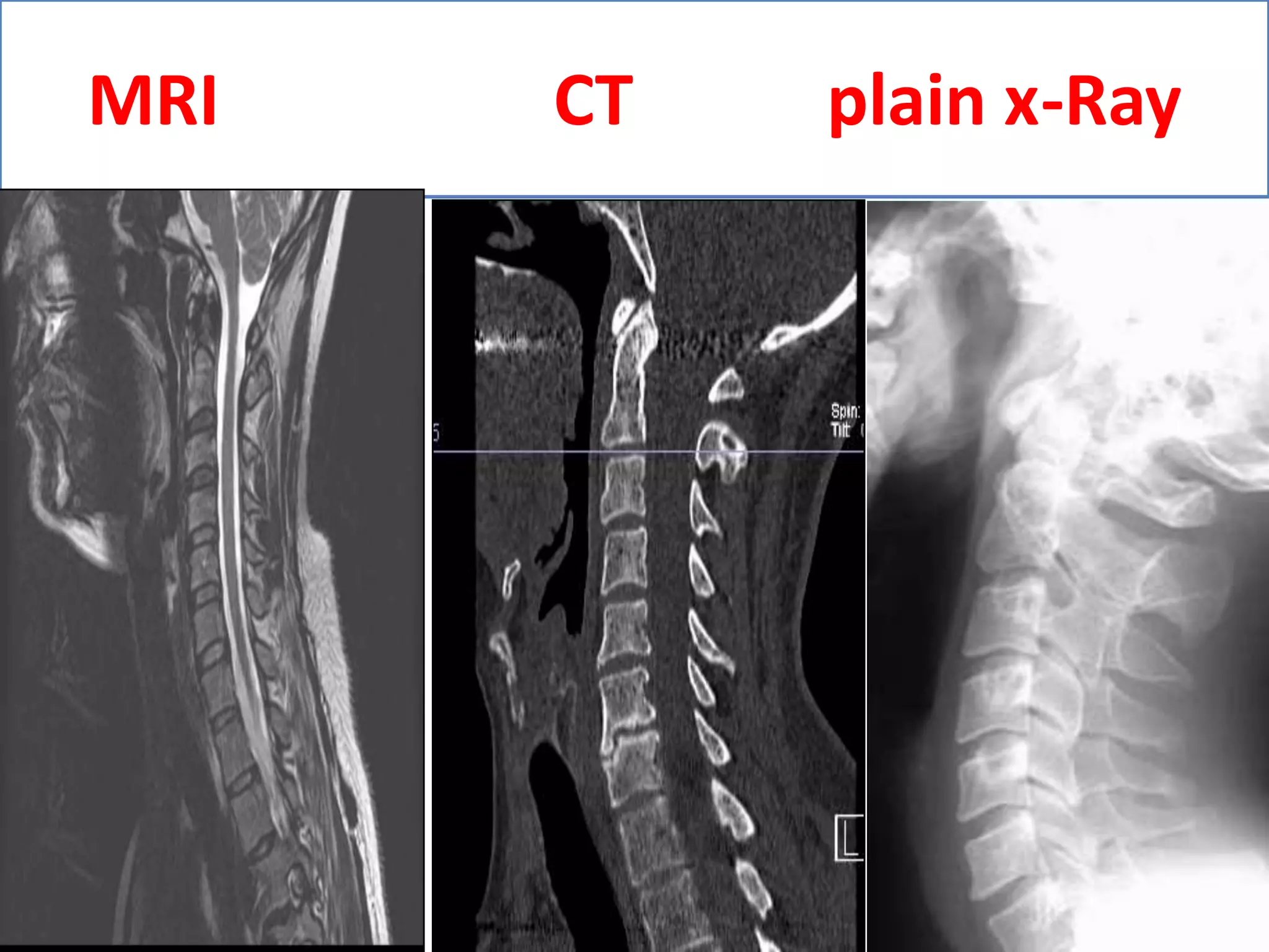
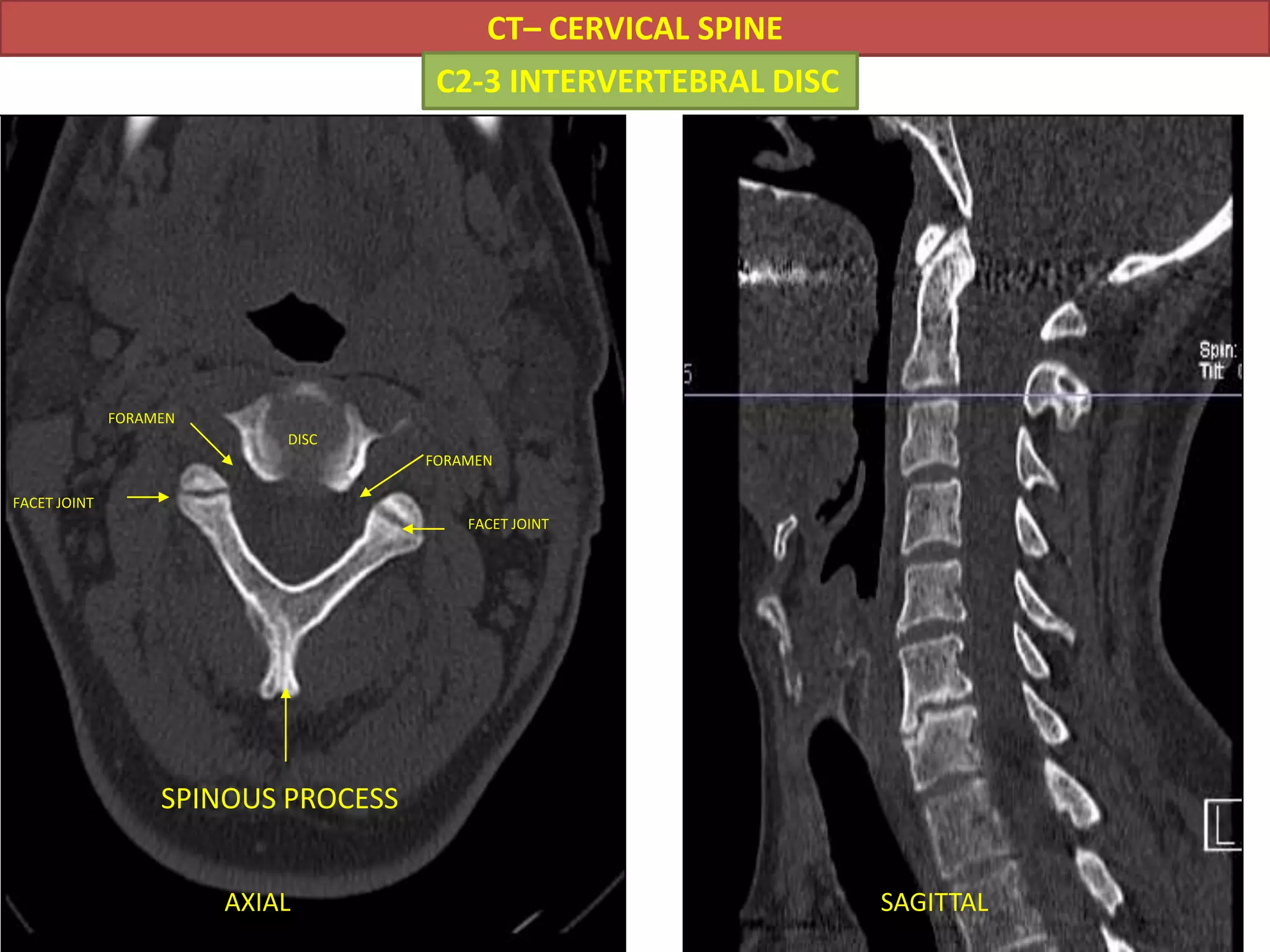
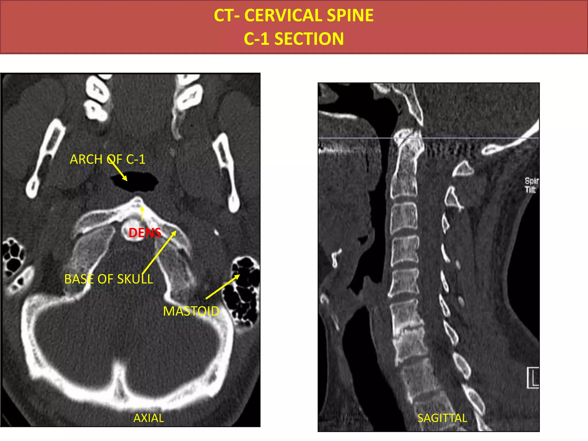
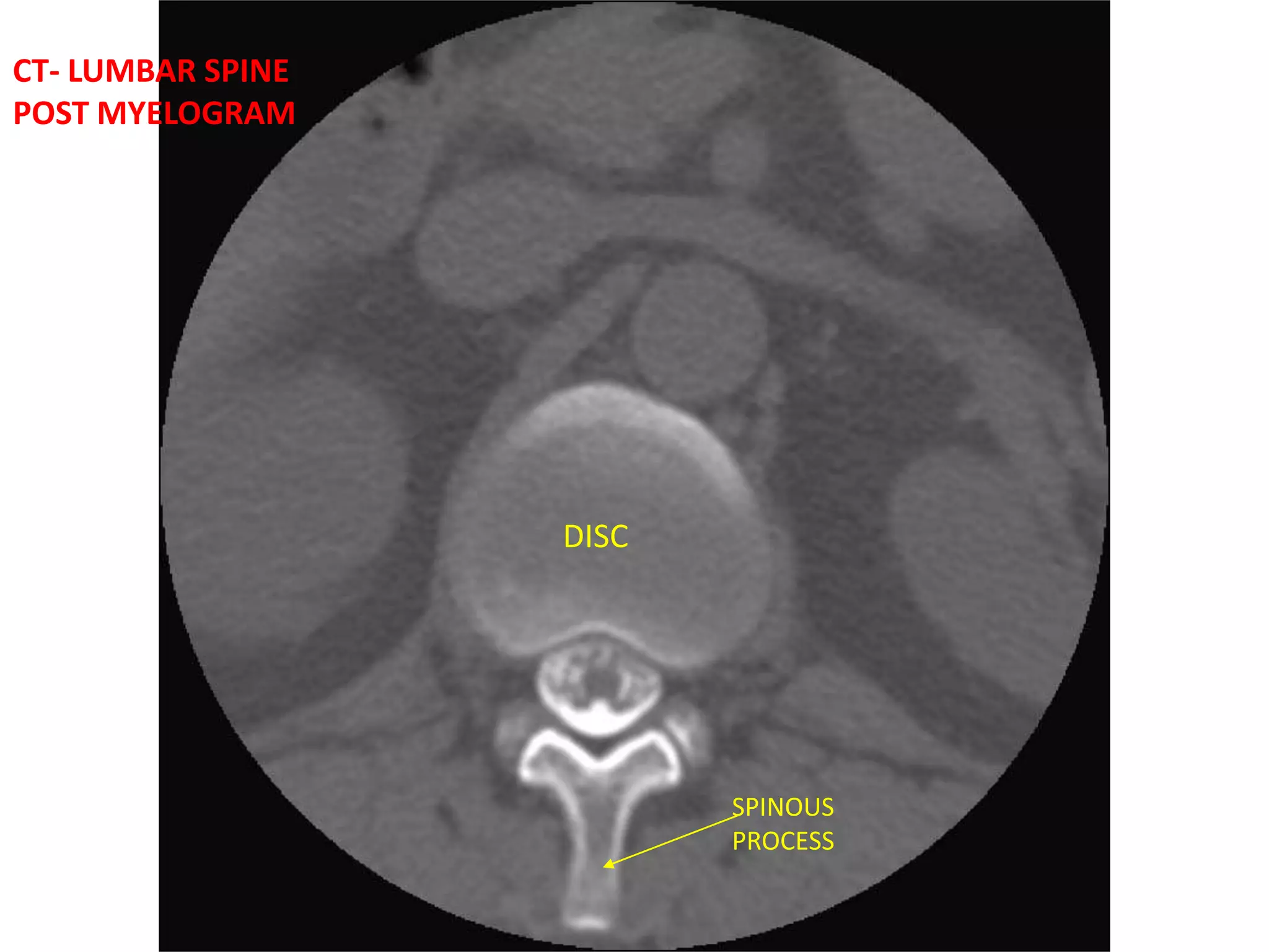
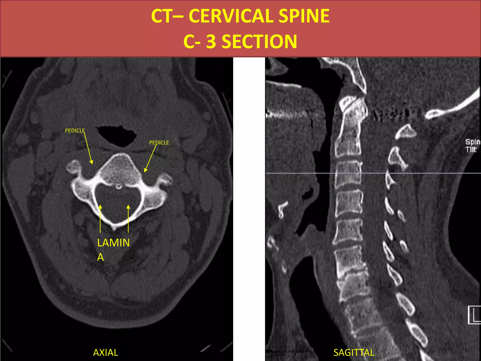
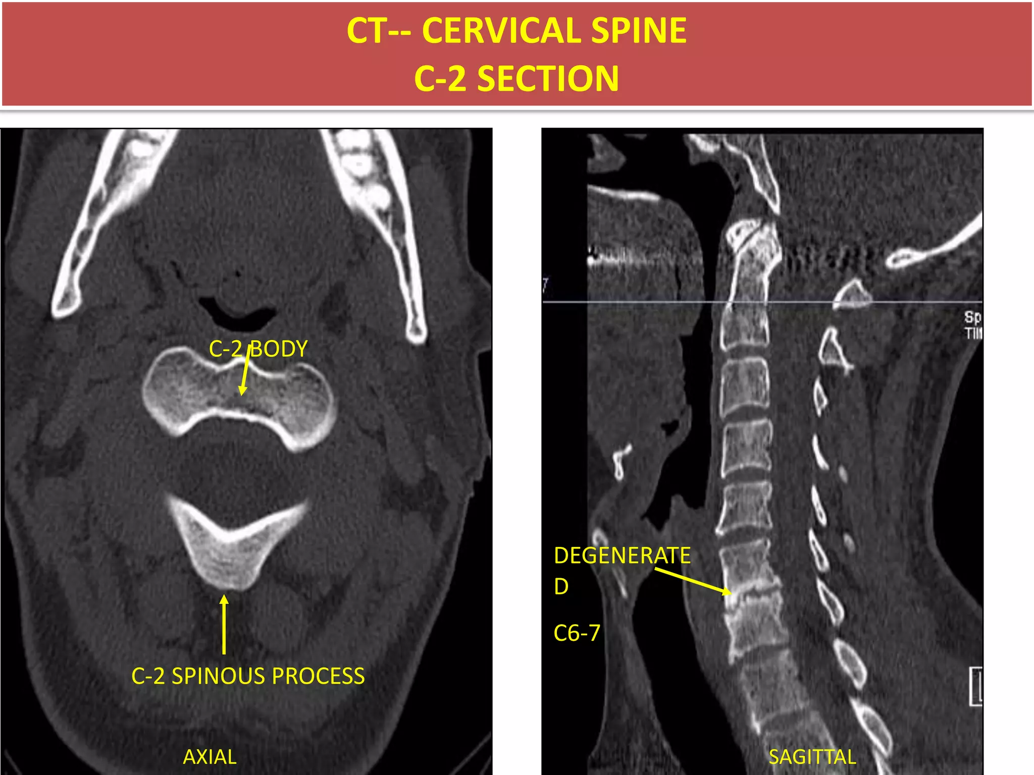
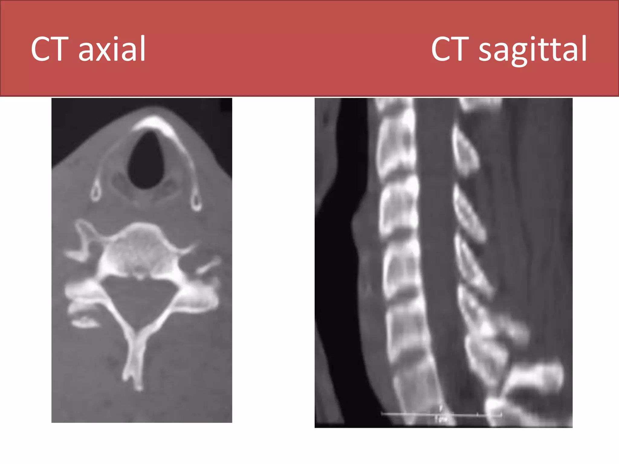
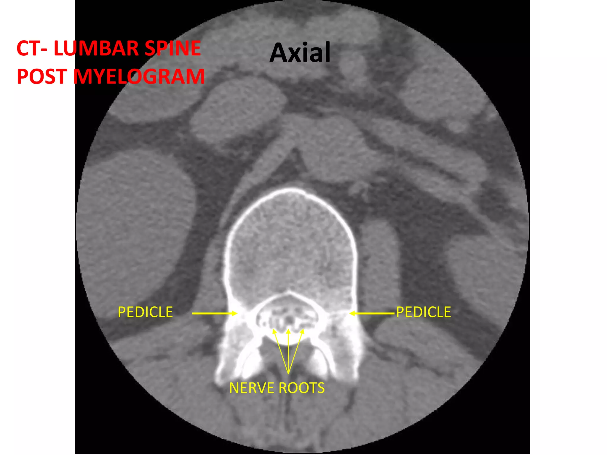
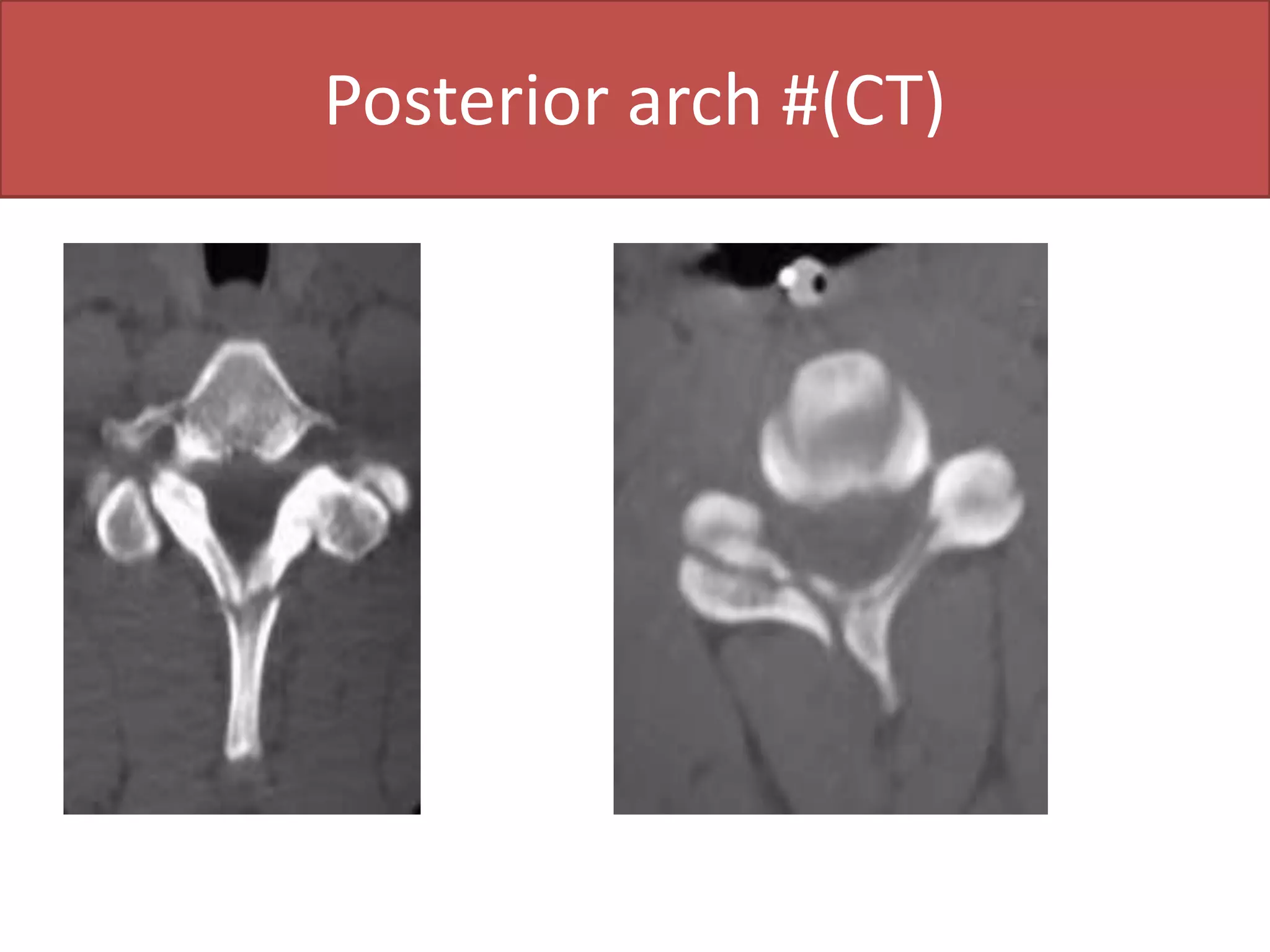
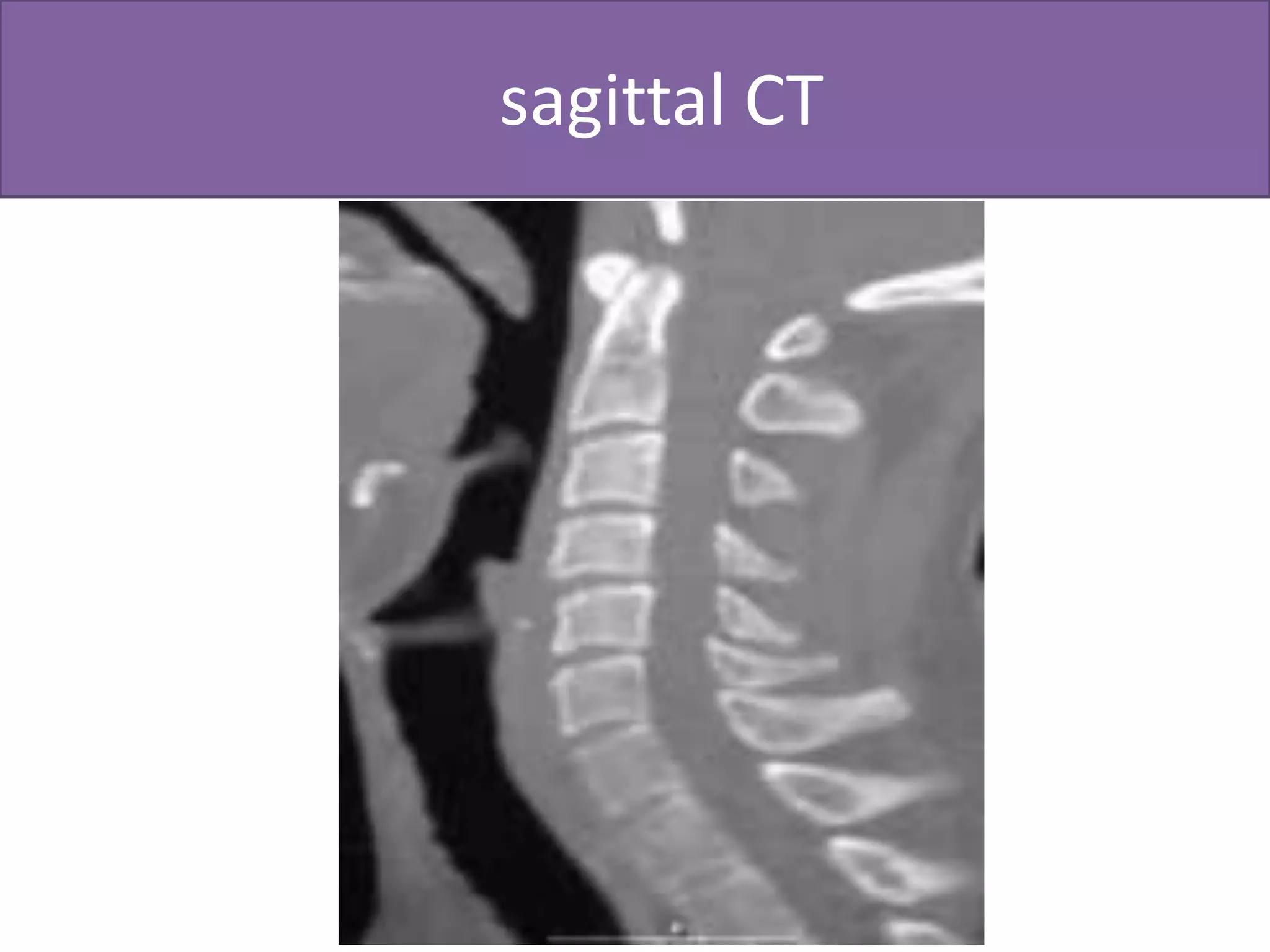
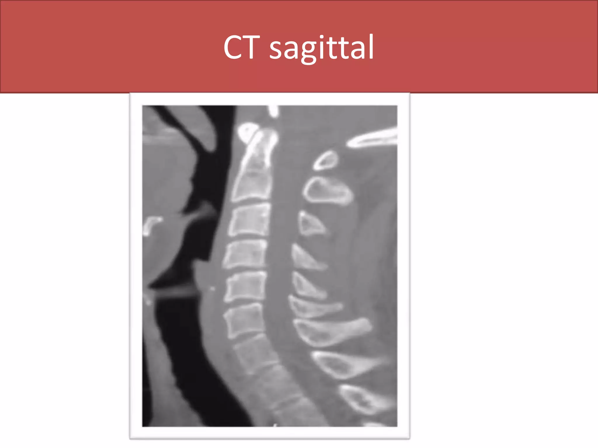
What are the indications for MRI & CT: Disc lesionsPost-operative spine(after surgery)TumorsAny degenerative diseaseTraumaCongenital abnormalities of the spine 
Preparations for CT & MRI:1-Fasting for 4-6 hours2-Contrast material Urographin ,telebrix 1-2 mg/kgAdministration of contrast:Yes: In post-operative lumbar spine, inflammatory disorders& neoplastic lesions.No:disc lesions, trauma.AnesthesiaUsed with children & uncooperative patients.
Patient position:     Is usually supine, sometimes side lying, & rarely prone.  How to see:Scanogram: primitive picture to detect the site of lumbar spine 
Computed tomography (CT):. Usual scanning.  . Axial slides 2-4 mm.	2 mm in cervical spine / 4 mm in lumbar spineHas bone & soft tissue window. CT screening:. Whole segment of the spine   5 mm in cervical spine, 8 mm in lumbar spine.. Selective Scanning   Every 3 mm especially in trauma, also in cervical disc lesions. N.B:If I want to see one vertebra e.g. L3 I have to take 1 vertebra above (L2 )& 1 below (L4).
CT Myelography:Is considered as intrathecal contrast injection with L- puncture needle.
We have 2 windows
Soft tissue & bone window.What are the structures I should evaluate in CT of lumbar spine:Lumbar spinal canal diameter normally 13 mm.Disc lesions.Others.facet, sacroiliac joints & paravertebral soft tissue.
Soft &bone window (CT) Lumbar
1. Lumbar spinal canal diameter:Spinal canal is bony structure, so we see it in bone window.The spinal canal must be closed (at the level of pedicles).We measure the AP diameter. Types of canal stenosis:1-Relative:11-12 mm & this doesn’t need operation but it needs operation if there’s disc.2- Absolute:      8 -10 mm & it must be operated. 
Disc lesions:We detect it in soft tissue window.		Posterior border of the disc is more important as it has relation to the disc.		The normal posterior border of the disc is CONCAVE.The abnormal is STRAIGHT OR CONVEX.
N.B:. Normally due to overload ,the disc of L5-S1 is CONVEX& the  abnormal is also convex,so to judge if it’s normal or no look at the next slide if: the posterior border of the disc is convex so it is ABNORMAL.. The angle of inclination in L5-S1 is more than 30 & the device accept up till 30 only so part of the slide will contain bone & part will contain disc.
Normal CT Lumbar
Disc herniation (CT axial)
MRI – LUMBAR SPINEMNERVE ROOTSFORAMENAXIAL VIEW
Manifestations of arthritis  in any joint: (Spondylosis in spine  and osteoarthritis of other joints)Osteophytic lipping.Narrow joint space.Subarticular bone sclerosisSub cortical pseudo cystic changes.Intra articular air.(vaccum phenomena)
Vacuum phenomena
CT of cervical spine
We have 2 types of joints:Neurocentral joint:Is the articulation between one vertebra above & one vertebra below which makes the shape of the body of the vertebral end plate .Facet joint:Is the articulation between the inferior lip of the transverse process of one  vertebra above  with the superior lip of the transverse process of the vertebra below, it’s called Hamburger’s Sandwich. Arthritis of the neurocentral or facet joint gives the same manifestations of nerve compression due to disc lesion.
Cervical disc in CT:We see it in soft tissue window .The disc in cervical spine is very narrow so every slide will contain both disc & bone, therefore there isn’t a slide of pure disc, So we choose the slide which contain more disc for assessment.. All posterior edges of cervical spine are normally convex.. See if there’s disc substance protruded than the bone.. Normal spinal cord picture is kidney shaped. 
Difference between cervical & lumbar spine in CT 
Stages of disc pathology (4 stages)
CT of the spine
CT axial  bone& soft tissue widow
CT machine
MRI                   CT           plain x-Ray
CT– CERVICAL SPINEC2-3 INTERVERTEBRAL DISC FORAMENDISCFORAMENFACET JOINTFACET JOINTSPINOUS PROCESSAXIALSAGITTAL
CT- CERVICAL SPINEC-1 SECTIONARCH OF C-1DENSBASE OF SKULLMASTOIDAXIALSAGITTAL
CT- LUMBAR SPINEPOST MYELOGRAM DISCSPINOUS PROCESS
CT– CERVICAL SPINEC- 3 SECTIONPEDICLEPEDICLELAMINAAXIALSAGITTAL
CT-- CERVICAL SPINEC-2 SECTIONC-2 BODYDEGENERATEDC6-7C-2 SPINOUS PROCESSAXIALSAGITTAL
CT axial                                 CT sagittal
CT- LUMBAR SPINEPOST MYELOGRAMAxialPEDICLEPEDICLENERVE ROOTS
Posterior arch #(CT)
Burst # (CT) axial
   sagittal CT
CT axial section (base of the skull)
CT- LUMBAR SPINEPOST MYELOGRAMFORAMENFORAMEN
CT of cervical spine(sagittal)
CT axial
CT of cervical spine (axial)
CT sagittal
CT sagittal tear drop # dislocationTear drop #  dislocation(plain)
CT-LUMBAR SPINEPOST MYELOGRAMTHECAL SACLAMINALAMINA

Ct&Mri

  • 1.
    What are theindications for MRI & CT: Disc lesionsPost-operative spine(after surgery)TumorsAny degenerative diseaseTraumaCongenital abnormalities of the spine 
  • 2.
    Preparations for CT& MRI:1-Fasting for 4-6 hours2-Contrast material Urographin ,telebrix 1-2 mg/kgAdministration of contrast:Yes: In post-operative lumbar spine, inflammatory disorders& neoplastic lesions.No:disc lesions, trauma.AnesthesiaUsed with children & uncooperative patients.
  • 3.
    Patient position: Is usually supine, sometimes side lying, & rarely prone.  How to see:Scanogram: primitive picture to detect the site of lumbar spine 
  • 4.
    Computed tomography (CT):.Usual scanning. . Axial slides 2-4 mm. 2 mm in cervical spine / 4 mm in lumbar spineHas bone & soft tissue window. CT screening:. Whole segment of the spine 5 mm in cervical spine, 8 mm in lumbar spine.. Selective Scanning Every 3 mm especially in trauma, also in cervical disc lesions. N.B:If I want to see one vertebra e.g. L3 I have to take 1 vertebra above (L2 )& 1 below (L4).
  • 7.
    CT Myelography:Is consideredas intrathecal contrast injection with L- puncture needle.
  • 8.
    We have 2windows
  • 9.
    Soft tissue &bone window.What are the structures I should evaluate in CT of lumbar spine:Lumbar spinal canal diameter normally 13 mm.Disc lesions.Others.facet, sacroiliac joints & paravertebral soft tissue.
  • 10.
  • 12.
    1. Lumbar spinalcanal diameter:Spinal canal is bony structure, so we see it in bone window.The spinal canal must be closed (at the level of pedicles).We measure the AP diameter. Types of canal stenosis:1-Relative:11-12 mm & this doesn’t need operation but it needs operation if there’s disc.2- Absolute: 8 -10 mm & it must be operated. 
  • 13.
    Disc lesions:We detectit in soft tissue window. Posterior border of the disc is more important as it has relation to the disc. The normal posterior border of the disc is CONCAVE.The abnormal is STRAIGHT OR CONVEX.
  • 14.
    N.B:. Normally dueto overload ,the disc of L5-S1 is CONVEX& the abnormal is also convex,so to judge if it’s normal or no look at the next slide if: the posterior border of the disc is convex so it is ABNORMAL.. The angle of inclination in L5-S1 is more than 30 & the device accept up till 30 only so part of the slide will contain bone & part will contain disc.
  • 17.
  • 18.
  • 19.
    MRI – LUMBARSPINEMNERVE ROOTSFORAMENAXIAL VIEW
  • 20.
    Manifestations of arthritis in any joint: (Spondylosis in spine and osteoarthritis of other joints)Osteophytic lipping.Narrow joint space.Subarticular bone sclerosisSub cortical pseudo cystic changes.Intra articular air.(vaccum phenomena)
  • 21.
  • 22.
  • 23.
    We have 2types of joints:Neurocentral joint:Is the articulation between one vertebra above & one vertebra below which makes the shape of the body of the vertebral end plate .Facet joint:Is the articulation between the inferior lip of the transverse process of one vertebra above with the superior lip of the transverse process of the vertebra below, it’s called Hamburger’s Sandwich. Arthritis of the neurocentral or facet joint gives the same manifestations of nerve compression due to disc lesion.
  • 24.
    Cervical disc inCT:We see it in soft tissue window .The disc in cervical spine is very narrow so every slide will contain both disc & bone, therefore there isn’t a slide of pure disc, So we choose the slide which contain more disc for assessment.. All posterior edges of cervical spine are normally convex.. See if there’s disc substance protruded than the bone.. Normal spinal cord picture is kidney shaped. 
  • 25.
    Difference between cervical& lumbar spine in CT 
  • 29.
    Stages of discpathology (4 stages)
  • 30.
  • 31.
    CT axial bone& soft tissue widow
  • 33.
  • 40.
    MRI CT plain x-Ray
  • 41.
    CT– CERVICAL SPINEC2-3INTERVERTEBRAL DISC FORAMENDISCFORAMENFACET JOINTFACET JOINTSPINOUS PROCESSAXIALSAGITTAL
  • 42.
    CT- CERVICAL SPINEC-1SECTIONARCH OF C-1DENSBASE OF SKULLMASTOIDAXIALSAGITTAL
  • 43.
    CT- LUMBAR SPINEPOSTMYELOGRAM DISCSPINOUS PROCESS
  • 44.
    CT– CERVICAL SPINEC-3 SECTIONPEDICLEPEDICLELAMINAAXIALSAGITTAL
  • 45.
    CT-- CERVICAL SPINEC-2SECTIONC-2 BODYDEGENERATEDC6-7C-2 SPINOUS PROCESSAXIALSAGITTAL
  • 46.
    CT axial CT sagittal
  • 47.
    CT- LUMBAR SPINEPOSTMYELOGRAMAxialPEDICLEPEDICLENERVE ROOTS
  • 48.
  • 49.
  • 50.
    sagittal CT
  • 51.
    CT axial section(base of the skull)
  • 52.
    CT- LUMBAR SPINEPOSTMYELOGRAMFORAMENFORAMEN
  • 53.
    CT of cervicalspine(sagittal)
  • 54.
  • 55.
    CT of cervicalspine (axial)
  • 56.
  • 57.
    CT sagittal teardrop # dislocationTear drop # dislocation(plain)
  • 58.
  • 59.
    CT- LUMBAR SPINEPOSTMYELOGRAMFACET JOINTS
  • 60.
    CT- LUMBAR SPINEPOSTMYELOGRAMPEDICLEPEDICLENERVE ROOTS