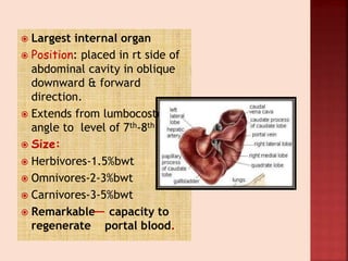
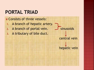
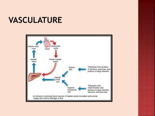
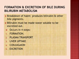
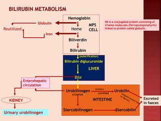
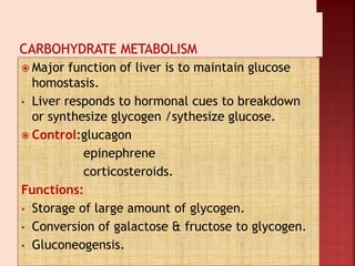
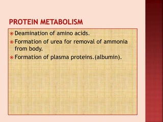
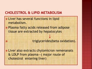
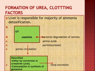
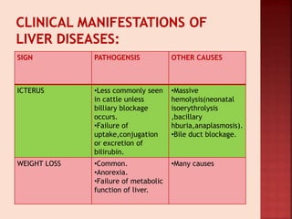
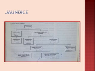
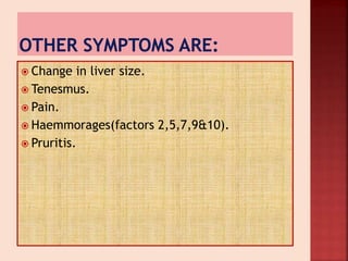
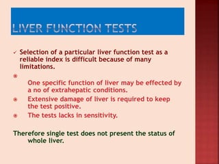
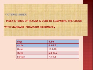
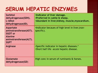
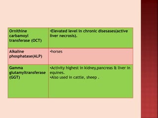
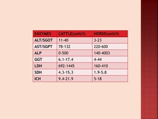
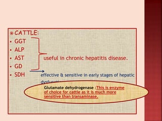
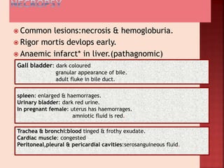
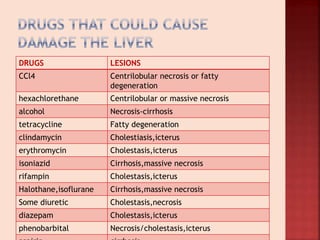
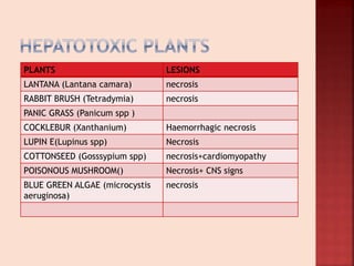
SlideShare a Scribd company logo
Dr SUMREEN KOUR
MVSc Scholar
 Largest internal organ
 Position: placed in rt side of
abdominal cavity in oblique
downward & forward
direction.
 Extends from lumbocosto
angle to level of 7th-8th rib.
 Size:
 Herbivores-1.5%bwt
 Omnivores-2-3%bwt
 Carnivores-3-5%bwt
 Remarkable capacity to
regenerate portal blood.
 Origin:
• derived from outpocketing of endoderm.
 Lobes:
 left lateral lobe,rt lateral lobe,left medial,rt
medial,quardate, caudate & papillary.
 Ligaments:
• Falciform ligament.
• Rt lateral ligament.
• Round ligament.
• Caudate ligament.
• Lesser omentum.
 Consists of three vessels:
1. A branch of hepatic artery.
2. A branch of portal vein. sinusoids
3. A tributary of bile duct.
central vein
hepatic vein
 Breakdown of haem produces bilirubin & other
bile pigments.
 Bilirubin must be made water soluble to be
excreted out.
 Occurs in 4 steps:
1. FORMATION.
2. PLASMA TRANSPORT
3. LIVER UPTAKE
4. CONJUGAION
5. EXCRETION
Hemoglobin
Heme
Biliverdin
Bilirubin
MPS
CELL
Globulin
Iron
Reutilized
Bilirubin diglucuronide
Bile
LIVER
Urobilinogen
Stercobilinogen
Urobilin
Stercobilin
Excreted
in faeces
INTESTINE
Enterohepatic
circulation
Urinary urobilinogen
KIDNEY
Hb is a conjugated protein consisting of
4 heme molecules (Ferroprotoporphyrin)
linked to protein called globulin.
(esterification)
(colorless)
(colon)
(oxidized)
 Major function of liver is to maintain glucose
homostasis.
• Liver responds to hormonal cues to breakdown
or synthesize glycogen /sythesize glucose.
 Control:glucagon
epinephrene
corticosteroids.
Functions:
• Storage of large amount of glycogen.
• Conversion of galactose & fructose to glycogen.
• Gluconeogensis.
 Deamination of amino acids.
 Formation of urea for removal of ammonia
from body.
 Formation of plasma proteins.(albumin).
 Liver has several functions in lipid
metabolism.
 Plasma fatty acids released from adipose
tissue are extracted by hepatocytes
 triglycerides(beta oxidation).
 Liver also extracts chylomicron remenanats
& LDLP from plasma—( major route of
cholestrol entering liver)
 Liver is responsible for majority of ammonia
detoxification.

 bacterial degradation of amines.
 amino acids
 purines(urease)
 portal circulation
 renal excretion
GIT
AMMONIA
Detoxified
•either by conversion in
urea(kreb cycle).
•Comsumption in synthesis of
glutamine
 Liver acts as store
house of various
vitamins.
 Liver also synthesize
clotting factors.
 Liver is site of vit-K
dependent activation
factors II, VII, IX, X
 Liver also stores iron as
ferritin.
 Hepatic cells contain
large amount of
apoferritin.
 When iron is in excess
amount+ apoferritin
ferritin (stored
in hepatic cells) & thus
acts as BLOOD IRON
BUFFER.
 Defencesive & detoxification:
 Kuffer cells(pahocytosis) of liver play impt role.
 Liver removes or excretes drugs,hormones & other substance.
 Drugs:sulphonamide
 pencillin
 ampicillin
 erythromycin
 Calcium from body is excreted by liver---bile—gut-faeces.
 Bile acids are composed of cholestrol,bile
salts & steriods.
 Main bile acids are:cholic acid
 Conjugated to taurine & glycine in liver so
they exist in ionised form as bile acids.
SIGN PATHOGENSIS OTHER CAUSES
ICTERUS •Less commonly seen
in cattle unless
billiary blockage
occurs.
•Failure of
uptake,conjugation
or excretion of
bilirubin.
•Massive
hemolysis(neonatal
isoerythrolysis
,bacillary
hburia,anaplasmosis).
•Bile duct blockage.
WEIGHT LOSS •Common.
•Anorexia.
•Failure of metabolic
function of liver.
•Many causes
DIARRHEA •Especially cattle with
chronic liver disease.
•Related to portal
hypotension &
increased hydrostatic
pressure.
•GIT disease.
•Systemic disease.
•Septicemia.
ASCITES •Calves with liver
cirrhosis.
•hypoalbuminemia,porta
l hypertension caused by
venous blackade
&increased hydrostatic
pressure & protein
leakage to peritoneum.
•Cardiac failure.
•Hypoproteinemia.
•Cushing syndrome.
CHANGE IN FECAL
COLOR
•Young animalsimple
digestive tracts-
stercobilin-metabolite
of bilirubin.
•Cholestasis-lighter fecal
colour.
DERMATITIS:
•Occurs because of hepatic Photosensitization(type 3).
•Caused by photodynamic substance-phylloerythrin.
•Normal end product of chlorophyll metabolism-phylloerythrin
•Formed by bacterial degredation of chlorophyll in GIT.
•
Normally phylloerthrin is secreted into intestine by billiary system &
excreted in faeces.
•
•Damage to this mechanism prevents excretion of phylloerythrin
allowing PS to enter circulation &accumulate in skin.
•This condition is exacebrated by hepatic parasites..
 These include:
• Hyperexcitability.
• Convulsions.
• Muscular tremors & weakness
• Dullness
• Compulsive walking.
• Head pressing.
• Circling.
• Coma.
 Causes:
 hypoglycemia
 accumulation of excess amino acids &
ammonia.
 Change in liver size.
 Tenesmus.
 Pain.
 Haemmorages(factors 2,5,7,9&10).
 Pruritis.
 Selection of a particular liver function test as a
reliable index is difficult because of many
limitations.

One specific function of liver may be effected by
a no of extrahepatic conditions.
 Extensive damage of liver is required to keep
the test positive.
 The tests lacks in sensitivity.
Therefore single test does not present the status of
whole liver.
dogs 5.8-6
cattle 8.4-9.8
Horse 15.2-18
sheep 6.8-10.7
buffalo 7.1-9.8
Conjugated bilirubin or soluble bilirubin or
Direct bilirubin when comes in contact with
Ehrlich`s diazo reagent, gets diazotized and
there upon produces “azobilirubin” the
color of which is red.
Unconjugated bilirubin is water insoluble and
therefore produces color only after addition
of methly alcohol to make it soluble.
Unconjugated bilirubin = Total bilirubin -
Conjugated bilirubin
species Conjugated(mg/dl) Total
bilirubin(mg/dl)
Dog 0.14 1.0-10.0
Cattle 0.18 1.9
Horse 0.10 0.2-6.0
Sheep 0.12 0-0.4
Interpretation:
Cattle:In generalized liver disease maximum bilirubin will not exceed
3.5 mg/dl due to large functional reserves of bovine liver for excretion
of bilirubin.
One of several Vit. K dependant
coagulation factors synthesized by liver
Species Normal clotting
time
Sheep 2 minutes
Dog 2-3 minutes
Cattle 7 minutes
Horse 11 minutes.
Interpretations: Prolonged time could be due to
-Decreased hepatic cell mass (extensive necrosis/
cirrhosis)
-Cholestasis (insufficient bile for absorption of fat soluble
vitamin)
 BROMOSULPHTHALEIN CLEARANCE TEST:
 In large animals this test is mostly used.
 In this test 500-1000mg of BSP is injection I/v.
blood sample is taken before the injection & 4 times 5-
12min following injection(5,7,9,11min).
samples are analysed for for color produced by BSP. & half
life determined by plotting graph.
HALF LIFE:
Normal horses-3.5min.
Ruminants-5min
Sorbitol
dehydrogenase(SDH).
L-iditol
dehydrogenase(ID).
•Indicator of liver damage.
•Preferred in cattle & sheep.
•Abundant in liver,kidney, muscle,myocardium.
Aspartate
aminotransferase(AST).
SGOT or
Alanine
aminotransferase(ALT).
SGPT
•Indicator becaues of high level in liver.(non-
specific).
Arginase •Specific indicator in hepatic diseases.*
•Short half life –acute hepatic disease.
Glutamate
dehydrogenase(GD)
High conc in serum of ruminants & horses.
Ornithine
carbamoyl
transferase (OCT)
•Elevated level in chronic disesases(active
liver necrosis).
Alkaline
phosphatase(ALP)
•horses
Gamma
glutamyltransferase
(GGT)
•Activity highest in kidney,pancreas & liver in
equines.
•Also used in cattle, sheep .
ENZYMES CATTLE(unit/l) HORSE(unit/l)
ALT/SGOT 11-40 3-23
AST/SGPT 78-132 220-600
ALP 0-500 140-4003
GGT 6.1-17.4 4-44
LDH 692-1445 160-410
SDH 4.3-15.3 1.9-5.8
ICH 9.4-21.9 5-18
 CATTLE:
 GGT
 ALP
 AST useful in chronic hepatitis disease.
 GD
 SDH effective & sensitive in early stages of hepatic
dysfunction.
Glutamate dehydrogenase :This is enzyme
of choice for cattle as it is much more
sensitive than transaminase.
Remove primary reason
Provide CHO rich diet
Restrict protein
Provide rest to animal
Water soluble vitamins
Provide vitamin K
Choline
Cholegogues:cholekinetcs&
cholerectics
Sedatives like Xylazine
metronidazole
steroids
Diuretics & laxatives
 Inflammation of hepatic cells.
 May be acute or chronic.
 INORGANIC POISONS:
• Copper , phosphorous,
arsenic, possibly
selenium.
 ORGANIC POISONS:
• Carbon tetrachloride,
• hexachloride,gossypol,cr
esols & coal tar
pitch,chloroform.
 POISONOUS PLANTS:
• Weeds: Senecio,
Crotolaria , Heliotropium.
• Pasture & cultivated
plants:lupins,Alsike
clover,water damaged
alfa-alfa hay.
• Trees & shrubs: lantana
camara,myoporum spp.
 FUNGUS:
• fusarium sp, Penicillium
rubrum, Aspergillus flavus.
 INSECTS : sawfly larvae
INFECTIOUS HEPATITIS PARASITIC CAUSES
 Virus of rift valley
fever.
 Chlymadia sp.
 histoplasmosis
 Liver flukes.
 Migrating ascaris.
 Sustained Inflammatory process within liver.
 ETIOLOGY:in domestic animal is not known bt
however toxins play major role.
 RED WATER DISEASE
 ICTEROHAEMOGLOBINURIA
o Bacterial disease- Anerobic Cl. Haemolyticum
(cl novyi type D).
o Soil borne anerobe growing bacteria in liver.
o Affects cattle.
o Highest incidence is seen in ruminants consuming forages in
area with snail.
o
 Spores of Cl. Haemolyticum are transported by blood &
lymph to body tissue & organs.
 Pre-requisite: Host
Presence of toxins
Inflammation/necrosis*.
Result in formation of hypoxic milieu favourable for
spore germination & growth of toxin producing
vegtative cells.
Migrating fluke are primary
factors leading to liver necrosis &
anerobic condition.
 Vegetative cells releases toxins(necrotoxin &
hemolytic )
 Causes necrosis of surrounding tissue & allowing
infection.
 There is development of organised thrombus in
subterminal branch of portal vein producing infarct
which is characteristic of this disease.
 Most of bacteria grow in this anerobic env
producing toxin.
 Causing:toxemia
vascular damage
intravascular haemolysis.
Principle toxin produced is is beta
toxin(phospholipase C) highly lethal.
 EARLY SIGNS:
• CNS depression.
• Anorexia.
• Segregation of affected afected
animal.
• Rumination & lactation ceazes.
• Initially tempt is 104-106’F but
declines to normal or below
before death.
• Breathing is
rapid,shallow,distresed.
• Blood tinged froth from
nostrils,mouth.
 LATE SIGNS:
 If animal survives after 24hrs,
• Wine coloured
urine(hemoglobiunuria)
• Anaemia.
• Icterus.
• Juglar vein pulsation.
• Blood is thin & coagulates
slowly..
• s/c odema of ventral areas
seen.
•Infarcts in liver
•Blackish kidney
 Common lesions:necrosis & hemogloburia.
 Rigor mortis devlops early.
 Anaemic infarct* in liver.(pathagnomic)
Gall bladder: dark coloured
granular appearance of bile.
adult fluke in bile duct.
spleen: enlarged & haemorrages.
Urinary bladder: dark red urine.
In pregnant female: uterus has haemorrages.
amniotic fluid is red.
Trachea & bronchi:blood tinged & frothy exudate.
Cardiac muscle: congested
Peritoneal,pleural & pericardial cavities:serosanguineous fluid.
leptospira All ages cattle on
pasture
•Acute fever
•Red coloured
milk
•Hburia
•May die 24-48hrs
Leptospira titre
Postpaturient
hburia
Lactating cows 4-
6wk pp
•Acute
•No changes in
milk
•No fever
•Die in 12-48hrs
•hburia
hypophosphatemia
babesiosis Enzootic area
Tick borne
Young animals
•Fever
•Jaundice
•Abortioncourse 2-
3wk
•hburia
Blood smear
Complement
fixation test
Chronic cu
poisoning
Long term oral
adm of medicine
Jaundice
No fever
hburia
Acute haemolytic
anaemia
 TREATMENT:
 Penicillin-20000I/M
or
Tetracycline-10mg/kgbwt
I/M
 Blood transfusion
 Vitamin & iron
supplementation.
 PREVENTION:
 Vaccination
 Commercial
bacterin/toxoids.
 Immunization 2dose 3-
4wk apart.
 Caused by :
 Fasciola hepatica in
ruminants.
 Cattle develop degree of
resistance to repeated
infections,but sheeps do
not.
 Fasciola magna:infection
do not become patent in
cattle as fluke are
surrounded by capsules.
 Dicrocoelium dendriticum
Young migrate to hepatic parenchyma
Bile duct doubles size
Adult cattle & sheep act as carrier.
IH:snails lymnaeid –infective
metacercaria
 ACUTE
 After entry of hepatic fasciolasis in liver parenchyma
clinical signs appear 5-6wks after ingestion of
metacercaria.
 Quiescent spores of Cl.novyi become activated in anerobic
condition created by Fasciola hepatica causing infectious
necrotic hepatitis.
 CHRONIC:
 Adult fluke in bile duct
 Causes: cholangitis
 biliary obstruction(due to high proline)
 fibrosis
 leakage of plasma protien across epithelium –
 hypoalbunemia

 anaemia*

 Increases susceptibility of Salmonella dublin
 NECROPSY:
 Acute:
 swollen liver
 Perforation & subscapular
& haemorrage
 Parenchyma is damaged
& friable.
 Chronic:
 Leaf like flukes &
thickened bile duct
 Calcification of bile duct
in cattle
 DIAGNOSIS:
 Fecal examination.
 ELISA & DOT ELISA
 TREATMENT:
• Albendazole @5mg/kgbwt-sheep
• @10mg/kg bwt-cattle
• Clorsulon
• @2mg/kgbwt-s/c
• Oxyclozanide
• 12mg/kgbwt
 Fascioloides magna:
• Parasite of deer.
• Can also infect grazing animals.
 Dicrocoelium dendriticum:
• IIH:snail-helicella spp
Cercaria passed in slime ball used as food by
ants--grazing animal swallow ant—bile duct.
 Seen in all ruminants.
 Etiology:Fusobacterium necrophorum(80-90%).
 Other causes: corynebacterium pyogenes
streptococcus
staphylococcus
bacteriodes.
 Liver is susceptible to abcesses because it recieves blood from
hepatic artery, portal system & umblical vein in fetus & neonates.
 Feed lot cattle mostly affected.
Pathopysiology & pathogenesis
1. Abscesses may rupture into vein or form thrombus in caudal
vena cava producing portal hypertension.
hydrostatic pressure & both ascites & diarrhoea can be seen.
2.Emboli from thrombus may be carried to lung.
Hematogenous pneumonia with sepsis & pulmonary
hypertension.
3. Enlarged absecces may obstruct bile duct,excretion of
bilirubin,bile acids & phylloerythrin.
 Weight loss.
 Decreased milk production.
 Fever & anorexia.
 Pain on moving ,lying down &
on physical examination.
 CVCT: 3symptoms:-
1. Sudden death from shock.
2. Epistaxis , hemoptysis &
anemia.
3. Severe dyspnea , open
mouth breathing & abnormal
lung sounds.
 Photosensitization & icterus.
 Peritonitis.
 Clinical pathology:
• Plasma fibrinogen is activated.
• Total plasma protein is elevated-globulin.
• In CVCT-globulin -9g/dl.
• Decreased PCV,hb,RBC count.
• Liver enzyme-GGT,SDH,AST.
 TREATMENT:
 OTC @ 75mg/head/dat.
 Tylosin @60-90mg/kg/head
 FAT COW SYNDROME
 Caused by mobilization of excessive quantities
fat from body depots to liver during period of
negative energy balance at parturient period.
 Cow that become obese before parturation are
more predisposed.
 Morbidity-50-90%.
 Fatty acids synthesises in liver & are stored as
triglycerides in extra-hepatic site.
during negative energy balance triglycerides in adipose
tissue are converted to glycerols & NEFA.
albumin bound NEFA acts as source of energy & gets
deposited in liver,spleen,mammary glands,muscles.
liver recieves much of NEFA & due to large blood
supply.
 In liver NEFA is back re-esterfied to tryglycerides
& remain in liver until they can be oxidized or
repackaged in envelope of –cholestrol
phospholipids
proteins.
which is very slow proccess leading to fatty liver.
 Increased glucose influences & favoures fatty
liver syndrome & opposite favours ketosis..
Clinical signs Clinical pathology & diagnosis
 Depression.
 Anorexia.
 Deblitation.
 Weakness.
 Decreased milk production
 ketonuria.
 Affected cow may show
signs:
 Milk
fever,ketosis,indigestion,LD
A,metritis,mastitis,salmonel
losis.
 Glucose:below 40mg/dl
 Ketone bodies:80-100mg/dl
 FFA: above 7mg/100ml
 OCT:above 200sigma U/ml
 SDH:above 500 sigma U/l
 AST:above 100 units/l
 NEFA:more 1000mEq/l
 Leukopenia denerative left shift
 PCV -.increased
 BUN- increased
 BSP test done
 Obesity.
 Large quantity of
omental fat.
 Liver
enlarged,rounded,swo
llen edges,pale
yellow in color,often
floats in water.
 Fatty infilteration of
hepatocytes
 Glu ,Ca, Mg salts are given.
 Symptomatic treatment-glucose given.
 Choline chloride(50%)-50g orally once-precursor of
lipoprotein.
 Inositol & methionine also given.
 Insulin @ 200-300IU.s/c twice daily.
 Increased NEFA level declines DMI.
 Oxidation of NEFA will lead to formation of ketone bodies.
 Low glucose level-low insulin level(inhibits fat mobilization
from adipose tissue).
 PROTEIN ENERGY MALNUTRITION & PREGNANCY
TOXEMIA OF BEEF COWS:
• Usually occuring during winter manifested by
weight loss,weakness, depression & sometime
inability to rise.
• Condition results because of negative energy
balance.
DRUGS LESIONS
CCl4 Centrilobular necrosis or fatty
degeneration
hexachlorethane Centrilobular or massive necrosis
alcohol Necrosis-cirrhosis
tetracycline Fatty degeneration
clindamycin Cholestiasis,icterus
erythromycin Cholestasis,icterus
isoniazid Cirrhosis,massive necrosis
rifampin Cholestasis,icterus
Halothane,isoflurane Cirrhosis,massive necrosis
Some diuretic Cholestasis,necrosis
diazepam Cholestasis,icterus
phenobarbital Necrosis/cholestasis,icterus
PLANTS LESIONS
LANTANA (Lantana camara) necrosis
RABBIT BRUSH (Tetradymia) necrosis
PANIC GRASS (Panicum spp )
COCKLEBUR (Xanthanium) Haemorrhagic necrosis
LUPIN E(Lupinus spp) Necrosis
COTTONSEED (Gosssypium spp) necrosis+cardiomyopathy
POISONOUS MUSHROOM() Necrosis+ CNS signs
BLUE GREEN ALGAE (microcystis
aeruginosa)
necrosis
 Causes secondary photosensitization.
 Plant contains toxin:lantadene A
 lantadene B
 Incidence maximum in summer.
 Clinical signs:
 Constipation
 Ataxia
 Incordination movements
 Photophobic
 Photosenstization-fissures on muzzle,ear tips
 Erythema,odema
 Depressed
 rumenpstasis
 Anorectic
 Jaundice.
 Faeces may contain blood.
 Biochemical findings:
 Elevated cojugated bilirubin.
 Necropsy:
 Yellow ,mottled liver
 hepatomegaly
 Treatment:
 Animal should be house fed.
 Laxatives i.e mineral oil.
 Liver tonics.
 Corticosteroids given.
 Secondary skin infection treatment.
 Common plants:
1. Senecia lacobaea
2. Senecia vulgaris
3. Senecia longilobus
4. Crotolaria spp.
 PA are poisonous plants containing 4-6 different
alkaloids.
after absorbtion via portal cirucaltion carried to liver
Pyrrole cross link with dsDNA.
antimomotic effect
Toxic pyrroles
 Hepatocytes become megalocytes*
 also inhibit protein synthesis.
hepatocyte death.
CLINICAL SIGNS:
Diarrhoes
Tenesmus
Prolapsed rectum
ascites
Clinical pathology & necropsy
Treatment
 Rise in GGT,ALP.
 Necropsy:
• Hepatic megalocytes.
• Biliary hyperplasia.
• Necrosis
• Fibrosis
• Venoocuslive disease.
 Provide low protein
diet.
 Herbicide spraying.
CHEMICAL SOURCE LIVER LESIONS
CCl4 Fumigant,solvent Fatty degeneration
Centrilobular necrosis
Cl-hydrocarbon Insecticide,solvent necrosis
phenol Wood
preservative,disinfectant
Haem centrilobular
necrosis
iron Iron supplements Portal necrosis
copper Toot bath,molluscides, Portal
vaculation+Hemolytic
crisis
phosphorus Fertilizers,rhodenticides Portal necrosis+fatty
degeneration
Tannic acid Skin astrigent Centrilobular necrosis
Paraquat/diquat herbicides Centrilobular necrosis
1. Telangiectasia.
2. Ischemia,hypoxia & cogestion.
3. Fetal liver damage.
4. Neoplasia.
5. Failure of drug metabolism & excretion.
 Other:
1. Cholelithiasis.
2. Cholangitis.
3. Cholecystitis.
TELANGIECTASIA (SAW DUST):
•Its focal degeneration in liver lobular circulation chr by red- brown
foci (1-5mm in dia).
• Hepatocytes are distorted & sinusoids are congested.
ISCHEMIA,HYPOXIA & CONGESTION:
•Can lead to death of hepatocytes.
•Chronic passive congestion causes grossly visible nutmeg liver.
FETAL LIVER DAMAGE:
•Damaged by infectious & toxic agents
•Aborted fetus of cattle-IBR.
FAILURE OF DRUG METABOLISM & EXCRETION:
•Delayed clearance with hepatic insufficiency.
•Chloremphenicol,tetracycline,erythromycin.
CHOLELITHIASIS
• Presence of
calculi in gall
bladder or
common bile
duct.
• Observed in
cattle.
• Ca, bile salts,
cholestrol etc.
CHOLANGITIS
• Inflammation of
biliary duct system.
• In cattle liver
flukes due to
irritation & toxins.
• Decreased milk
production,
hypophagia,
tachycardia,anorexi
a,diarrhoea.
CHOLECYSTITIS
• Inflammation of
gall bladder.
• Bacterial
infection with E
coli,
salmonella,are
observed.
sumreen2707@gmail.com

More Related Content

What's hot

Angels presenting chronic patellar luxation in cattle.by pavul
Angels presenting chronic patellar luxation in cattle.by pavulAngels presenting chronic patellar luxation in cattle.by pavul
Angels presenting chronic patellar luxation in cattle.by pavul
Pavulraj Selvaraj
 
Recent advanes in handling of dystocia
Recent advanes in handling of dystociaRecent advanes in handling of dystocia
Recent advanes in handling of dystocia
rahulvet27
 
Canine Distemper
Canine DistemperCanine Distemper
Canine Distemper
Ganes Adhikari
 
Lecture 8 anestrus in domestic animals
Lecture 8 anestrus in domestic animalsLecture 8 anestrus in domestic animals
Lecture 8 anestrus in domestic animals
DrGovindNarayanPuroh
 
Downer's cow syndrome
Downer's cow syndromeDowner's cow syndrome
Downer's cow syndrome
saurabh gupta
 
Canine ehrlichiosis
Canine ehrlichiosisCanine ehrlichiosis
Canine ehrlichiosis
Dr. Ram Chander Tiwari
 
Management of renal disease in dog
Management of renal disease in dogManagement of renal disease in dog
Management of renal disease in dog
Vikash Babu Rajput
 
Lamness in cattle
Lamness in cattleLamness in cattle
Lamness in cattle
Dr Dhavalkumar F. Chaudhary
 
Milk fever
Milk feverMilk fever
Milk fever
Dr-Mohamed Ghanem
 
Presetation on rumen impaction lactic acidosis final osr
Presetation on rumen impaction lactic acidosis final osrPresetation on rumen impaction lactic acidosis final osr
Presetation on rumen impaction lactic acidosis final osr
Harshit Saxena
 
Etiopathogenesis, therapy, prevention and control of milk fever in dairy cattle
Etiopathogenesis, therapy, prevention and control of milk fever in dairy cattleEtiopathogenesis, therapy, prevention and control of milk fever in dairy cattle
Etiopathogenesis, therapy, prevention and control of milk fever in dairy cattle
Radhika Vaidya
 
Seizure in animals
Seizure in animalsSeizure in animals
Seizure in animals
Radhika Vaidya
 
Lameness, Hoof, and Leg Issues in Dairy Cattle- Ernest Hovingh
Lameness, Hoof, and Leg Issues in Dairy Cattle- Ernest HovinghLameness, Hoof, and Leg Issues in Dairy Cattle- Ernest Hovingh
Lameness, Hoof, and Leg Issues in Dairy Cattle- Ernest Hovingh
DAIReXNET
 
Pyometra in cow
Pyometra in cowPyometra in cow
Pyometra in cow
Aynul Haque
 
Affection of horn
Affection of hornAffection of horn
Affection of horn
Bikas Puri
 
Vet obst lecture 7 Causes of dystocia in farm animals
Vet obst lecture 7 Causes of dystocia in farm animalsVet obst lecture 7 Causes of dystocia in farm animals
Vet obst lecture 7 Causes of dystocia in farm animals
DrGovindNarayanPuroh
 
Ccpp
CcppCcpp

What's hot (20)

Angels presenting chronic patellar luxation in cattle.by pavul
Angels presenting chronic patellar luxation in cattle.by pavulAngels presenting chronic patellar luxation in cattle.by pavul
Angels presenting chronic patellar luxation in cattle.by pavul
 
Recent advanes in handling of dystocia
Recent advanes in handling of dystociaRecent advanes in handling of dystocia
Recent advanes in handling of dystocia
 
Canine Distemper
Canine DistemperCanine Distemper
Canine Distemper
 
Lecture 8 anestrus in domestic animals
Lecture 8 anestrus in domestic animalsLecture 8 anestrus in domestic animals
Lecture 8 anestrus in domestic animals
 
Rumen fluid examination
Rumen fluid examinationRumen fluid examination
Rumen fluid examination
 
Downer's cow syndrome
Downer's cow syndromeDowner's cow syndrome
Downer's cow syndrome
 
Calf diseases 4
Calf diseases 4Calf diseases 4
Calf diseases 4
 
Canine ehrlichiosis
Canine ehrlichiosisCanine ehrlichiosis
Canine ehrlichiosis
 
Management of renal disease in dog
Management of renal disease in dogManagement of renal disease in dog
Management of renal disease in dog
 
Lamness in cattle
Lamness in cattleLamness in cattle
Lamness in cattle
 
Milk fever
Milk feverMilk fever
Milk fever
 
Presetation on rumen impaction lactic acidosis final osr
Presetation on rumen impaction lactic acidosis final osrPresetation on rumen impaction lactic acidosis final osr
Presetation on rumen impaction lactic acidosis final osr
 
Etiopathogenesis, therapy, prevention and control of milk fever in dairy cattle
Etiopathogenesis, therapy, prevention and control of milk fever in dairy cattleEtiopathogenesis, therapy, prevention and control of milk fever in dairy cattle
Etiopathogenesis, therapy, prevention and control of milk fever in dairy cattle
 
Seizure in animals
Seizure in animalsSeizure in animals
Seizure in animals
 
Lameness, Hoof, and Leg Issues in Dairy Cattle- Ernest Hovingh
Lameness, Hoof, and Leg Issues in Dairy Cattle- Ernest HovinghLameness, Hoof, and Leg Issues in Dairy Cattle- Ernest Hovingh
Lameness, Hoof, and Leg Issues in Dairy Cattle- Ernest Hovingh
 
Pyometra in cow
Pyometra in cowPyometra in cow
Pyometra in cow
 
Affection of horn
Affection of hornAffection of horn
Affection of horn
 
Vet obst lecture 7 Causes of dystocia in farm animals
Vet obst lecture 7 Causes of dystocia in farm animalsVet obst lecture 7 Causes of dystocia in farm animals
Vet obst lecture 7 Causes of dystocia in farm animals
 
Fluid therapy in animals
Fluid therapy in animalsFluid therapy in animals
Fluid therapy in animals
 
Ccpp
CcppCcpp
Ccpp
 

Similar to Liver diseases in cattle

Liver ppt
Liver pptLiver ppt
Liver ppt
KHAN AYAZ
 
INTERGRATED RESPONSE TO A MEAL V. INTESTINAL PHASE (ii)
INTERGRATED RESPONSE TO A MEAL V. INTESTINAL PHASE (ii)INTERGRATED RESPONSE TO A MEAL V. INTESTINAL PHASE (ii)
INTERGRATED RESPONSE TO A MEAL V. INTESTINAL PHASE (ii)
SAMOEINESH
 
liver, pancreas and gall bladder physiology and function and disorders and nu...
liver, pancreas and gall bladder physiology and function and disorders and nu...liver, pancreas and gall bladder physiology and function and disorders and nu...
liver, pancreas and gall bladder physiology and function and disorders and nu...
MeghanaMeghu11
 
Screening of hepatoprotective drugs
Screening of hepatoprotective drugsScreening of hepatoprotective drugs
Screening of hepatoprotective drugs
DipanjaliKamthe
 
LIVER AND GALL BLADDER.pptx DIGESTIVE SYSTEM
LIVER AND GALL BLADDER.pptx DIGESTIVE SYSTEMLIVER AND GALL BLADDER.pptx DIGESTIVE SYSTEM
LIVER AND GALL BLADDER.pptx DIGESTIVE SYSTEM
Vigneswaran31
 
LIVER (2).pptx
LIVER (2).pptxLIVER (2).pptx
LIVER (2).pptx
RaushanKhan3
 
dental management of hepatic patient.pptx
dental management of hepatic patient.pptxdental management of hepatic patient.pptx
dental management of hepatic patient.pptx
SoadAbdElMoniem
 
Dental management of hepatic patients .pptx
Dental management of hepatic patients .pptxDental management of hepatic patients .pptx
Dental management of hepatic patients .pptx
Soad abdelmoniem
 
Accessory Organs of Mammalian Digestive System
Accessory Organs of Mammalian Digestive SystemAccessory Organs of Mammalian Digestive System
Accessory Organs of Mammalian Digestive System
Garry D. Lasaga
 
Gastrointestinal.ppt
Gastrointestinal.pptGastrointestinal.ppt
Gastrointestinal.pptShama
 
Structure and functions of liver
Structure and functions of liverStructure and functions of liver
Structure and functions of liver
K.M.College of Pharmacy, Madurai
 
liverfunctiontest111.pptx
liverfunctiontest111.pptxliverfunctiontest111.pptx
liverfunctiontest111.pptx
ShubhamkumarMaurya7
 
Liver & Liver Diseases
Liver &  Liver DiseasesLiver &  Liver Diseases
Liver & Liver Diseases
MdNazmulIslamTanmoy
 
A review of liver anatomy and physiology for anesthesiologists
A review of liver anatomy and physiology for anesthesiologistsA review of liver anatomy and physiology for anesthesiologists
A review of liver anatomy and physiology for anesthesiologistsArun Shetty
 
07 Blood.ppt
07 Blood.ppt07 Blood.ppt
07 Blood.ppt
Shubhangi kharat
 
characteristics of urine
characteristics of urinecharacteristics of urine
characteristics of urine
sana sana
 
Bile: a review of the biliary system
Bile: a review of the biliary systemBile: a review of the biliary system
Bile: a review of the biliary system
Lyndon Woytuck
 
Hepatitis C
Hepatitis CHepatitis C
Hepatitis C
Mohammed Aljaber
 

Similar to Liver diseases in cattle (20)

Liver ppt
Liver pptLiver ppt
Liver ppt
 
INTERGRATED RESPONSE TO A MEAL V. INTESTINAL PHASE (ii)
INTERGRATED RESPONSE TO A MEAL V. INTESTINAL PHASE (ii)INTERGRATED RESPONSE TO A MEAL V. INTESTINAL PHASE (ii)
INTERGRATED RESPONSE TO A MEAL V. INTESTINAL PHASE (ii)
 
liver, pancreas and gall bladder physiology and function and disorders and nu...
liver, pancreas and gall bladder physiology and function and disorders and nu...liver, pancreas and gall bladder physiology and function and disorders and nu...
liver, pancreas and gall bladder physiology and function and disorders and nu...
 
Screening of hepatoprotective drugs
Screening of hepatoprotective drugsScreening of hepatoprotective drugs
Screening of hepatoprotective drugs
 
LIVER AND GALL BLADDER.pptx DIGESTIVE SYSTEM
LIVER AND GALL BLADDER.pptx DIGESTIVE SYSTEMLIVER AND GALL BLADDER.pptx DIGESTIVE SYSTEM
LIVER AND GALL BLADDER.pptx DIGESTIVE SYSTEM
 
LIVER (2).pptx
LIVER (2).pptxLIVER (2).pptx
LIVER (2).pptx
 
JAUNDICE & LIVER FUNCTION TESTS
JAUNDICE & LIVER FUNCTION TESTSJAUNDICE & LIVER FUNCTION TESTS
JAUNDICE & LIVER FUNCTION TESTS
 
dental management of hepatic patient.pptx
dental management of hepatic patient.pptxdental management of hepatic patient.pptx
dental management of hepatic patient.pptx
 
Dental management of hepatic patients .pptx
Dental management of hepatic patients .pptxDental management of hepatic patients .pptx
Dental management of hepatic patients .pptx
 
Accessory Organs of Mammalian Digestive System
Accessory Organs of Mammalian Digestive SystemAccessory Organs of Mammalian Digestive System
Accessory Organs of Mammalian Digestive System
 
Gastrointestinal.ppt
Gastrointestinal.pptGastrointestinal.ppt
Gastrointestinal.ppt
 
Structure and functions of liver
Structure and functions of liverStructure and functions of liver
Structure and functions of liver
 
liverfunctiontest111.pptx
liverfunctiontest111.pptxliverfunctiontest111.pptx
liverfunctiontest111.pptx
 
Liver Disease
Liver DiseaseLiver Disease
Liver Disease
 
Liver & Liver Diseases
Liver &  Liver DiseasesLiver &  Liver Diseases
Liver & Liver Diseases
 
A review of liver anatomy and physiology for anesthesiologists
A review of liver anatomy and physiology for anesthesiologistsA review of liver anatomy and physiology for anesthesiologists
A review of liver anatomy and physiology for anesthesiologists
 
07 Blood.ppt
07 Blood.ppt07 Blood.ppt
07 Blood.ppt
 
characteristics of urine
characteristics of urinecharacteristics of urine
characteristics of urine
 
Bile: a review of the biliary system
Bile: a review of the biliary systemBile: a review of the biliary system
Bile: a review of the biliary system
 
Hepatitis C
Hepatitis CHepatitis C
Hepatitis C
 

Recently uploaded

The Normal Electrocardiogram - Part I of II
The Normal Electrocardiogram - Part I of IIThe Normal Electrocardiogram - Part I of II
The Normal Electrocardiogram - Part I of II
MedicoseAcademics
 
How to Give Better Lectures: Some Tips for Doctors
How to Give Better Lectures: Some Tips for DoctorsHow to Give Better Lectures: Some Tips for Doctors
How to Give Better Lectures: Some Tips for Doctors
LanceCatedral
 
Pharynx and Clinical Correlations BY Dr.Rabia Inam Gandapore.pptx
Pharynx and Clinical Correlations BY Dr.Rabia Inam Gandapore.pptxPharynx and Clinical Correlations BY Dr.Rabia Inam Gandapore.pptx
Pharynx and Clinical Correlations BY Dr.Rabia Inam Gandapore.pptx
Dr. Rabia Inam Gandapore
 
ANATOMY AND PHYSIOLOGY OF URINARY SYSTEM.pptx
ANATOMY AND PHYSIOLOGY OF URINARY SYSTEM.pptxANATOMY AND PHYSIOLOGY OF URINARY SYSTEM.pptx
ANATOMY AND PHYSIOLOGY OF URINARY SYSTEM.pptx
Swetaba Besh
 
Lung Cancer: Artificial Intelligence, Synergetics, Complex System Analysis, S...
Lung Cancer: Artificial Intelligence, Synergetics, Complex System Analysis, S...Lung Cancer: Artificial Intelligence, Synergetics, Complex System Analysis, S...
Lung Cancer: Artificial Intelligence, Synergetics, Complex System Analysis, S...
Oleg Kshivets
 
Are There Any Natural Remedies To Treat Syphilis.pdf
Are There Any Natural Remedies To Treat Syphilis.pdfAre There Any Natural Remedies To Treat Syphilis.pdf
Are There Any Natural Remedies To Treat Syphilis.pdf
Little Cross Family Clinic
 
263778731218 Abortion Clinic /Pills In Harare ,
263778731218 Abortion Clinic /Pills In Harare ,263778731218 Abortion Clinic /Pills In Harare ,
263778731218 Abortion Clinic /Pills In Harare ,
sisternakatoto
 
heat stroke and heat exhaustion in children
heat stroke and heat exhaustion in childrenheat stroke and heat exhaustion in children
heat stroke and heat exhaustion in children
SumeraAhmad5
 
NVBDCP.pptx Nation vector borne disease control program
NVBDCP.pptx Nation vector borne disease control programNVBDCP.pptx Nation vector borne disease control program
NVBDCP.pptx Nation vector borne disease control program
Sapna Thakur
 
Ophthalmology Clinical Tests for OSCE exam
Ophthalmology Clinical Tests for OSCE examOphthalmology Clinical Tests for OSCE exam
Ophthalmology Clinical Tests for OSCE exam
KafrELShiekh University
 
Superficial & Deep Fascia of the NECK.pptx
Superficial & Deep Fascia of the NECK.pptxSuperficial & Deep Fascia of the NECK.pptx
Superficial & Deep Fascia of the NECK.pptx
Dr. Rabia Inam Gandapore
 
Triangles of Neck and Clinical Correlation by Dr. RIG.pptx
Triangles of Neck and Clinical Correlation by Dr. RIG.pptxTriangles of Neck and Clinical Correlation by Dr. RIG.pptx
Triangles of Neck and Clinical Correlation by Dr. RIG.pptx
Dr. Rabia Inam Gandapore
 
Charaka Samhita Sutra sthana Chapter 15 Upakalpaniyaadhyaya
Charaka Samhita Sutra sthana Chapter 15 UpakalpaniyaadhyayaCharaka Samhita Sutra sthana Chapter 15 Upakalpaniyaadhyaya
Charaka Samhita Sutra sthana Chapter 15 Upakalpaniyaadhyaya
Dr KHALID B.M
 
basicmodesofventilation2022-220313203758.pdf
basicmodesofventilation2022-220313203758.pdfbasicmodesofventilation2022-220313203758.pdf
basicmodesofventilation2022-220313203758.pdf
aljamhori teaching hospital
 
ARTHROLOGY PPT NCISM SYLLABUS AYURVEDA STUDENTS
ARTHROLOGY PPT NCISM SYLLABUS AYURVEDA STUDENTSARTHROLOGY PPT NCISM SYLLABUS AYURVEDA STUDENTS
ARTHROLOGY PPT NCISM SYLLABUS AYURVEDA STUDENTS
Dr. Vinay Pareek
 
Evaluation of antidepressant activity of clitoris ternatea in animals
Evaluation of antidepressant activity of clitoris ternatea in animalsEvaluation of antidepressant activity of clitoris ternatea in animals
Evaluation of antidepressant activity of clitoris ternatea in animals
Shweta
 
ARTIFICIAL INTELLIGENCE IN HEALTHCARE.pdf
ARTIFICIAL INTELLIGENCE IN  HEALTHCARE.pdfARTIFICIAL INTELLIGENCE IN  HEALTHCARE.pdf
ARTIFICIAL INTELLIGENCE IN HEALTHCARE.pdf
Anujkumaranit
 
ACUTE SCROTUM.....pdf. ACUTE SCROTAL CONDITIOND
ACUTE SCROTUM.....pdf. ACUTE SCROTAL CONDITIONDACUTE SCROTUM.....pdf. ACUTE SCROTAL CONDITIOND
ACUTE SCROTUM.....pdf. ACUTE SCROTAL CONDITIOND
DR SETH JOTHAM
 
Cervical & Brachial Plexus By Dr. RIG.pptx
Cervical & Brachial Plexus By Dr. RIG.pptxCervical & Brachial Plexus By Dr. RIG.pptx
Cervical & Brachial Plexus By Dr. RIG.pptx
Dr. Rabia Inam Gandapore
 
KDIGO 2024 guidelines for diabetologists
KDIGO 2024 guidelines for diabetologistsKDIGO 2024 guidelines for diabetologists
KDIGO 2024 guidelines for diabetologists
د.محمود نجيب
 

Recently uploaded (20)

The Normal Electrocardiogram - Part I of II
The Normal Electrocardiogram - Part I of IIThe Normal Electrocardiogram - Part I of II
The Normal Electrocardiogram - Part I of II
 
How to Give Better Lectures: Some Tips for Doctors
How to Give Better Lectures: Some Tips for DoctorsHow to Give Better Lectures: Some Tips for Doctors
How to Give Better Lectures: Some Tips for Doctors
 
Pharynx and Clinical Correlations BY Dr.Rabia Inam Gandapore.pptx
Pharynx and Clinical Correlations BY Dr.Rabia Inam Gandapore.pptxPharynx and Clinical Correlations BY Dr.Rabia Inam Gandapore.pptx
Pharynx and Clinical Correlations BY Dr.Rabia Inam Gandapore.pptx
 
ANATOMY AND PHYSIOLOGY OF URINARY SYSTEM.pptx
ANATOMY AND PHYSIOLOGY OF URINARY SYSTEM.pptxANATOMY AND PHYSIOLOGY OF URINARY SYSTEM.pptx
ANATOMY AND PHYSIOLOGY OF URINARY SYSTEM.pptx
 
Lung Cancer: Artificial Intelligence, Synergetics, Complex System Analysis, S...
Lung Cancer: Artificial Intelligence, Synergetics, Complex System Analysis, S...Lung Cancer: Artificial Intelligence, Synergetics, Complex System Analysis, S...
Lung Cancer: Artificial Intelligence, Synergetics, Complex System Analysis, S...
 
Are There Any Natural Remedies To Treat Syphilis.pdf
Are There Any Natural Remedies To Treat Syphilis.pdfAre There Any Natural Remedies To Treat Syphilis.pdf
Are There Any Natural Remedies To Treat Syphilis.pdf
 
263778731218 Abortion Clinic /Pills In Harare ,
263778731218 Abortion Clinic /Pills In Harare ,263778731218 Abortion Clinic /Pills In Harare ,
263778731218 Abortion Clinic /Pills In Harare ,
 
heat stroke and heat exhaustion in children
heat stroke and heat exhaustion in childrenheat stroke and heat exhaustion in children
heat stroke and heat exhaustion in children
 
NVBDCP.pptx Nation vector borne disease control program
NVBDCP.pptx Nation vector borne disease control programNVBDCP.pptx Nation vector borne disease control program
NVBDCP.pptx Nation vector borne disease control program
 
Ophthalmology Clinical Tests for OSCE exam
Ophthalmology Clinical Tests for OSCE examOphthalmology Clinical Tests for OSCE exam
Ophthalmology Clinical Tests for OSCE exam
 
Superficial & Deep Fascia of the NECK.pptx
Superficial & Deep Fascia of the NECK.pptxSuperficial & Deep Fascia of the NECK.pptx
Superficial & Deep Fascia of the NECK.pptx
 
Triangles of Neck and Clinical Correlation by Dr. RIG.pptx
Triangles of Neck and Clinical Correlation by Dr. RIG.pptxTriangles of Neck and Clinical Correlation by Dr. RIG.pptx
Triangles of Neck and Clinical Correlation by Dr. RIG.pptx
 
Charaka Samhita Sutra sthana Chapter 15 Upakalpaniyaadhyaya
Charaka Samhita Sutra sthana Chapter 15 UpakalpaniyaadhyayaCharaka Samhita Sutra sthana Chapter 15 Upakalpaniyaadhyaya
Charaka Samhita Sutra sthana Chapter 15 Upakalpaniyaadhyaya
 
basicmodesofventilation2022-220313203758.pdf
basicmodesofventilation2022-220313203758.pdfbasicmodesofventilation2022-220313203758.pdf
basicmodesofventilation2022-220313203758.pdf
 
ARTHROLOGY PPT NCISM SYLLABUS AYURVEDA STUDENTS
ARTHROLOGY PPT NCISM SYLLABUS AYURVEDA STUDENTSARTHROLOGY PPT NCISM SYLLABUS AYURVEDA STUDENTS
ARTHROLOGY PPT NCISM SYLLABUS AYURVEDA STUDENTS
 
Evaluation of antidepressant activity of clitoris ternatea in animals
Evaluation of antidepressant activity of clitoris ternatea in animalsEvaluation of antidepressant activity of clitoris ternatea in animals
Evaluation of antidepressant activity of clitoris ternatea in animals
 
ARTIFICIAL INTELLIGENCE IN HEALTHCARE.pdf
ARTIFICIAL INTELLIGENCE IN  HEALTHCARE.pdfARTIFICIAL INTELLIGENCE IN  HEALTHCARE.pdf
ARTIFICIAL INTELLIGENCE IN HEALTHCARE.pdf
 
ACUTE SCROTUM.....pdf. ACUTE SCROTAL CONDITIOND
ACUTE SCROTUM.....pdf. ACUTE SCROTAL CONDITIONDACUTE SCROTUM.....pdf. ACUTE SCROTAL CONDITIOND
ACUTE SCROTUM.....pdf. ACUTE SCROTAL CONDITIOND
 
Cervical & Brachial Plexus By Dr. RIG.pptx
Cervical & Brachial Plexus By Dr. RIG.pptxCervical & Brachial Plexus By Dr. RIG.pptx
Cervical & Brachial Plexus By Dr. RIG.pptx
 
KDIGO 2024 guidelines for diabetologists
KDIGO 2024 guidelines for diabetologistsKDIGO 2024 guidelines for diabetologists
KDIGO 2024 guidelines for diabetologists
 

Liver diseases in cattle

  • 2.  Largest internal organ  Position: placed in rt side of abdominal cavity in oblique downward & forward direction.  Extends from lumbocosto angle to level of 7th-8th rib.  Size:  Herbivores-1.5%bwt  Omnivores-2-3%bwt  Carnivores-3-5%bwt  Remarkable capacity to regenerate portal blood.
  • 3.  Origin: • derived from outpocketing of endoderm.  Lobes:  left lateral lobe,rt lateral lobe,left medial,rt medial,quardate, caudate & papillary.  Ligaments: • Falciform ligament. • Rt lateral ligament. • Round ligament. • Caudate ligament. • Lesser omentum.
  • 4.  Consists of three vessels: 1. A branch of hepatic artery. 2. A branch of portal vein. sinusoids 3. A tributary of bile duct. central vein hepatic vein
  • 5.
  • 6.
  • 7.  Breakdown of haem produces bilirubin & other bile pigments.  Bilirubin must be made water soluble to be excreted out.  Occurs in 4 steps: 1. FORMATION. 2. PLASMA TRANSPORT 3. LIVER UPTAKE 4. CONJUGAION 5. EXCRETION
  • 8. Hemoglobin Heme Biliverdin Bilirubin MPS CELL Globulin Iron Reutilized Bilirubin diglucuronide Bile LIVER Urobilinogen Stercobilinogen Urobilin Stercobilin Excreted in faeces INTESTINE Enterohepatic circulation Urinary urobilinogen KIDNEY Hb is a conjugated protein consisting of 4 heme molecules (Ferroprotoporphyrin) linked to protein called globulin. (esterification) (colorless) (colon) (oxidized)
  • 9.  Major function of liver is to maintain glucose homostasis. • Liver responds to hormonal cues to breakdown or synthesize glycogen /sythesize glucose.  Control:glucagon epinephrene corticosteroids. Functions: • Storage of large amount of glycogen. • Conversion of galactose & fructose to glycogen. • Gluconeogensis.
  • 10.  Deamination of amino acids.  Formation of urea for removal of ammonia from body.  Formation of plasma proteins.(albumin).
  • 11.  Liver has several functions in lipid metabolism.  Plasma fatty acids released from adipose tissue are extracted by hepatocytes  triglycerides(beta oxidation).  Liver also extracts chylomicron remenanats & LDLP from plasma—( major route of cholestrol entering liver)
  • 12.  Liver is responsible for majority of ammonia detoxification.   bacterial degradation of amines.  amino acids  purines(urease)  portal circulation  renal excretion GIT AMMONIA Detoxified •either by conversion in urea(kreb cycle). •Comsumption in synthesis of glutamine
  • 13.  Liver acts as store house of various vitamins.  Liver also synthesize clotting factors.  Liver is site of vit-K dependent activation factors II, VII, IX, X  Liver also stores iron as ferritin.  Hepatic cells contain large amount of apoferritin.  When iron is in excess amount+ apoferritin ferritin (stored in hepatic cells) & thus acts as BLOOD IRON BUFFER.
  • 14.  Defencesive & detoxification:  Kuffer cells(pahocytosis) of liver play impt role.  Liver removes or excretes drugs,hormones & other substance.  Drugs:sulphonamide  pencillin  ampicillin  erythromycin  Calcium from body is excreted by liver---bile—gut-faeces.
  • 15.  Bile acids are composed of cholestrol,bile salts & steriods.  Main bile acids are:cholic acid  Conjugated to taurine & glycine in liver so they exist in ionised form as bile acids.
  • 16. SIGN PATHOGENSIS OTHER CAUSES ICTERUS •Less commonly seen in cattle unless billiary blockage occurs. •Failure of uptake,conjugation or excretion of bilirubin. •Massive hemolysis(neonatal isoerythrolysis ,bacillary hburia,anaplasmosis). •Bile duct blockage. WEIGHT LOSS •Common. •Anorexia. •Failure of metabolic function of liver. •Many causes
  • 17. DIARRHEA •Especially cattle with chronic liver disease. •Related to portal hypotension & increased hydrostatic pressure. •GIT disease. •Systemic disease. •Septicemia. ASCITES •Calves with liver cirrhosis. •hypoalbuminemia,porta l hypertension caused by venous blackade &increased hydrostatic pressure & protein leakage to peritoneum. •Cardiac failure. •Hypoproteinemia. •Cushing syndrome. CHANGE IN FECAL COLOR •Young animalsimple digestive tracts- stercobilin-metabolite of bilirubin. •Cholestasis-lighter fecal colour.
  • 18. DERMATITIS: •Occurs because of hepatic Photosensitization(type 3). •Caused by photodynamic substance-phylloerythrin. •Normal end product of chlorophyll metabolism-phylloerythrin •Formed by bacterial degredation of chlorophyll in GIT. • Normally phylloerthrin is secreted into intestine by billiary system & excreted in faeces. • •Damage to this mechanism prevents excretion of phylloerythrin allowing PS to enter circulation &accumulate in skin. •This condition is exacebrated by hepatic parasites..
  • 19.
  • 20.  These include: • Hyperexcitability. • Convulsions. • Muscular tremors & weakness • Dullness • Compulsive walking. • Head pressing. • Circling. • Coma.
  • 21.  Causes:  hypoglycemia  accumulation of excess amino acids & ammonia.
  • 22.
  • 23.  Change in liver size.  Tenesmus.  Pain.  Haemmorages(factors 2,5,7,9&10).  Pruritis.
  • 24.  Selection of a particular liver function test as a reliable index is difficult because of many limitations.  One specific function of liver may be effected by a no of extrahepatic conditions.  Extensive damage of liver is required to keep the test positive.  The tests lacks in sensitivity. Therefore single test does not present the status of whole liver.
  • 25. dogs 5.8-6 cattle 8.4-9.8 Horse 15.2-18 sheep 6.8-10.7 buffalo 7.1-9.8
  • 26. Conjugated bilirubin or soluble bilirubin or Direct bilirubin when comes in contact with Ehrlich`s diazo reagent, gets diazotized and there upon produces “azobilirubin” the color of which is red. Unconjugated bilirubin is water insoluble and therefore produces color only after addition of methly alcohol to make it soluble. Unconjugated bilirubin = Total bilirubin - Conjugated bilirubin
  • 27. species Conjugated(mg/dl) Total bilirubin(mg/dl) Dog 0.14 1.0-10.0 Cattle 0.18 1.9 Horse 0.10 0.2-6.0 Sheep 0.12 0-0.4 Interpretation: Cattle:In generalized liver disease maximum bilirubin will not exceed 3.5 mg/dl due to large functional reserves of bovine liver for excretion of bilirubin.
  • 28. One of several Vit. K dependant coagulation factors synthesized by liver Species Normal clotting time Sheep 2 minutes Dog 2-3 minutes Cattle 7 minutes Horse 11 minutes. Interpretations: Prolonged time could be due to -Decreased hepatic cell mass (extensive necrosis/ cirrhosis) -Cholestasis (insufficient bile for absorption of fat soluble vitamin)
  • 29.  BROMOSULPHTHALEIN CLEARANCE TEST:  In large animals this test is mostly used.  In this test 500-1000mg of BSP is injection I/v. blood sample is taken before the injection & 4 times 5- 12min following injection(5,7,9,11min). samples are analysed for for color produced by BSP. & half life determined by plotting graph. HALF LIFE: Normal horses-3.5min. Ruminants-5min
  • 30. Sorbitol dehydrogenase(SDH). L-iditol dehydrogenase(ID). •Indicator of liver damage. •Preferred in cattle & sheep. •Abundant in liver,kidney, muscle,myocardium. Aspartate aminotransferase(AST). SGOT or Alanine aminotransferase(ALT). SGPT •Indicator becaues of high level in liver.(non- specific). Arginase •Specific indicator in hepatic diseases.* •Short half life –acute hepatic disease. Glutamate dehydrogenase(GD) High conc in serum of ruminants & horses.
  • 31. Ornithine carbamoyl transferase (OCT) •Elevated level in chronic disesases(active liver necrosis). Alkaline phosphatase(ALP) •horses Gamma glutamyltransferase (GGT) •Activity highest in kidney,pancreas & liver in equines. •Also used in cattle, sheep .
  • 32. ENZYMES CATTLE(unit/l) HORSE(unit/l) ALT/SGOT 11-40 3-23 AST/SGPT 78-132 220-600 ALP 0-500 140-4003 GGT 6.1-17.4 4-44 LDH 692-1445 160-410 SDH 4.3-15.3 1.9-5.8 ICH 9.4-21.9 5-18
  • 33.  CATTLE:  GGT  ALP  AST useful in chronic hepatitis disease.  GD  SDH effective & sensitive in early stages of hepatic dysfunction. Glutamate dehydrogenase :This is enzyme of choice for cattle as it is much more sensitive than transaminase.
  • 34. Remove primary reason Provide CHO rich diet Restrict protein Provide rest to animal Water soluble vitamins Provide vitamin K Choline
  • 36.
  • 37.  Inflammation of hepatic cells.  May be acute or chronic.
  • 38.  INORGANIC POISONS: • Copper , phosphorous, arsenic, possibly selenium.  ORGANIC POISONS: • Carbon tetrachloride, • hexachloride,gossypol,cr esols & coal tar pitch,chloroform.  POISONOUS PLANTS: • Weeds: Senecio, Crotolaria , Heliotropium. • Pasture & cultivated plants:lupins,Alsike clover,water damaged alfa-alfa hay. • Trees & shrubs: lantana camara,myoporum spp.  FUNGUS: • fusarium sp, Penicillium rubrum, Aspergillus flavus.  INSECTS : sawfly larvae
  • 39. INFECTIOUS HEPATITIS PARASITIC CAUSES  Virus of rift valley fever.  Chlymadia sp.  histoplasmosis  Liver flukes.  Migrating ascaris.
  • 40.  Sustained Inflammatory process within liver.  ETIOLOGY:in domestic animal is not known bt however toxins play major role.
  • 41.  RED WATER DISEASE  ICTEROHAEMOGLOBINURIA o Bacterial disease- Anerobic Cl. Haemolyticum (cl novyi type D). o Soil borne anerobe growing bacteria in liver. o Affects cattle. o Highest incidence is seen in ruminants consuming forages in area with snail. o
  • 42.  Spores of Cl. Haemolyticum are transported by blood & lymph to body tissue & organs.  Pre-requisite: Host Presence of toxins Inflammation/necrosis*. Result in formation of hypoxic milieu favourable for spore germination & growth of toxin producing vegtative cells. Migrating fluke are primary factors leading to liver necrosis & anerobic condition.
  • 43.  Vegetative cells releases toxins(necrotoxin & hemolytic )  Causes necrosis of surrounding tissue & allowing infection.  There is development of organised thrombus in subterminal branch of portal vein producing infarct which is characteristic of this disease.  Most of bacteria grow in this anerobic env producing toxin.
  • 44.  Causing:toxemia vascular damage intravascular haemolysis. Principle toxin produced is is beta toxin(phospholipase C) highly lethal.
  • 45.  EARLY SIGNS: • CNS depression. • Anorexia. • Segregation of affected afected animal. • Rumination & lactation ceazes. • Initially tempt is 104-106’F but declines to normal or below before death. • Breathing is rapid,shallow,distresed. • Blood tinged froth from nostrils,mouth.  LATE SIGNS:  If animal survives after 24hrs, • Wine coloured urine(hemoglobiunuria) • Anaemia. • Icterus. • Juglar vein pulsation. • Blood is thin & coagulates slowly.. • s/c odema of ventral areas seen.
  • 47.  Common lesions:necrosis & hemogloburia.  Rigor mortis devlops early.  Anaemic infarct* in liver.(pathagnomic) Gall bladder: dark coloured granular appearance of bile. adult fluke in bile duct. spleen: enlarged & haemorrages. Urinary bladder: dark red urine. In pregnant female: uterus has haemorrages. amniotic fluid is red. Trachea & bronchi:blood tinged & frothy exudate. Cardiac muscle: congested Peritoneal,pleural & pericardial cavities:serosanguineous fluid.
  • 48. leptospira All ages cattle on pasture •Acute fever •Red coloured milk •Hburia •May die 24-48hrs Leptospira titre Postpaturient hburia Lactating cows 4- 6wk pp •Acute •No changes in milk •No fever •Die in 12-48hrs •hburia hypophosphatemia babesiosis Enzootic area Tick borne Young animals •Fever •Jaundice •Abortioncourse 2- 3wk •hburia Blood smear Complement fixation test Chronic cu poisoning Long term oral adm of medicine Jaundice No fever hburia Acute haemolytic anaemia
  • 49.  TREATMENT:  Penicillin-20000I/M or Tetracycline-10mg/kgbwt I/M  Blood transfusion  Vitamin & iron supplementation.  PREVENTION:  Vaccination  Commercial bacterin/toxoids.  Immunization 2dose 3- 4wk apart.
  • 50.  Caused by :  Fasciola hepatica in ruminants.  Cattle develop degree of resistance to repeated infections,but sheeps do not.  Fasciola magna:infection do not become patent in cattle as fluke are surrounded by capsules.  Dicrocoelium dendriticum
  • 51. Young migrate to hepatic parenchyma Bile duct doubles size Adult cattle & sheep act as carrier. IH:snails lymnaeid –infective metacercaria
  • 52.  ACUTE  After entry of hepatic fasciolasis in liver parenchyma clinical signs appear 5-6wks after ingestion of metacercaria.  Quiescent spores of Cl.novyi become activated in anerobic condition created by Fasciola hepatica causing infectious necrotic hepatitis.
  • 53.  CHRONIC:  Adult fluke in bile duct  Causes: cholangitis  biliary obstruction(due to high proline)  fibrosis  leakage of plasma protien across epithelium –  hypoalbunemia   anaemia*   Increases susceptibility of Salmonella dublin
  • 54.  NECROPSY:  Acute:  swollen liver  Perforation & subscapular & haemorrage  Parenchyma is damaged & friable.  Chronic:  Leaf like flukes & thickened bile duct  Calcification of bile duct in cattle
  • 55.
  • 56.  DIAGNOSIS:  Fecal examination.  ELISA & DOT ELISA  TREATMENT: • Albendazole @5mg/kgbwt-sheep • @10mg/kg bwt-cattle • Clorsulon • @2mg/kgbwt-s/c • Oxyclozanide • 12mg/kgbwt
  • 57.  Fascioloides magna: • Parasite of deer. • Can also infect grazing animals.  Dicrocoelium dendriticum: • IIH:snail-helicella spp Cercaria passed in slime ball used as food by ants--grazing animal swallow ant—bile duct.
  • 58.  Seen in all ruminants.  Etiology:Fusobacterium necrophorum(80-90%).  Other causes: corynebacterium pyogenes streptococcus staphylococcus bacteriodes.  Liver is susceptible to abcesses because it recieves blood from hepatic artery, portal system & umblical vein in fetus & neonates.  Feed lot cattle mostly affected.
  • 60. 1. Abscesses may rupture into vein or form thrombus in caudal vena cava producing portal hypertension. hydrostatic pressure & both ascites & diarrhoea can be seen. 2.Emboli from thrombus may be carried to lung. Hematogenous pneumonia with sepsis & pulmonary hypertension. 3. Enlarged absecces may obstruct bile duct,excretion of bilirubin,bile acids & phylloerythrin.
  • 61.  Weight loss.  Decreased milk production.  Fever & anorexia.  Pain on moving ,lying down & on physical examination.  CVCT: 3symptoms:- 1. Sudden death from shock. 2. Epistaxis , hemoptysis & anemia. 3. Severe dyspnea , open mouth breathing & abnormal lung sounds.  Photosensitization & icterus.  Peritonitis.
  • 62.  Clinical pathology: • Plasma fibrinogen is activated. • Total plasma protein is elevated-globulin. • In CVCT-globulin -9g/dl. • Decreased PCV,hb,RBC count. • Liver enzyme-GGT,SDH,AST.  TREATMENT:  OTC @ 75mg/head/dat.  Tylosin @60-90mg/kg/head
  • 63.  FAT COW SYNDROME  Caused by mobilization of excessive quantities fat from body depots to liver during period of negative energy balance at parturient period.  Cow that become obese before parturation are more predisposed.  Morbidity-50-90%.
  • 64.  Fatty acids synthesises in liver & are stored as triglycerides in extra-hepatic site. during negative energy balance triglycerides in adipose tissue are converted to glycerols & NEFA. albumin bound NEFA acts as source of energy & gets deposited in liver,spleen,mammary glands,muscles. liver recieves much of NEFA & due to large blood supply.
  • 65.  In liver NEFA is back re-esterfied to tryglycerides & remain in liver until they can be oxidized or repackaged in envelope of –cholestrol phospholipids proteins. which is very slow proccess leading to fatty liver.  Increased glucose influences & favoures fatty liver syndrome & opposite favours ketosis..
  • 66. Clinical signs Clinical pathology & diagnosis  Depression.  Anorexia.  Deblitation.  Weakness.  Decreased milk production  ketonuria.  Affected cow may show signs:  Milk fever,ketosis,indigestion,LD A,metritis,mastitis,salmonel losis.  Glucose:below 40mg/dl  Ketone bodies:80-100mg/dl  FFA: above 7mg/100ml  OCT:above 200sigma U/ml  SDH:above 500 sigma U/l  AST:above 100 units/l  NEFA:more 1000mEq/l  Leukopenia denerative left shift  PCV -.increased  BUN- increased  BSP test done
  • 67.  Obesity.  Large quantity of omental fat.  Liver enlarged,rounded,swo llen edges,pale yellow in color,often floats in water.  Fatty infilteration of hepatocytes
  • 68.  Glu ,Ca, Mg salts are given.  Symptomatic treatment-glucose given.  Choline chloride(50%)-50g orally once-precursor of lipoprotein.  Inositol & methionine also given.  Insulin @ 200-300IU.s/c twice daily.
  • 69.  Increased NEFA level declines DMI.  Oxidation of NEFA will lead to formation of ketone bodies.  Low glucose level-low insulin level(inhibits fat mobilization from adipose tissue).
  • 70.  PROTEIN ENERGY MALNUTRITION & PREGNANCY TOXEMIA OF BEEF COWS: • Usually occuring during winter manifested by weight loss,weakness, depression & sometime inability to rise. • Condition results because of negative energy balance.
  • 71. DRUGS LESIONS CCl4 Centrilobular necrosis or fatty degeneration hexachlorethane Centrilobular or massive necrosis alcohol Necrosis-cirrhosis tetracycline Fatty degeneration clindamycin Cholestiasis,icterus erythromycin Cholestasis,icterus isoniazid Cirrhosis,massive necrosis rifampin Cholestasis,icterus Halothane,isoflurane Cirrhosis,massive necrosis Some diuretic Cholestasis,necrosis diazepam Cholestasis,icterus phenobarbital Necrosis/cholestasis,icterus
  • 72. PLANTS LESIONS LANTANA (Lantana camara) necrosis RABBIT BRUSH (Tetradymia) necrosis PANIC GRASS (Panicum spp ) COCKLEBUR (Xanthanium) Haemorrhagic necrosis LUPIN E(Lupinus spp) Necrosis COTTONSEED (Gosssypium spp) necrosis+cardiomyopathy POISONOUS MUSHROOM() Necrosis+ CNS signs BLUE GREEN ALGAE (microcystis aeruginosa) necrosis
  • 73.  Causes secondary photosensitization.  Plant contains toxin:lantadene A  lantadene B  Incidence maximum in summer.  Clinical signs:  Constipation  Ataxia  Incordination movements  Photophobic  Photosenstization-fissures on muzzle,ear tips  Erythema,odema  Depressed  rumenpstasis  Anorectic  Jaundice.  Faeces may contain blood.
  • 74.  Biochemical findings:  Elevated cojugated bilirubin.  Necropsy:  Yellow ,mottled liver  hepatomegaly  Treatment:  Animal should be house fed.  Laxatives i.e mineral oil.  Liver tonics.  Corticosteroids given.  Secondary skin infection treatment.
  • 75.  Common plants: 1. Senecia lacobaea 2. Senecia vulgaris 3. Senecia longilobus 4. Crotolaria spp.
  • 76.  PA are poisonous plants containing 4-6 different alkaloids. after absorbtion via portal cirucaltion carried to liver Pyrrole cross link with dsDNA. antimomotic effect Toxic pyrroles
  • 77.  Hepatocytes become megalocytes*  also inhibit protein synthesis. hepatocyte death. CLINICAL SIGNS: Diarrhoes Tenesmus Prolapsed rectum ascites
  • 78. Clinical pathology & necropsy Treatment  Rise in GGT,ALP.  Necropsy: • Hepatic megalocytes. • Biliary hyperplasia. • Necrosis • Fibrosis • Venoocuslive disease.  Provide low protein diet.  Herbicide spraying.
  • 79. CHEMICAL SOURCE LIVER LESIONS CCl4 Fumigant,solvent Fatty degeneration Centrilobular necrosis Cl-hydrocarbon Insecticide,solvent necrosis phenol Wood preservative,disinfectant Haem centrilobular necrosis iron Iron supplements Portal necrosis copper Toot bath,molluscides, Portal vaculation+Hemolytic crisis phosphorus Fertilizers,rhodenticides Portal necrosis+fatty degeneration Tannic acid Skin astrigent Centrilobular necrosis Paraquat/diquat herbicides Centrilobular necrosis
  • 80. 1. Telangiectasia. 2. Ischemia,hypoxia & cogestion. 3. Fetal liver damage. 4. Neoplasia. 5. Failure of drug metabolism & excretion.  Other: 1. Cholelithiasis. 2. Cholangitis. 3. Cholecystitis.
  • 81. TELANGIECTASIA (SAW DUST): •Its focal degeneration in liver lobular circulation chr by red- brown foci (1-5mm in dia). • Hepatocytes are distorted & sinusoids are congested. ISCHEMIA,HYPOXIA & CONGESTION: •Can lead to death of hepatocytes. •Chronic passive congestion causes grossly visible nutmeg liver. FETAL LIVER DAMAGE: •Damaged by infectious & toxic agents •Aborted fetus of cattle-IBR.
  • 82. FAILURE OF DRUG METABOLISM & EXCRETION: •Delayed clearance with hepatic insufficiency. •Chloremphenicol,tetracycline,erythromycin.
  • 83. CHOLELITHIASIS • Presence of calculi in gall bladder or common bile duct. • Observed in cattle. • Ca, bile salts, cholestrol etc. CHOLANGITIS • Inflammation of biliary duct system. • In cattle liver flukes due to irritation & toxins. • Decreased milk production, hypophagia, tachycardia,anorexi a,diarrhoea. CHOLECYSTITIS • Inflammation of gall bladder. • Bacterial infection with E coli, salmonella,are observed.