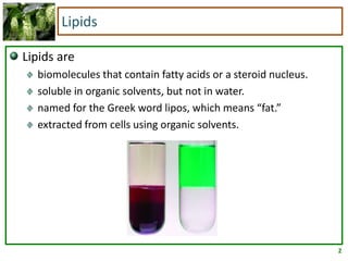
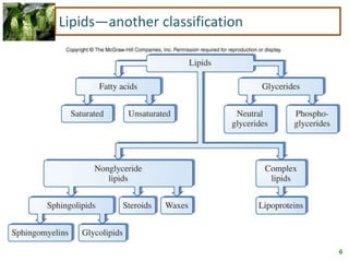
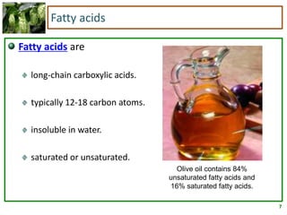
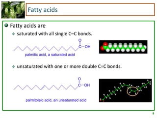
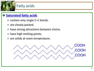
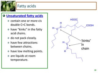
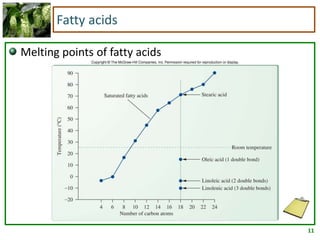
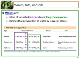
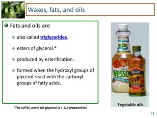
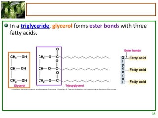
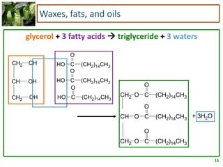
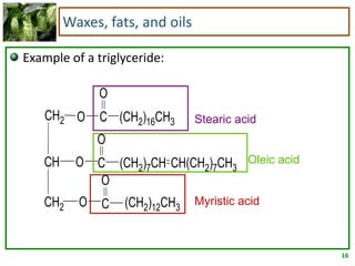
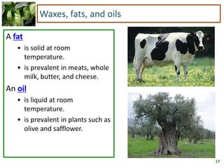
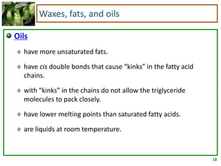
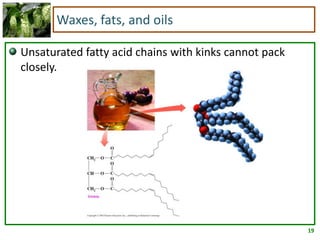
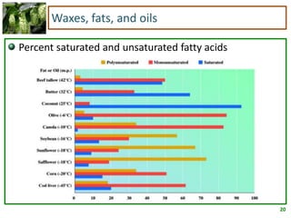
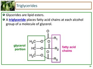
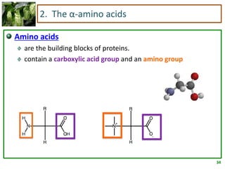
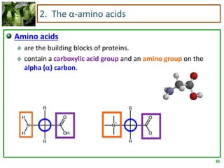
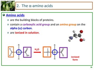
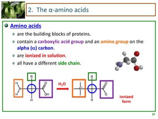
1
Chapter 17
Lipids and Their Functions
in Biological Systems
2
Lipids
Lipids are
biomolecules that contain fatty acids or a steroid nucleus.
soluble in organic solvents, but not in water.
named for the Greek word lipos, which means “fat.”
extracted from cells using organic solvents.
3
Lipids—one classification
The types of lipids containing fatty acids are
waxes
fats and oils (triacylglycerols)
glycerophospholipids
prostaglandins
The types of lipids that do not contain fatty acids are
steroids
4
Lipids—one classification
5
Lipids—another classification
Four main groups
Fatty acids
Saturated
Unsaturated
Glycerides: glycerol-containing lipids
Nonglyceride lipids
Sphingolipids
Steroids
Waxes
Complex lipids: lipoproteins
6
Lipids—another classification
7
Fatty acids
Fatty acids are
long-chain carboxylic acids.
typically 12-18 carbon atoms.
insoluble in water.
saturated or unsaturated.
Olive oil contains 84%
unsaturated fatty acids and
16% saturated fatty acids.
8
Fatty acids
Fatty acids are
saturated with all single C–C bonds.
unsaturated with one or more double C=C bonds.
9
Fatty acids
Saturated fatty acids
contain only single C–C bonds.
are closely packed.
have strong attractions between chains.
have high melting points.
are solids at room temperature.
COOH
COOH
COOH
10
COOH
H
H C
C
HOOC
H
H
C
C
Fatty acids
Unsaturated fatty acids
contain one or more cis
double C=C bonds.
have “kinks” in the fatty
acid chains.
do not pack closely.
have few attractions
between chains.
have low melting points.
are liquids at room
temperature.
“kinks”
in
chain
11
Fatty acids
Melting points of fatty acids
12
Waxes, fats, and oils
Waxes are:
esters of saturated fatty acids and long-chain alcohols.
coatings that prevent loss of water by leaves of plants.
13
Waxes, fats, and oils
Fats and oils are
also called triglycerides.
esters of glycerol.*
produced by esterification.
formed when the hydroxyl groups of
glycerol react with the carboxyl
groups of fatty acids.
*The IUPAC name for glycerol is 1,2,3-propanetriol.
14
In a triglyceride, glycerol forms ester bonds with three
fatty acids.
15
glycerol + 3 fatty acids  triglyceride + 3 waters
Waxes, fats, and oils
OHCH2
OH
OHCH2
CH
O
(CH2)14CH3CHO
O
(CH2)14CH3CHO
O
(CH2)14CH3CHO
+ 3H2O
O
O
C (CH2)14CH3
CH O
O
C (CH2)14CH3
CH2 O
O
C (CH2)14CH3
CH2
16
(CH2)12CH3
O
C
CH(CH2)7CH3(CH2)7CH
O
C
O
(CH2)16CH3C
O
O
OCH2
CH2
CH
Waxes, fats, and oils
Example of a triglyceride:
Stearic acid
Oleic acid
Myristic acid
17
Waxes, fats, and oils
A fat
• is solid at room
temperature.
• is prevalent in meats, whole
milk, butter, and cheese.
An oil
• is liquid at room
temperature.
• is prevalent in plants such as
olive and safflower.
18
Waxes, fats, and oils
Oils
have more unsaturated fats.
have cis double bonds that cause “kinks” in the fatty acid
chains.
with “kinks” in the chains do not allow the triglyceride
molecules to pack closely.
have lower melting points than saturated fatty acids.
are liquids at room temperature.
19
Waxes, fats, and oils
Unsaturated fatty acid chains with kinks cannot pack
closely.
20
Waxes, fats, and oils
Percent saturated and unsaturated fatty acids
21
Chemical reactions of fatty acids
In hydrogenation1, double bonds in unsaturated fatty
acids react with H2 in the presence of a Ni or Pt catalyst.
CH(CH2)7CH3(CH2)5CH
O
C
CH(CH2)7CH3(CH2)5CH
O
C
CH(CH2)7CH3(CH2)5CH
O
C
O
O
OCH2
CH2
CH
O
(CH2)14CH3C
O
(CH2)14CH3C
O
(CH2)14CH3C
O
O
OCH2
CH2
CH
+ 3 H2
Ni
glyceryl tripalmitoleate glyceryl tripalmitate
1See Chapter 11, pages 363-365
22
Chemical reactions of fatty acids
Unsaturated fatty acids can be
cis with bulky groups on same side of C=C.
trans with bulky groups on opposite sides of C=C.
23
Chemical reactions of fatty acids
Most naturally occurring
fatty acids have cis double
bonds.
During hydrogenation, some
cis double bonds are
converted to trans double
bonds.
In the body, trans fatty acids
behave like saturated fatty
acids.
Why would trans fatty acids
behave like saturated fatty
acids, while cis fatty acids do
not?
24
Chemical reactions of fatty acids
In hydrolysis2, ester bonds are split by water in the
presence of an acid, a base, or an enzyme.
OCH2
OCH
OCH2
OHCH2
OHCH
OHCH2 O
(CH2)14CH3CHO
H2O
O
(CH2)14CH3C
O
(CH2)14CH3C
O
(CH2)14CH3C
H+
+
+
2See Chapter 14, pages 472-473
25
Chemical reactions of fatty acids
O
O
C (CH2)14CH3
CH O
O
C (CH2)14CH3
CH2 O
O
C (CH2)14CH3
CH2
+ 3NaOH
Na+ -
O
O
C (CH2)14CH33
OH
CH OH
CH2 OH
CH2
+
“soap”
In saponification, a fatty acid or
triglyceride reacts with a strong base
to form an alcohol or glycerol, and
the salt(s) of fatty acid(s).
26
Triglycerides
Glycerides are lipid esters.
A triglyceride places fatty acid chains at each alcohol
group of a molecule of glycerol.
glycerol
portion
fatty acid
chains
27
Triglycerides
Triglycerides undergo three basic reactions, identical to
those studied in carboxylic acids.
Triglyceride
Glycerol
Fatty Acids
Glycerol
Fatty Acid Salts
More saturated
triglyceride
H2O, H+
NaOH
H2, Ni
28
Glycerophospholipids
Glycerophospholipids are
the most abundant lipids in cell membranes.
composed of glycerol, two fatty acids, phosphate, and an
amino alcohol.
Glycerol
PO4
Amino
alcohol
Fatty acid
Fatty acid
example
29
Cholesterol and steroid hormones
A steroid nucleus consists of
three cyclohexane rings, and
one cyclopentane ring.
The rings are fused (joined along one side).
30
Cholesterol and steroid hormones
Cholesterol
is the most abundant steroid in the body.
has methyl (-CH3) groups, an alkyl chain, and an -OH attached to
the steroid nucleus.
31
Cholesterol and steroid hormones
A steroid is any natural or synthetic compound based on
the four-fused-ring structure in cholesterol.
testosterone
cortisone
progesterone
32
Chapter 18
Protein Structure and Function
33
2. The α-amino acids
Amino acids
are the building blocks of proteins.
contain a carboxylic acid group
34
2. The α-amino acids
Amino acids
are the building blocks of proteins.
contain a carboxylic acid group and an amino group
35
2. The α-amino acids
Amino acids
are the building blocks of proteins.
contain a carboxylic acid group and an amino group on the
alpha () carbon.
36
2. The α-amino acids
Amino acids
are the building blocks of proteins.
contain a carboxylic acid group and an amino group on the
alpha () carbon.
are ionized in solution.
H2O
ionized
form
37
2. The α-amino acids
Amino acids
are the building blocks of proteins.
contain a carboxylic acid group and an amino group on the
alpha () carbon.
are ionized in solution.
all have a different side chain.
H2O
ionized
form
38
2. The α-amino acids
Examples of amino acids
glycine
alanine
39
2. The α-amino acids
All but one of the amino acids have a chiral carbon. The
α carbon is attached to
a carboxylate group (COO-),
a protonated amino group (-NH3
+),
a hydrogen atom, and
a side chain (R group).
Glycine has R = H, which gives it two hydrogens attached
to the α carbon and no chiral carbon.
40
2. The α-amino acids
Each amino acid (except glycine) can exist as one of two
stereoisomers.
Only L- isomers of amino acids are found in proteins.
Recall that mainly D- isomers of monosaccharides exist in
nature.
As for monosaccharides, Fischer projections for amino
acids have the most oxidized group at the top.
L-alanine D-alanine L-cysteine D-cysteine
41
2. The α-amino acids
Amino acids are classified based on the nature of their
side chains.
Nonpolar amino acids are hydrophobic and have hydrocarbon
side chains.
Polar amino acids are hydrophilic and have polar or ionic side
chains.
Acidic amino acids are hydrophilic and have acidic (carboxylic
acid) side chains.
Basic amino acids are hydrophilic and have amino side chains.
42
2. The α-amino acids
Nonpolar: The R group is H, alkyl, or aromatic.
glycine alanine valine
methionine prolinephenylalanine
leucine isoleucine
tryptophan
43
2. The α-amino acids
Polar: the R group is an alcohol, thiol, or amide
serine threonine tyrosine
cysteineasparagin
e
glutamine
44
2. The α-amino acids
Acidic: The R group is a carboxylic acid.
aspartate glutamate
45
2. The α-amino acids
Basic: The R group is an amine.
histidine argininelysine
46
3. The peptide bond
A peptide bond is an amide bond that forms between
the carboxyl group of one amino acid and the amino
group of the next amino acid.
glycine alanine
+ H2O
47
3. The peptide bond
Naming peptides
For small peptides, the general name “peptide” is preceded by
a prefix indicating how many amino acids were condensed to
form the peptide.
The peptide formed on the previous slide is a dipeptide.
The end of the peptide with the free NH3
+ group is called the N-
terminal amino acid.
The end of the peptide with the free –COO- group is called the
C-terminal amino acid.
48
3. The peptide bond
Naming peptides (cont.)
The root name of a peptide is the name of the C-terminal
amino acid, which uses its entire name.
For all other amino acids in the peptide, the ending –ine is
changed to –yl.
The amino acids are named in order starting with the N-
terminal amino acid.
A peptide composed of aspartine, glutamine, and serine
(in that order) would be named:
aspartyl-glutamyl-serine
49
3. The peptide bond
The structures of small peptides are based on a repeating
backbone:
N—C—C—N—C—C—N—C—C
α-amino group (N)
α-carbon (always attached to H and R)
α-carboxyl group (C)
50
3. The peptide bond
Draw the structure of aspartyl-glutamyl-serine.
This is a tripeptide, so the backbone will have three repeats.
The left end is the N-terminal amino acid and the right end is
the C-terminal amino acid.
The carboxyl carbons all have a carbonyl and the α carbons all
have a hydrogen.
51
3. The peptide bond
Draw the structure of aspartyl-glutamyl-serine. (cont.)
Each α carbon has the R group characteristic of the particular
amino acid.
aspartame
glutamate
serine
52
4. The primary structure of proteins
The primary structure of a protein is the amino acid
sequence of the protein’s peptide backbone.
There are 20 amino acids which can be arranged in varying
orders.
The length of the peptide backbone can vary.
53
4. The primary structure of proteins
Primary structure is the amino acid sequence of the
polypeptide chain.
It is the result of covalent bonding (peptide bonds) between
amino acids
Each protein has a different primary structure with
different amino acids in different places along the chain
CH3
SH
CH2
CH3
S
CH2
CH2CH O
O-
CCH
H
N
O
CCH
H
N
O
CCH
H
N
O
CCHH3N
CH3
CH3CH
Ala─Leu─Cys─Met
54
4. The primary structure of proteins
Insulin was the first protein to have its primary
structure determined.
It has a primary structure of two polypeptide chains
linked by disulfide bonds.
One chain (A) has 21 amino acids and the other (B) has
30 amino acids.
55
5. The secondary structure of proteins
When the primary sequence of the polypeptide folds into
regularly repeating structures, secondary structure is
formed.
Secondary structure results from hydrogen bonding between
the amide hydrogens and carbonyl oxygens of the peptide
bonds.
56
5. The secondary structure of proteins
The α-helix is the most common type
of secondary structure.
Features:
a three-dimensional spatial arrangement
of amino acids in a polypeptide chain.
held by H bonds between the H of –N-H
group and the O of C=O of the fourth
amino acid down the chain.
a corkscrew shape that looks like a coiled
“telephone cord”.
57
5. The secondary structure of proteins
Top view of the α-helix, looking down into the “barrel.”
The side chains (-R) point out.
58
5. The secondary structure of proteins
The second most common secondary structure is the β-
pleated sheet.
The β-pleated sheet consists of polypeptide chains arranged
side by side with hydrogen bonds between chains.
Side chains (-R) are above and below the sheet.
This structure is typical of fibrous materials like silk.
59
6. The tertiary structure of proteins
Soluble proteins are usually
globular proteins.
A third level of structure,
tertiary structure, is added to
the primary and secondary
structures.
Areas of α-helix and β-pleated
secondary structure are folded in
on themselves and held in place
by the forces responsible for
tertiary structure.
60
6. The tertiary structure of proteins
Crosslinks in tertiary structures involve attractions and
repulsions between the side chains (-R) of the amino
acids in the polypeptide chain.
Hydrophobic interactions: attractions between nonpolar
groups.
Hydrophilic interactions: attractions between polar groups and
water.
Salt bridges: ionic interactions between acidic and basic amino
acids.
Hydrogen bonds: between H and oxygen or nitrogen.
Disulfide bonds: covalent links between sulfur atoms of two
cysteine amino acids.
61
6. The tertiary structure of proteins
salt bridge
hydrophobic
interactions
hydrogen
bonds
disulfide
bonds
62
7. The quaternary structure of proteins
Quaternary structure is the arrangement of subunits or
peptides that form a larger protein.
A subunit is a polypeptide chain having primary, secondary, and
tertiary structural features that is a part of a larger protein.
Quaternary structure is maintained by the same forces which
are active in maintaining tertiary structure.
Hemoglobin consists of four polypeptide chains as
subunits.
63
8. Overview of protein structure
Primary structure:
Amino acid sequence
Results from formation of covalent
peptide bonds between amino acids
Secondary structure:
Includes α-helix and β-sheet
Hydrogen bonding between amide
hydrogens and carbonyl oxygens of the
peptide bonds
64
8. Overview of protein structure
Tertiary structure:
Overall folding of the entire
polypeptide chain
Interactions between different
amino acid side chains
Quaternary structure:
Concerned with topological, spatial
arrangement of two or more
polypeptide chains
Involves both disulfide bridges and
noncovalent interactions
65
10. Denaturation of proteins
Denaturation involves the disruption of bonds in the
secondary, tertiary and quaternary protein structures.
Denaturation is the loss of organized structure of a globular
protein.
Denaturation does not alter primary structure.
Causes of denaturation:
heat and organic compounds that break apart H bonds and
disrupt hydrophobic interactions.
acids and bases that break H bonds between polar R groups
and disrupt ionic bonds.
heavy metal ions that react with S-S bonds to form solids.
agitation such as whipping that stretches peptide chains until
interactive forces are broken.
66
10. Denaturation of proteins
Heat: As the temperature rises, molecules move and
vibrate more. The weaker hydrogen bonds are the first
to break.
pH: Amino acids include basic (amino) and acidic
(carboxylate) groups. An excess of H+ or OH- changes
ionic interactions involving these groups.
Organic solvents: Alcohols disrupt hydrogen bonding
because they take part in it themselves. The nonpolar
portions of alcohols disrupt nonpolar interactions.
Denaturation of Protein by Strong Acid
67
10. Denaturation of proteins
Detergents: The hydrophobic region of detergents
disrupts hydrophobic interactions in proteins.
Heavy metals: Metal cations such as mercury or lead can
bond with negative side chains and disrupt their
interactions. They can also bind to sulfur and disrupt
disulfide bonds.
Mechanical stress: Shaking or whipping can disrupt the
intermolecular forces that maintain the conformation of
the protein.

Lipids and Proteins

  • 1.
    1 Chapter 17 Lipids andTheir Functions in Biological Systems
  • 2.
    2 Lipids Lipids are biomolecules thatcontain fatty acids or a steroid nucleus. soluble in organic solvents, but not in water. named for the Greek word lipos, which means “fat.” extracted from cells using organic solvents.
  • 3.
    3 Lipids—one classification The typesof lipids containing fatty acids are waxes fats and oils (triacylglycerols) glycerophospholipids prostaglandins The types of lipids that do not contain fatty acids are steroids
  • 4.
  • 5.
    5 Lipids—another classification Four maingroups Fatty acids Saturated Unsaturated Glycerides: glycerol-containing lipids Nonglyceride lipids Sphingolipids Steroids Waxes Complex lipids: lipoproteins
  • 6.
  • 7.
    7 Fatty acids Fatty acidsare long-chain carboxylic acids. typically 12-18 carbon atoms. insoluble in water. saturated or unsaturated. Olive oil contains 84% unsaturated fatty acids and 16% saturated fatty acids.
  • 8.
    8 Fatty acids Fatty acidsare saturated with all single C–C bonds. unsaturated with one or more double C=C bonds.
  • 9.
    9 Fatty acids Saturated fattyacids contain only single C–C bonds. are closely packed. have strong attractions between chains. have high melting points. are solids at room temperature. COOH COOH COOH
  • 10.
    10 COOH H H C C HOOC H H C C Fatty acids Unsaturatedfatty acids contain one or more cis double C=C bonds. have “kinks” in the fatty acid chains. do not pack closely. have few attractions between chains. have low melting points. are liquids at room temperature. “kinks” in chain
  • 11.
  • 12.
    12 Waxes, fats, andoils Waxes are: esters of saturated fatty acids and long-chain alcohols. coatings that prevent loss of water by leaves of plants.
  • 13.
    13 Waxes, fats, andoils Fats and oils are also called triglycerides. esters of glycerol.* produced by esterification. formed when the hydroxyl groups of glycerol react with the carboxyl groups of fatty acids. *The IUPAC name for glycerol is 1,2,3-propanetriol.
  • 14.
    14 In a triglyceride,glycerol forms ester bonds with three fatty acids.
  • 15.
    15 glycerol + 3fatty acids  triglyceride + 3 waters Waxes, fats, and oils OHCH2 OH OHCH2 CH O (CH2)14CH3CHO O (CH2)14CH3CHO O (CH2)14CH3CHO + 3H2O O O C (CH2)14CH3 CH O O C (CH2)14CH3 CH2 O O C (CH2)14CH3 CH2
  • 16.
    16 (CH2)12CH3 O C CH(CH2)7CH3(CH2)7CH O C O (CH2)16CH3C O O OCH2 CH2 CH Waxes, fats, andoils Example of a triglyceride: Stearic acid Oleic acid Myristic acid
  • 17.
    17 Waxes, fats, andoils A fat • is solid at room temperature. • is prevalent in meats, whole milk, butter, and cheese. An oil • is liquid at room temperature. • is prevalent in plants such as olive and safflower.
  • 18.
    18 Waxes, fats, andoils Oils have more unsaturated fats. have cis double bonds that cause “kinks” in the fatty acid chains. with “kinks” in the chains do not allow the triglyceride molecules to pack closely. have lower melting points than saturated fatty acids. are liquids at room temperature.
  • 19.
    19 Waxes, fats, andoils Unsaturated fatty acid chains with kinks cannot pack closely.
  • 20.
    20 Waxes, fats, andoils Percent saturated and unsaturated fatty acids
  • 21.
    21 Chemical reactions offatty acids In hydrogenation1, double bonds in unsaturated fatty acids react with H2 in the presence of a Ni or Pt catalyst. CH(CH2)7CH3(CH2)5CH O C CH(CH2)7CH3(CH2)5CH O C CH(CH2)7CH3(CH2)5CH O C O O OCH2 CH2 CH O (CH2)14CH3C O (CH2)14CH3C O (CH2)14CH3C O O OCH2 CH2 CH + 3 H2 Ni glyceryl tripalmitoleate glyceryl tripalmitate 1See Chapter 11, pages 363-365
  • 22.
    22 Chemical reactions offatty acids Unsaturated fatty acids can be cis with bulky groups on same side of C=C. trans with bulky groups on opposite sides of C=C.
  • 23.
    23 Chemical reactions offatty acids Most naturally occurring fatty acids have cis double bonds. During hydrogenation, some cis double bonds are converted to trans double bonds. In the body, trans fatty acids behave like saturated fatty acids. Why would trans fatty acids behave like saturated fatty acids, while cis fatty acids do not?
  • 24.
    24 Chemical reactions offatty acids In hydrolysis2, ester bonds are split by water in the presence of an acid, a base, or an enzyme. OCH2 OCH OCH2 OHCH2 OHCH OHCH2 O (CH2)14CH3CHO H2O O (CH2)14CH3C O (CH2)14CH3C O (CH2)14CH3C H+ + + 2See Chapter 14, pages 472-473
  • 25.
    25 Chemical reactions offatty acids O O C (CH2)14CH3 CH O O C (CH2)14CH3 CH2 O O C (CH2)14CH3 CH2 + 3NaOH Na+ - O O C (CH2)14CH33 OH CH OH CH2 OH CH2 + “soap” In saponification, a fatty acid or triglyceride reacts with a strong base to form an alcohol or glycerol, and the salt(s) of fatty acid(s).
  • 26.
    26 Triglycerides Glycerides are lipidesters. A triglyceride places fatty acid chains at each alcohol group of a molecule of glycerol. glycerol portion fatty acid chains
  • 27.
    27 Triglycerides Triglycerides undergo threebasic reactions, identical to those studied in carboxylic acids. Triglyceride Glycerol Fatty Acids Glycerol Fatty Acid Salts More saturated triglyceride H2O, H+ NaOH H2, Ni
  • 28.
    28 Glycerophospholipids Glycerophospholipids are the mostabundant lipids in cell membranes. composed of glycerol, two fatty acids, phosphate, and an amino alcohol. Glycerol PO4 Amino alcohol Fatty acid Fatty acid example
  • 29.
    29 Cholesterol and steroidhormones A steroid nucleus consists of three cyclohexane rings, and one cyclopentane ring. The rings are fused (joined along one side).
  • 30.
    30 Cholesterol and steroidhormones Cholesterol is the most abundant steroid in the body. has methyl (-CH3) groups, an alkyl chain, and an -OH attached to the steroid nucleus.
  • 31.
    31 Cholesterol and steroidhormones A steroid is any natural or synthetic compound based on the four-fused-ring structure in cholesterol. testosterone cortisone progesterone
  • 32.
  • 33.
    33 2. The α-aminoacids Amino acids are the building blocks of proteins. contain a carboxylic acid group
  • 34.
    34 2. The α-aminoacids Amino acids are the building blocks of proteins. contain a carboxylic acid group and an amino group
  • 35.
    35 2. The α-aminoacids Amino acids are the building blocks of proteins. contain a carboxylic acid group and an amino group on the alpha () carbon.
  • 36.
    36 2. The α-aminoacids Amino acids are the building blocks of proteins. contain a carboxylic acid group and an amino group on the alpha () carbon. are ionized in solution. H2O ionized form
  • 37.
    37 2. The α-aminoacids Amino acids are the building blocks of proteins. contain a carboxylic acid group and an amino group on the alpha () carbon. are ionized in solution. all have a different side chain. H2O ionized form
  • 38.
    38 2. The α-aminoacids Examples of amino acids glycine alanine
  • 39.
    39 2. The α-aminoacids All but one of the amino acids have a chiral carbon. The α carbon is attached to a carboxylate group (COO-), a protonated amino group (-NH3 +), a hydrogen atom, and a side chain (R group). Glycine has R = H, which gives it two hydrogens attached to the α carbon and no chiral carbon.
  • 40.
    40 2. The α-aminoacids Each amino acid (except glycine) can exist as one of two stereoisomers. Only L- isomers of amino acids are found in proteins. Recall that mainly D- isomers of monosaccharides exist in nature. As for monosaccharides, Fischer projections for amino acids have the most oxidized group at the top. L-alanine D-alanine L-cysteine D-cysteine
  • 41.
    41 2. The α-aminoacids Amino acids are classified based on the nature of their side chains. Nonpolar amino acids are hydrophobic and have hydrocarbon side chains. Polar amino acids are hydrophilic and have polar or ionic side chains. Acidic amino acids are hydrophilic and have acidic (carboxylic acid) side chains. Basic amino acids are hydrophilic and have amino side chains.
  • 42.
    42 2. The α-aminoacids Nonpolar: The R group is H, alkyl, or aromatic. glycine alanine valine methionine prolinephenylalanine leucine isoleucine tryptophan
  • 43.
    43 2. The α-aminoacids Polar: the R group is an alcohol, thiol, or amide serine threonine tyrosine cysteineasparagin e glutamine
  • 44.
    44 2. The α-aminoacids Acidic: The R group is a carboxylic acid. aspartate glutamate
  • 45.
    45 2. The α-aminoacids Basic: The R group is an amine. histidine argininelysine
  • 46.
    46 3. The peptidebond A peptide bond is an amide bond that forms between the carboxyl group of one amino acid and the amino group of the next amino acid. glycine alanine + H2O
  • 47.
    47 3. The peptidebond Naming peptides For small peptides, the general name “peptide” is preceded by a prefix indicating how many amino acids were condensed to form the peptide. The peptide formed on the previous slide is a dipeptide. The end of the peptide with the free NH3 + group is called the N- terminal amino acid. The end of the peptide with the free –COO- group is called the C-terminal amino acid.
  • 48.
    48 3. The peptidebond Naming peptides (cont.) The root name of a peptide is the name of the C-terminal amino acid, which uses its entire name. For all other amino acids in the peptide, the ending –ine is changed to –yl. The amino acids are named in order starting with the N- terminal amino acid. A peptide composed of aspartine, glutamine, and serine (in that order) would be named: aspartyl-glutamyl-serine
  • 49.
    49 3. The peptidebond The structures of small peptides are based on a repeating backbone: N—C—C—N—C—C—N—C—C α-amino group (N) α-carbon (always attached to H and R) α-carboxyl group (C)
  • 50.
    50 3. The peptidebond Draw the structure of aspartyl-glutamyl-serine. This is a tripeptide, so the backbone will have three repeats. The left end is the N-terminal amino acid and the right end is the C-terminal amino acid. The carboxyl carbons all have a carbonyl and the α carbons all have a hydrogen.
  • 51.
    51 3. The peptidebond Draw the structure of aspartyl-glutamyl-serine. (cont.) Each α carbon has the R group characteristic of the particular amino acid. aspartame glutamate serine
  • 52.
    52 4. The primarystructure of proteins The primary structure of a protein is the amino acid sequence of the protein’s peptide backbone. There are 20 amino acids which can be arranged in varying orders. The length of the peptide backbone can vary.
  • 53.
    53 4. The primarystructure of proteins Primary structure is the amino acid sequence of the polypeptide chain. It is the result of covalent bonding (peptide bonds) between amino acids Each protein has a different primary structure with different amino acids in different places along the chain CH3 SH CH2 CH3 S CH2 CH2CH O O- CCH H N O CCH H N O CCH H N O CCHH3N CH3 CH3CH Ala─Leu─Cys─Met
  • 54.
    54 4. The primarystructure of proteins Insulin was the first protein to have its primary structure determined. It has a primary structure of two polypeptide chains linked by disulfide bonds. One chain (A) has 21 amino acids and the other (B) has 30 amino acids.
  • 55.
    55 5. The secondarystructure of proteins When the primary sequence of the polypeptide folds into regularly repeating structures, secondary structure is formed. Secondary structure results from hydrogen bonding between the amide hydrogens and carbonyl oxygens of the peptide bonds.
  • 56.
    56 5. The secondarystructure of proteins The α-helix is the most common type of secondary structure. Features: a three-dimensional spatial arrangement of amino acids in a polypeptide chain. held by H bonds between the H of –N-H group and the O of C=O of the fourth amino acid down the chain. a corkscrew shape that looks like a coiled “telephone cord”.
  • 57.
    57 5. The secondarystructure of proteins Top view of the α-helix, looking down into the “barrel.” The side chains (-R) point out.
  • 58.
    58 5. The secondarystructure of proteins The second most common secondary structure is the β- pleated sheet. The β-pleated sheet consists of polypeptide chains arranged side by side with hydrogen bonds between chains. Side chains (-R) are above and below the sheet. This structure is typical of fibrous materials like silk.
  • 59.
    59 6. The tertiarystructure of proteins Soluble proteins are usually globular proteins. A third level of structure, tertiary structure, is added to the primary and secondary structures. Areas of α-helix and β-pleated secondary structure are folded in on themselves and held in place by the forces responsible for tertiary structure.
  • 60.
    60 6. The tertiarystructure of proteins Crosslinks in tertiary structures involve attractions and repulsions between the side chains (-R) of the amino acids in the polypeptide chain. Hydrophobic interactions: attractions between nonpolar groups. Hydrophilic interactions: attractions between polar groups and water. Salt bridges: ionic interactions between acidic and basic amino acids. Hydrogen bonds: between H and oxygen or nitrogen. Disulfide bonds: covalent links between sulfur atoms of two cysteine amino acids.
  • 61.
    61 6. The tertiarystructure of proteins salt bridge hydrophobic interactions hydrogen bonds disulfide bonds
  • 62.
    62 7. The quaternarystructure of proteins Quaternary structure is the arrangement of subunits or peptides that form a larger protein. A subunit is a polypeptide chain having primary, secondary, and tertiary structural features that is a part of a larger protein. Quaternary structure is maintained by the same forces which are active in maintaining tertiary structure. Hemoglobin consists of four polypeptide chains as subunits.
  • 63.
    63 8. Overview ofprotein structure Primary structure: Amino acid sequence Results from formation of covalent peptide bonds between amino acids Secondary structure: Includes α-helix and β-sheet Hydrogen bonding between amide hydrogens and carbonyl oxygens of the peptide bonds
  • 64.
    64 8. Overview ofprotein structure Tertiary structure: Overall folding of the entire polypeptide chain Interactions between different amino acid side chains Quaternary structure: Concerned with topological, spatial arrangement of two or more polypeptide chains Involves both disulfide bridges and noncovalent interactions
  • 65.
    65 10. Denaturation ofproteins Denaturation involves the disruption of bonds in the secondary, tertiary and quaternary protein structures. Denaturation is the loss of organized structure of a globular protein. Denaturation does not alter primary structure. Causes of denaturation: heat and organic compounds that break apart H bonds and disrupt hydrophobic interactions. acids and bases that break H bonds between polar R groups and disrupt ionic bonds. heavy metal ions that react with S-S bonds to form solids. agitation such as whipping that stretches peptide chains until interactive forces are broken.
  • 66.
    66 10. Denaturation ofproteins Heat: As the temperature rises, molecules move and vibrate more. The weaker hydrogen bonds are the first to break. pH: Amino acids include basic (amino) and acidic (carboxylate) groups. An excess of H+ or OH- changes ionic interactions involving these groups. Organic solvents: Alcohols disrupt hydrogen bonding because they take part in it themselves. The nonpolar portions of alcohols disrupt nonpolar interactions. Denaturation of Protein by Strong Acid
  • 67.
    67 10. Denaturation ofproteins Detergents: The hydrophobic region of detergents disrupts hydrophobic interactions in proteins. Heavy metals: Metal cations such as mercury or lead can bond with negative side chains and disrupt their interactions. They can also bind to sulfur and disrupt disulfide bonds. Mechanical stress: Shaking or whipping can disrupt the intermolecular forces that maintain the conformation of the protein.

Editor's Notes

  • #24 A trans fatty acid is more linear than a cis fatty acid (see slide 23), so the molecules can pack more closely together than they can in a cis fatty acid. Closer molecules mean stronger intermolecular forces and higher melting points.