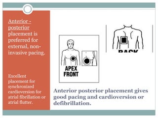
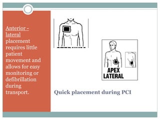
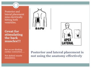
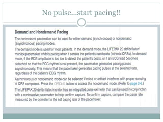
E X T E R N A L P A C I N G
Lifepak 20
Noninvasive Pacing Noninvasive pacing is primarily used for emergency treatment
of symptomatic bradycardia. Electrical current is passed from an external pulse
generator via a conducting cable and externally applied, self-adhesive electrodes
through the chest wall and heart. Other terms for noninvasive pacing are
transcutaneous, transchest or transthoracic pacing
Who can do noninvasive pacing?
 Noninvasive pacing is comparatively easy to perform and
requires minimal training. It can be initiated by nurses,
paramedics, physicians and other emergency cardiac care
providers. Requiring very little set-up time, it generally does
not include any of the complications associated with invasive
techniques.
 It is the least expensive pacing approach and may be
used for standby pacing, reducing the need for
prophylactic placement of a transvenous catheter.
 It is especially useful for patients at high risk for infection or
bleeding. The major disadvantage of noninvasive pacing is
discomfort. Current is applied across the chest which results
in cutaneous nerve stimulation as well as skeletal muscle
stimulation.
Standby if needed
 Noninvasive pacing should be used on standby in
situations when the patient is clinically stable yet may
quickly decompensate or become unstable.
 Patients who may benefit from standby pacing include:
cardiac patients undergoing surgery, patients with acute
MI and signs of early heart block, patients needing
surgery for permanent pacemaker implantation,
pulse generator change, or lead wire
replacement, patients undergoing cardiac
catheterization or angioplasty, and those with risk
of developing post-cardioversion bradycardias
Seems like we have been here before
Do these patients
 sound familiar???
How about pacemaker dependent?
 Patients having pacer or defibrillator generator
changed and needing pacing should be set up for
possible external pacing should asystole be
prolonged during generator change or the lead
falling out.
 Stuff happens.
 We monitor to make the procedure safer.
When do you need to pace?
 Conditions in which non-invasive pacing is most often indicated
include:
 Absolute bradycardia (HR < 30 bpm) with evidence of very low perfusion
or frank shock (often associated with acute Myocardial infarction or cardiac
ischemia involving the SA or AV nodes).
 Unstable or relative bradycardia as defined by these signs of inadequate
perfusion:
 Altered mental status with poor perfusion
 Clinical signs of shock
 Severe shortness of breath/pulmonary edema
 Severe chest pain (consistent with ischemia)
 Inadequate skin perfusion with diaphoresis
 Cyanotic on 100% oxygen with adequate ventilation
 Asystole with a short time (< 10 minutes) since collapse, particularly if the
asystole occurred shortly after defibrillation or medication administration
If the doctor is at bedside
 Pace when Dr Ghali tells you too!!
 But won’t he be impressed if you are ready to go
without him telling you??
 Nobody wants to know why we were not ready to
pace the patient……………………………….
Monitor
electrode
placement
Place three wire monitor
cable with electrodes
perpendicular to the
Medi-Trace Cadence
pads.
Both arms and the right
side works well.
Select lead II or III
A good R wave is needed
to sense spontaneous
electical activity.
Apply electrodes properly
 Electrodes
Non-invasive pacing can cause discomfort for patients
and can be quite painful. Pain is a function of the current
delivered per unit of skin surface area. Electrodes with a
large surface area minimize pain sensation. Most
commercially available electrodes are 80-100 cm2. Non-
invasive pacemakers perform best with electrodes
designed to function with that specific cardiac
resuscitation system.
 Proper application of the external pacemaker electrodes
is simple but critical. Proper skin contact is an important
factor in reducing resistance and improving capture.
Anterior posterior placement gives
good pacing and cardioversion or
defibrillation.
Anterior -
posterior
placement is
preferred for
external, non-
invasive pacing.
Excellent
placement for
synchronized
cardioversion for
atrial fibrillation or
atrial flutter.
Heart symbol on anterior apex.. Patch to right
posterior
Quick placement during PCI
Anterior -
lateral
placement
requires little
patient
movement and
allows for easy
monitoring or
defibrillation
during
transport.
Vector through RA and RV and LV
Posterior and lateral placement is
not using the anatomy effectively
Posterior and
lateral placement
miss electrically
hitting both
ventricles.
Great for
stimulating
the back
muscles!!!
But we are thinking
cardiac contraction…..
Not skeletal muscle
stimulation.
Vector through LA and posterior LV
Identification of mechanical capture
 Mechanical capture manifests with signs of
improving cardiac output such as an increased level
of consciousness or blood pressure. The clinician
must monitor and assess for both electrical and
mechanical capture of the myocardium. The
electrical activity of the external pacer shows up
clearly on the monitor as large complexes at the rate
you have selected. While pacing capture on the
monitor is an important sign, the appearance of
these complexes does not mean that the
patient’s myocardium is mechanically
captured and cardiac output is occurring.
Do vital signs improve??
 The clinician must confirm that the heart is pumping
and the cardiovascular system is generating blood
flow by monitoring the pulse and not assume that
the electrical activity of the pacer means that the
heart is being paced
 Is the pulse oximeter giving a waveform??
 Does the patient have a palpable pulse??
Pace controls behind the door
Option button left of pacer controls
Roll to pacing and choose demand or nondemand
No pulse…start pacing!!
Prepare for worst possible … be glad if you do not
need the external pacing
 1 - hook em up
 2 -place 3 wire electrodes and place Medi-Trace
patches……. AP works well….anterior lateral works
too!
 3- Select pacer….set rate….. Check r wave
sensing….turn up the juice to theraputic level.
 4- 40mV to 100 mV usually works
 ****Check vital signs…. Pulse… pulse oximetry****
Pause is 25% pacing..press pacer to shut off
How much juice??
 Current Requirements
 Human studies have shown that the average current
necessary for external pacing is about 65-100
milliamperes (mA) in unstable bradycardias and
about 50-70 mA in hemodynamically stable
patients and volunteers.
 The clinician increases the current until the pacemaker
“captures” the myocardium, taking over the pacemaker
functions of the heart and resulting in a characteristic
pacemaker rhythm. The clinician then confirms the
presence of a pulse following each pacemaker spike. The
force of skeletal muscle contraction, not the electrical
current, determines the patient’s level of discomfort
during non-invasive pacing
Does not cure sudden death
 Non-invasive pacing is not likely to be
effective in situations of prolonged duration
of cardiac arrest.
 The outcome of prolonged bradycardia/asystolic
cardiac arrest is poor, even with non-invasive pacing.
Indiscriminate pacing of this rhythm is unwarranted,
particularly as a late effort in the resuscitation.
Pacing of bradycardia/asystole of short duration,
especially post counter-shock bradycardia/asystole,
is more likely to be useful
Happy patient.. happy doc..happy nurse
Intuitive procedure
Lifepak manual

Lifepak 20 TTP

  • 1.
    E X TE R N A L P A C I N G Lifepak 20 Noninvasive Pacing Noninvasive pacing is primarily used for emergency treatment of symptomatic bradycardia. Electrical current is passed from an external pulse generator via a conducting cable and externally applied, self-adhesive electrodes through the chest wall and heart. Other terms for noninvasive pacing are transcutaneous, transchest or transthoracic pacing
  • 2.
    Who can dononinvasive pacing?  Noninvasive pacing is comparatively easy to perform and requires minimal training. It can be initiated by nurses, paramedics, physicians and other emergency cardiac care providers. Requiring very little set-up time, it generally does not include any of the complications associated with invasive techniques.  It is the least expensive pacing approach and may be used for standby pacing, reducing the need for prophylactic placement of a transvenous catheter.  It is especially useful for patients at high risk for infection or bleeding. The major disadvantage of noninvasive pacing is discomfort. Current is applied across the chest which results in cutaneous nerve stimulation as well as skeletal muscle stimulation.
  • 3.
    Standby if needed Noninvasive pacing should be used on standby in situations when the patient is clinically stable yet may quickly decompensate or become unstable.  Patients who may benefit from standby pacing include: cardiac patients undergoing surgery, patients with acute MI and signs of early heart block, patients needing surgery for permanent pacemaker implantation, pulse generator change, or lead wire replacement, patients undergoing cardiac catheterization or angioplasty, and those with risk of developing post-cardioversion bradycardias
  • 4.
    Seems like wehave been here before Do these patients  sound familiar???
  • 5.
    How about pacemakerdependent?  Patients having pacer or defibrillator generator changed and needing pacing should be set up for possible external pacing should asystole be prolonged during generator change or the lead falling out.  Stuff happens.  We monitor to make the procedure safer.
  • 6.
    When do youneed to pace?  Conditions in which non-invasive pacing is most often indicated include:  Absolute bradycardia (HR < 30 bpm) with evidence of very low perfusion or frank shock (often associated with acute Myocardial infarction or cardiac ischemia involving the SA or AV nodes).  Unstable or relative bradycardia as defined by these signs of inadequate perfusion:  Altered mental status with poor perfusion  Clinical signs of shock  Severe shortness of breath/pulmonary edema  Severe chest pain (consistent with ischemia)  Inadequate skin perfusion with diaphoresis  Cyanotic on 100% oxygen with adequate ventilation  Asystole with a short time (< 10 minutes) since collapse, particularly if the asystole occurred shortly after defibrillation or medication administration
  • 7.
    If the doctoris at bedside  Pace when Dr Ghali tells you too!!  But won’t he be impressed if you are ready to go without him telling you??  Nobody wants to know why we were not ready to pace the patient……………………………….
  • 8.
    Monitor electrode placement Place three wiremonitor cable with electrodes perpendicular to the Medi-Trace Cadence pads. Both arms and the right side works well. Select lead II or III A good R wave is needed to sense spontaneous electical activity.
  • 9.
    Apply electrodes properly Electrodes Non-invasive pacing can cause discomfort for patients and can be quite painful. Pain is a function of the current delivered per unit of skin surface area. Electrodes with a large surface area minimize pain sensation. Most commercially available electrodes are 80-100 cm2. Non- invasive pacemakers perform best with electrodes designed to function with that specific cardiac resuscitation system.  Proper application of the external pacemaker electrodes is simple but critical. Proper skin contact is an important factor in reducing resistance and improving capture.
  • 10.
    Anterior posterior placementgives good pacing and cardioversion or defibrillation. Anterior - posterior placement is preferred for external, non- invasive pacing. Excellent placement for synchronized cardioversion for atrial fibrillation or atrial flutter.
  • 11.
    Heart symbol onanterior apex.. Patch to right posterior
  • 12.
    Quick placement duringPCI Anterior - lateral placement requires little patient movement and allows for easy monitoring or defibrillation during transport.
  • 13.
    Vector through RAand RV and LV
  • 14.
    Posterior and lateralplacement is not using the anatomy effectively Posterior and lateral placement miss electrically hitting both ventricles. Great for stimulating the back muscles!!! But we are thinking cardiac contraction….. Not skeletal muscle stimulation.
  • 15.
    Vector through LAand posterior LV
  • 16.
    Identification of mechanicalcapture  Mechanical capture manifests with signs of improving cardiac output such as an increased level of consciousness or blood pressure. The clinician must monitor and assess for both electrical and mechanical capture of the myocardium. The electrical activity of the external pacer shows up clearly on the monitor as large complexes at the rate you have selected. While pacing capture on the monitor is an important sign, the appearance of these complexes does not mean that the patient’s myocardium is mechanically captured and cardiac output is occurring.
  • 17.
    Do vital signsimprove??  The clinician must confirm that the heart is pumping and the cardiovascular system is generating blood flow by monitoring the pulse and not assume that the electrical activity of the pacer means that the heart is being paced  Is the pulse oximeter giving a waveform??  Does the patient have a palpable pulse??
  • 18.
  • 19.
    Option button leftof pacer controls
  • 20.
    Roll to pacingand choose demand or nondemand
  • 21.
  • 22.
    Prepare for worstpossible … be glad if you do not need the external pacing  1 - hook em up  2 -place 3 wire electrodes and place Medi-Trace patches……. AP works well….anterior lateral works too!  3- Select pacer….set rate….. Check r wave sensing….turn up the juice to theraputic level.  4- 40mV to 100 mV usually works  ****Check vital signs…. Pulse… pulse oximetry****
  • 23.
    Pause is 25%pacing..press pacer to shut off
  • 24.
    How much juice?? Current Requirements  Human studies have shown that the average current necessary for external pacing is about 65-100 milliamperes (mA) in unstable bradycardias and about 50-70 mA in hemodynamically stable patients and volunteers.  The clinician increases the current until the pacemaker “captures” the myocardium, taking over the pacemaker functions of the heart and resulting in a characteristic pacemaker rhythm. The clinician then confirms the presence of a pulse following each pacemaker spike. The force of skeletal muscle contraction, not the electrical current, determines the patient’s level of discomfort during non-invasive pacing
  • 25.
    Does not curesudden death  Non-invasive pacing is not likely to be effective in situations of prolonged duration of cardiac arrest.  The outcome of prolonged bradycardia/asystolic cardiac arrest is poor, even with non-invasive pacing. Indiscriminate pacing of this rhythm is unwarranted, particularly as a late effort in the resuscitation. Pacing of bradycardia/asystole of short duration, especially post counter-shock bradycardia/asystole, is more likely to be useful
  • 26.
    Happy patient.. happydoc..happy nurse
  • 27.
  • 28.