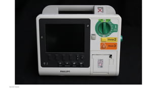
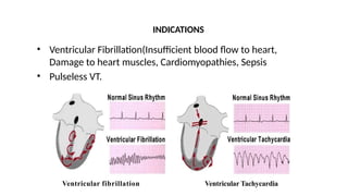
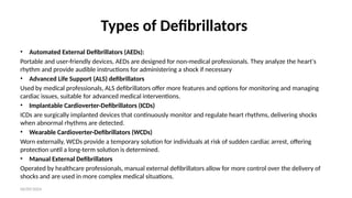
06/09/2024
DEFIBRILLATION
06/09/2024
Defibrillation
•Defibrillation is a process in which an electronic device
called defibrillator sends an electric shock to the heart to
stop an extremely rapid, irregular heartbeat, and restore
the normal heart rhythm.
•Defibrillators are lifesaving devices that apply an electric
shock to establish a more normal cardiac rhythm in
patients who are experiencing ventricular fibrillation (VF)
or another shockable rhythm.
06/09/2024
06/09/2024
Purposes
• Defibrillation is performed to correct life- threatening
fibrillations of the heart, which could result in cardiac arrest.
• It should be performed immediately after identifying that the
patient is experiencing a cardiac emergency, has no pulse,
and is unresponsive.
Ventricular fibrillation
INDICATIONS
• Ventricular Fibrillation(Insufficient blood flow to heart,
Damage to heart muscles, Cardiomyopathies, Sepsis
• Pulseless VT.
Ventricular Tachycardia
06/09/2024
Types of Defibrillators
• Automated External Defibrillators (AEDs):
Portable and user-friendly devices, AEDs are designed for non-medical professionals. They analyze the heart's
rhythm and provide audible instructions for administering a shock if necessary
• Advanced Life Support (ALS) defibrillators
Used by medical professionals, ALS defibrillators offer more features and options for monitoring and managing
cardiac issues, suitable for advanced medical interventions.
• Implantable Cardioverter-Defibrillators (ICDs)
ICDs are surgically implanted devices that continuously monitor and regulate heart rhythms, delivering shocks
when abnormal rhythms are detected.
• Wearable Cardioverter-Defibrillators (WCDs)
Worn externally, WCDs provide a temporary solution for individuals at risk of sudden cardiac arrest, offering
protection until a long-term solution is determined.
• Manual External Defibrillators
Operated by healthcare professionals, manual external defibrillators allow for more control over the delivery of
shocks and are used in more complex medical situations.
• Defibrillators should be portable, battery
operated, small size.
• Energy in defibrillators usually stored
in large capacitors.
• Equipment's:
• Direct Current Defibrillator with
paddles
• Interface material (Gel pads)
• Resuscitative Equipment's.
External Defibrillator
06/09/2024
Placement of Pads(Electrodes)
• One pad is placed at the third intercostal space to the right of
the sternum; the other is placed at the fifth intercostal space
on the left midaxillary line.
• Be sure that no one is touching the bed or the client during
shock.
• Placement directly over breast tissue should be avoided.
06/09/2024
Procedure
1. Expose anterior chest and remove jewelry in the area.
2. Start CPR immediately
3. Apply interface material to the patient (gel pads) or to the
paddles (gel, paste). The electrode paddles should be in firm
contact with the patient's skin.
4. Remove oxygen immediately from area.
06/09/2024
Cont.….
5. A second person should turn on the defibrillator to the prescribed
setting. The American Heart Association recommends that initial
defibrillation should be 200 to 300 joules of delivered energy.
– A second attempt at same level should be given if first attempt is
unsuccessful.
– A third attempt with an increase of energy level to 360 joules should be
attempted.
– Allow only approximately 5 seconds between successive attempts to
assess rhythm and pulse.
06/09/2024
Cont.….
6. Grasp the paddles only by the insulated handles.
7. Charge the paddles. Once paddles are charged, give the
command “ I clear, you clear, All clear”.
8.Push the discharge buttons on both paddles simultaneously
while placing about 25 pounds (11.33 kg) of pressure onto each
paddle.
06/09/2024
Cont.….
9.Remove the paddles from the patient immediately after
the shock is administered.
10. Resume CPR efforts until stable rhythm, spontaneous
respirations, pulse, and blood pressure return.
06/09/2024
Nursing Responsibilities
1. After the patient is defibrillated and rhythm is restored,
antiarrhythmics are usually given to prevent recurrent episodes.
2. Continue with intensive monitoring and care.
•Attach the external and internal paddles if the monitor reads, "No
paddles."
•Check to ensure that the leads are securely attached if the monitor
reads, "No leads.“
•Connect the unit to AC power if the message reads, "Low battery."
•Verify that the Energy Select control settings are correct if the
defibrillator does not charge.
•Change the electrodes and make sure that the electrodes adapter cable
is properly connected if you receive a message of "PACER FAILURE."
Restart the pacer.
•Close the recorder door and the paper roll if the monitor message
reads, "Check recorder”.
Nursing Responsibilities
06/09/2024
Summary
• Defibrillation is a process of delivering electric shock to the
heart, with the help of a defibrillator, which enables the heart's
natural pacemaker to regain control.
• A defibrillation strategy that enables rhythm analysis
to recognize ventricular fibrillation, defibrillator
charging and optimally timed shock delivery with
minimal or no interruptions to chest compressions
should improve the chances of shock success.
06/09/2024
CARDIAC PACING
06/09/2024
Normal conductive system of the heart
06/09/2024
Definition of cardiac pacing
It is an electric device that delivers direct electrical stimulation
to stimulate the myocardium to depolarize ,initiating a
mechanical contraction.
06/09/2024
Clinical Indication
1. Symptomatic bradycardia
2. Symptomatic heart block
• 2nd
degree heart block
• 3rd
or complete heart block
• Bifasicular or transfasicular bundle branch blocks.
3. Prophylaxis
06/09/2024
Pacemaker Design
1. Pulse generator
2. leads
06/09/2024
Pacemaker Design
Pulse generator
• In permanent pacemaker is encapsulated in a metal can ,to
protect the generator from electromagnetic interference
06/09/2024
Pacemaker Design
Pulse generator
• Temporary pacing system
generator is externally
contained in a small box
06/09/2024
Pacemaker Design
Pulse generator
• Transcutanus external pacing system house the
generator in a piece of equipment similar to
portable ECG monitor.
06/09/2024
Pacemaker Design
Pacemaker lead
1. Single chamber (unipolar) pacemaker
• Lead placed in atrium or ventricle
• Produce large spic on the ECG
• Sensing and pacing in the chamber where
the lead is located
• More likely to be affected by
electromechanically interference
06/09/2024
Single chamber (unipolar
06/09/2024
06/09/2024
Pacemaker Design
2. Dual-chamber (bipolar) pacemaker
• One Lead located in the atrium and one in
the ventricle
• Sensing and pacing in both chambers
mimicking the normal heart function
• Produce in visible spic in the ECG
• Less affected by electromechanical
interference.
06/09/2024
Dual-chamber (bipolar) pacemaker
06/09/2024
06/09/2024
Pacemaker function
1. Pacing function
2. Sensing function
3. Capture function
06/09/2024
Pacing function
Atrial pacing:
stimulation of RT atrium produce spic on ECG
preceding P wave
06/09/2024
Pacing function
Ventricle pacing :
stimulation of RT or LT ventricle produce a spic on ECG
preceding QRS complex.
06/09/2024
Pacing function
AVpacing:
direct stimulation of RT atrium and either ventricles mimic
normal heart conduction
06/09/2024
Sensing function
Sensing :
Ability of the cardiac pace maker to see intrinsic cardiac activity
when it occurs.
06/09/2024
Sensing function
Demand:
• pacing stimulation delivered only if the heart rate falls below the preset limit.
Fixed:
• no ability to sense. constantly delivers the preset stimulus at preset rate.
Triggered:
delivers stimuli in response to (sensing )cardiac event.
06/09/2024
Capture function
Capture:
Ability of the pacemaker to generate a response from the heart
(contraction) after electrical stimulation.
06/09/2024
Capture function
1. Electrical capture :
indicated by P or QRS following and corresponding to a
pacemaker spike.
2. Mechanical capture:
palpable pulse corresponding to the electrical event.
06/09/2024
Pacing types
• Permanent
• Temporary
• biventricular
06/09/2024
Types of pacing
1. Permanent pacemaker
• Used to treat chronic heart condition
• Surgically placed transvenuosly under local anesthesia
• Pulse generator placed in a pocket subcutaneously ,can be
adjusted externally
06/09/2024
Permanent pacemaker
06/09/2024
Types of pacing
2. Temporary pacemaker
• Placed during emergencies
• Indicated for pts’ high degree
heart block or unstable
bradycardia
• Can be placed transvenosly,
epicardially,transcutanusly or
transthorasicly
06/09/2024
3. Biventricular pacemaker
• Used in sever heart failure
• Utilize three leads in right
atrium, right ventricle and
left ventricle to coordinate
ventricular coordination
and improve cardiac
out put
Types of pacing
06/09/2024
Equipments
• Transvenous pacing catheter
• EKG machine
• Pacemaker generator with
battery and cable
• Emergency crash cart
• Lidocaine
• Defibrillator
• (2) 5cc syringe with 22 and
25 gauge needles
• External Pacer
• Sterile gown, gloves, mask
06/09/2024
INSERTION SITES
• Left Subclavian (most reliable)
• Internal jugular (lower incidence of pneumothorax)
• Femoral vein
• Brachial vein
06/09/2024
INSERTION PROCEDURE
• Check that patient has a patent IV, and the
defibrillator, emergency cart and appropriate
medications are available.
• obtain consent (time permitting).
• Obtain vital signs and ECG rhythm strip prior to
insertion. Connect to 12 lead EKG and continuously
monitor before, during and after
06/09/2024
INSERTION PROCEDURE
• Anesthetize the area locally.
• Prepare the external temporary generator:
• Portable Chest X-ray is required to confirm placement.
06/09/2024
Applying transcutaneous pacing
• Anterior/posterior:
• Anterior/anterior:
• Module on stand by. minimal out put
• Connect pacing to external module
• Increase milliamp until a pacing spike and corresponding QRS
are seen. palpate pulse
06/09/2024
Complication
• Movement and dislocation of the lead
• Injury
• Bleeding and hematoma
• Ventricular ectopy or VT from wall stimulation
• Infection
• Cardiac tamponad
06/09/2024
Patient education
Specific instruction include
• Not to lift items over 1.4kg or perform difficult arm
maneuver.
• Avoid excessive stretching or bending excessive.
• Avoid sport,tennis, until advised by doctor.
• Sexual activity can be resumed when desired
06/09/2024
Patient education
4. Pacemaker failure
• Teach patient to check own pulse at least weekly for 1 min
• Report slowing on the pulse less or greater than the setting
rate
• Report sign and symptom as
palpitation ,fatigue ,dizziness ,prolonged hiccups
• Wear identification bracelet and carry a pacemaker
identification cared.
06/09/2024
Patient education
5. Electromagnetic interference
• Caution patient that EMI could interfere with pacemaker function.
• Explain that high energy radar, TV and radio transmeters, MRI, large motors
may affect the pacemaker function.
• Teach patient to move 4-6 m away from source and check pulse. it should
return to normal.
06/09/2024
Patient education
• Most pacemaker equipped with internal
filters to prevent interaction with cell
phone.
• Tell patient that antitheft devices and
airport security alarms may affect
pacemaker and trigger security alarm.
• Household and kitchen appliance will not
affect pacemaker.
06/09/2024
Patient education
6. Care of pacemaker site.
• Wear loose-fitting
clothes around pacemaker
• Watch sign and symptom
of infection
• Keep incision site clean
and dry. not to scrub site
• Advise well balanced diet.
06/09/2024
References
• A Guide to Defibrillation By Pacific Medical Training retrieved from
https://www.heartsaver.co.nz/news/types-of-defibrillators/
• Defibrillator, External, Manual retrieved from
https://www.who.int/medical_devices/innovation/defibrillator_manual.pdf
• Nolan JP, Soar J. Defibrillation in clinical practice. Curr Opin Crit Care. 2009
Jun;15(3):209-15. doi: 10.1097/MCC.0b013e32832931cb. PMID: 19262373.
• Jing-Jing Liu et. al. Effectiveness of combining manual external defibrillator and
automated external defibrillator training for third-year nurse students, 6 March 2015
retrieved from https://www.sciencedirect.com/science/article/pii/S2352013215000125
• Grzegorz Cebula Paweł Krawczyk et. al. (2012) Manual defibrillation using paddles—
Which is the best technique? retrieved from
https://www.resuscitationjournal.com/article/S0300-9572(12)00023-8/pdf

Defibrilator & Pacemaker BY Sachin Dwivedi.pptx

  • 1.
  • 2.
    06/09/2024 Defibrillation •Defibrillation is aprocess in which an electronic device called defibrillator sends an electric shock to the heart to stop an extremely rapid, irregular heartbeat, and restore the normal heart rhythm. •Defibrillators are lifesaving devices that apply an electric shock to establish a more normal cardiac rhythm in patients who are experiencing ventricular fibrillation (VF) or another shockable rhythm.
  • 3.
  • 4.
    06/09/2024 Purposes • Defibrillation isperformed to correct life- threatening fibrillations of the heart, which could result in cardiac arrest. • It should be performed immediately after identifying that the patient is experiencing a cardiac emergency, has no pulse, and is unresponsive.
  • 5.
    Ventricular fibrillation INDICATIONS • VentricularFibrillation(Insufficient blood flow to heart, Damage to heart muscles, Cardiomyopathies, Sepsis • Pulseless VT. Ventricular Tachycardia
  • 6.
    06/09/2024 Types of Defibrillators •Automated External Defibrillators (AEDs): Portable and user-friendly devices, AEDs are designed for non-medical professionals. They analyze the heart's rhythm and provide audible instructions for administering a shock if necessary • Advanced Life Support (ALS) defibrillators Used by medical professionals, ALS defibrillators offer more features and options for monitoring and managing cardiac issues, suitable for advanced medical interventions. • Implantable Cardioverter-Defibrillators (ICDs) ICDs are surgically implanted devices that continuously monitor and regulate heart rhythms, delivering shocks when abnormal rhythms are detected. • Wearable Cardioverter-Defibrillators (WCDs) Worn externally, WCDs provide a temporary solution for individuals at risk of sudden cardiac arrest, offering protection until a long-term solution is determined. • Manual External Defibrillators Operated by healthcare professionals, manual external defibrillators allow for more control over the delivery of shocks and are used in more complex medical situations.
  • 7.
    • Defibrillators shouldbe portable, battery operated, small size. • Energy in defibrillators usually stored in large capacitors. • Equipment's: • Direct Current Defibrillator with paddles • Interface material (Gel pads) • Resuscitative Equipment's. External Defibrillator
  • 8.
    06/09/2024 Placement of Pads(Electrodes) •One pad is placed at the third intercostal space to the right of the sternum; the other is placed at the fifth intercostal space on the left midaxillary line. • Be sure that no one is touching the bed or the client during shock. • Placement directly over breast tissue should be avoided.
  • 9.
    06/09/2024 Procedure 1. Expose anteriorchest and remove jewelry in the area. 2. Start CPR immediately 3. Apply interface material to the patient (gel pads) or to the paddles (gel, paste). The electrode paddles should be in firm contact with the patient's skin. 4. Remove oxygen immediately from area.
  • 10.
    06/09/2024 Cont.…. 5. A secondperson should turn on the defibrillator to the prescribed setting. The American Heart Association recommends that initial defibrillation should be 200 to 300 joules of delivered energy. – A second attempt at same level should be given if first attempt is unsuccessful. – A third attempt with an increase of energy level to 360 joules should be attempted. – Allow only approximately 5 seconds between successive attempts to assess rhythm and pulse.
  • 11.
    06/09/2024 Cont.…. 6. Grasp thepaddles only by the insulated handles. 7. Charge the paddles. Once paddles are charged, give the command “ I clear, you clear, All clear”. 8.Push the discharge buttons on both paddles simultaneously while placing about 25 pounds (11.33 kg) of pressure onto each paddle.
  • 12.
    06/09/2024 Cont.…. 9.Remove the paddlesfrom the patient immediately after the shock is administered. 10. Resume CPR efforts until stable rhythm, spontaneous respirations, pulse, and blood pressure return.
  • 13.
    06/09/2024 Nursing Responsibilities 1. Afterthe patient is defibrillated and rhythm is restored, antiarrhythmics are usually given to prevent recurrent episodes. 2. Continue with intensive monitoring and care.
  • 14.
    •Attach the externaland internal paddles if the monitor reads, "No paddles." •Check to ensure that the leads are securely attached if the monitor reads, "No leads.“ •Connect the unit to AC power if the message reads, "Low battery." •Verify that the Energy Select control settings are correct if the defibrillator does not charge. •Change the electrodes and make sure that the electrodes adapter cable is properly connected if you receive a message of "PACER FAILURE." Restart the pacer. •Close the recorder door and the paper roll if the monitor message reads, "Check recorder”. Nursing Responsibilities
  • 15.
    06/09/2024 Summary • Defibrillation isa process of delivering electric shock to the heart, with the help of a defibrillator, which enables the heart's natural pacemaker to regain control. • A defibrillation strategy that enables rhythm analysis to recognize ventricular fibrillation, defibrillator charging and optimally timed shock delivery with minimal or no interruptions to chest compressions should improve the chances of shock success.
  • 16.
  • 17.
  • 18.
    06/09/2024 Definition of cardiacpacing It is an electric device that delivers direct electrical stimulation to stimulate the myocardium to depolarize ,initiating a mechanical contraction.
  • 19.
    06/09/2024 Clinical Indication 1. Symptomaticbradycardia 2. Symptomatic heart block • 2nd degree heart block • 3rd or complete heart block • Bifasicular or transfasicular bundle branch blocks. 3. Prophylaxis
  • 20.
  • 21.
    06/09/2024 Pacemaker Design Pulse generator •In permanent pacemaker is encapsulated in a metal can ,to protect the generator from electromagnetic interference
  • 22.
    06/09/2024 Pacemaker Design Pulse generator •Temporary pacing system generator is externally contained in a small box
  • 23.
    06/09/2024 Pacemaker Design Pulse generator •Transcutanus external pacing system house the generator in a piece of equipment similar to portable ECG monitor.
  • 24.
    06/09/2024 Pacemaker Design Pacemaker lead 1.Single chamber (unipolar) pacemaker • Lead placed in atrium or ventricle • Produce large spic on the ECG • Sensing and pacing in the chamber where the lead is located • More likely to be affected by electromechanically interference
  • 25.
  • 26.
  • 27.
    06/09/2024 Pacemaker Design 2. Dual-chamber(bipolar) pacemaker • One Lead located in the atrium and one in the ventricle • Sensing and pacing in both chambers mimicking the normal heart function • Produce in visible spic in the ECG • Less affected by electromechanical interference.
  • 28.
  • 29.
  • 30.
    06/09/2024 Pacemaker function 1. Pacingfunction 2. Sensing function 3. Capture function
  • 31.
    06/09/2024 Pacing function Atrial pacing: stimulationof RT atrium produce spic on ECG preceding P wave
  • 32.
    06/09/2024 Pacing function Ventricle pacing: stimulation of RT or LT ventricle produce a spic on ECG preceding QRS complex.
  • 33.
    06/09/2024 Pacing function AVpacing: direct stimulationof RT atrium and either ventricles mimic normal heart conduction
  • 34.
    06/09/2024 Sensing function Sensing : Abilityof the cardiac pace maker to see intrinsic cardiac activity when it occurs.
  • 35.
    06/09/2024 Sensing function Demand: • pacingstimulation delivered only if the heart rate falls below the preset limit. Fixed: • no ability to sense. constantly delivers the preset stimulus at preset rate. Triggered: delivers stimuli in response to (sensing )cardiac event.
  • 36.
    06/09/2024 Capture function Capture: Ability ofthe pacemaker to generate a response from the heart (contraction) after electrical stimulation.
  • 37.
    06/09/2024 Capture function 1. Electricalcapture : indicated by P or QRS following and corresponding to a pacemaker spike. 2. Mechanical capture: palpable pulse corresponding to the electrical event.
  • 38.
    06/09/2024 Pacing types • Permanent •Temporary • biventricular
  • 39.
    06/09/2024 Types of pacing 1.Permanent pacemaker • Used to treat chronic heart condition • Surgically placed transvenuosly under local anesthesia • Pulse generator placed in a pocket subcutaneously ,can be adjusted externally
  • 40.
  • 41.
    06/09/2024 Types of pacing 2.Temporary pacemaker • Placed during emergencies • Indicated for pts’ high degree heart block or unstable bradycardia • Can be placed transvenosly, epicardially,transcutanusly or transthorasicly
  • 42.
    06/09/2024 3. Biventricular pacemaker •Used in sever heart failure • Utilize three leads in right atrium, right ventricle and left ventricle to coordinate ventricular coordination and improve cardiac out put Types of pacing
  • 43.
    06/09/2024 Equipments • Transvenous pacingcatheter • EKG machine • Pacemaker generator with battery and cable • Emergency crash cart • Lidocaine • Defibrillator • (2) 5cc syringe with 22 and 25 gauge needles • External Pacer • Sterile gown, gloves, mask
  • 44.
    06/09/2024 INSERTION SITES • LeftSubclavian (most reliable) • Internal jugular (lower incidence of pneumothorax) • Femoral vein • Brachial vein
  • 45.
    06/09/2024 INSERTION PROCEDURE • Checkthat patient has a patent IV, and the defibrillator, emergency cart and appropriate medications are available. • obtain consent (time permitting). • Obtain vital signs and ECG rhythm strip prior to insertion. Connect to 12 lead EKG and continuously monitor before, during and after
  • 46.
    06/09/2024 INSERTION PROCEDURE • Anesthetizethe area locally. • Prepare the external temporary generator: • Portable Chest X-ray is required to confirm placement.
  • 47.
    06/09/2024 Applying transcutaneous pacing •Anterior/posterior: • Anterior/anterior: • Module on stand by. minimal out put • Connect pacing to external module • Increase milliamp until a pacing spike and corresponding QRS are seen. palpate pulse
  • 48.
    06/09/2024 Complication • Movement anddislocation of the lead • Injury • Bleeding and hematoma • Ventricular ectopy or VT from wall stimulation • Infection • Cardiac tamponad
  • 49.
    06/09/2024 Patient education Specific instructioninclude • Not to lift items over 1.4kg or perform difficult arm maneuver. • Avoid excessive stretching or bending excessive. • Avoid sport,tennis, until advised by doctor. • Sexual activity can be resumed when desired
  • 50.
    06/09/2024 Patient education 4. Pacemakerfailure • Teach patient to check own pulse at least weekly for 1 min • Report slowing on the pulse less or greater than the setting rate • Report sign and symptom as palpitation ,fatigue ,dizziness ,prolonged hiccups • Wear identification bracelet and carry a pacemaker identification cared.
  • 51.
    06/09/2024 Patient education 5. Electromagneticinterference • Caution patient that EMI could interfere with pacemaker function. • Explain that high energy radar, TV and radio transmeters, MRI, large motors may affect the pacemaker function. • Teach patient to move 4-6 m away from source and check pulse. it should return to normal.
  • 52.
    06/09/2024 Patient education • Mostpacemaker equipped with internal filters to prevent interaction with cell phone. • Tell patient that antitheft devices and airport security alarms may affect pacemaker and trigger security alarm. • Household and kitchen appliance will not affect pacemaker.
  • 53.
    06/09/2024 Patient education 6. Careof pacemaker site. • Wear loose-fitting clothes around pacemaker • Watch sign and symptom of infection • Keep incision site clean and dry. not to scrub site • Advise well balanced diet.
  • 54.
    06/09/2024 References • A Guideto Defibrillation By Pacific Medical Training retrieved from https://www.heartsaver.co.nz/news/types-of-defibrillators/ • Defibrillator, External, Manual retrieved from https://www.who.int/medical_devices/innovation/defibrillator_manual.pdf • Nolan JP, Soar J. Defibrillation in clinical practice. Curr Opin Crit Care. 2009 Jun;15(3):209-15. doi: 10.1097/MCC.0b013e32832931cb. PMID: 19262373. • Jing-Jing Liu et. al. Effectiveness of combining manual external defibrillator and automated external defibrillator training for third-year nurse students, 6 March 2015 retrieved from https://www.sciencedirect.com/science/article/pii/S2352013215000125 • Grzegorz Cebula Paweł Krawczyk et. al. (2012) Manual defibrillation using paddles— Which is the best technique? retrieved from https://www.resuscitationjournal.com/article/S0300-9572(12)00023-8/pdf