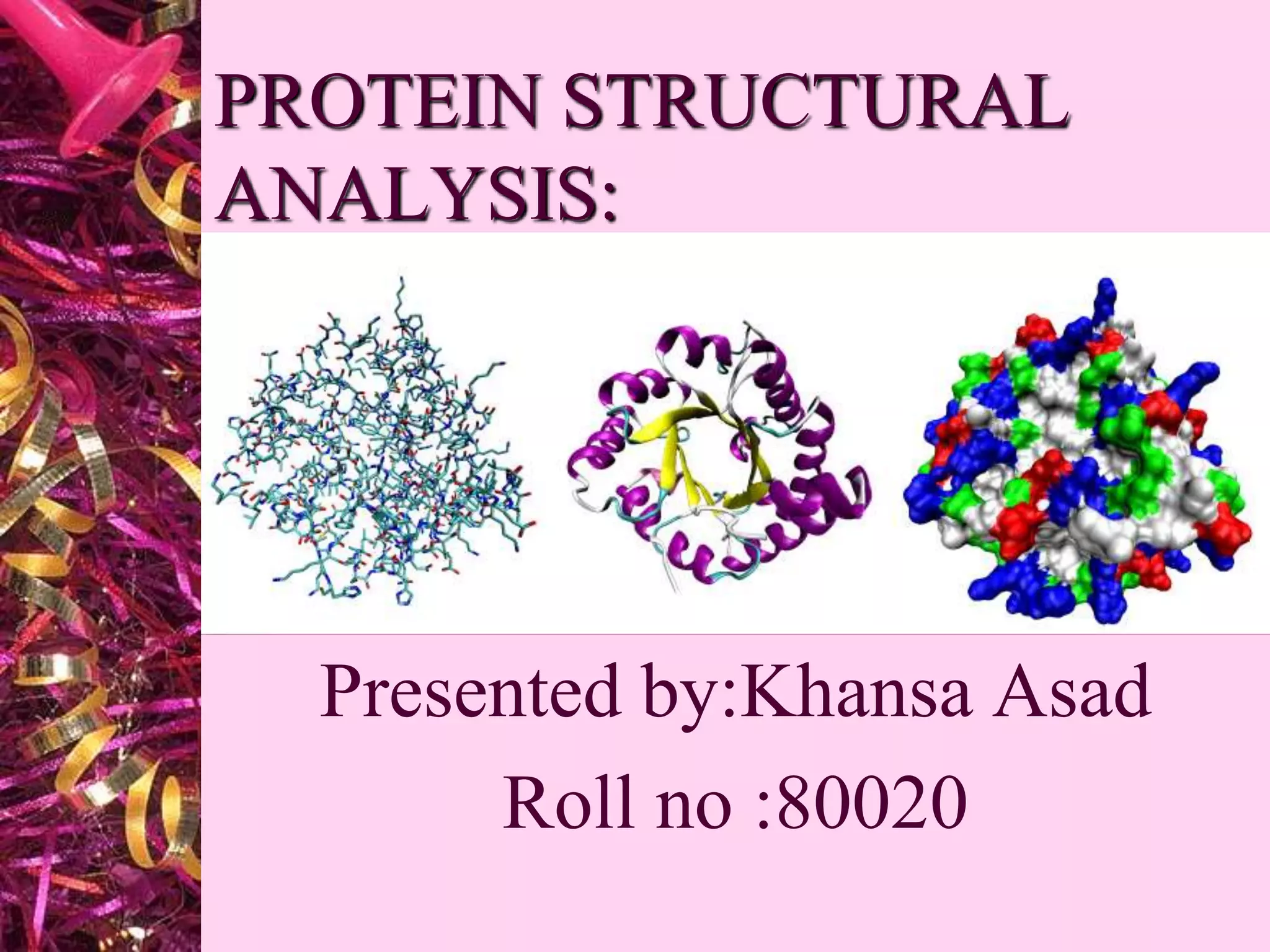
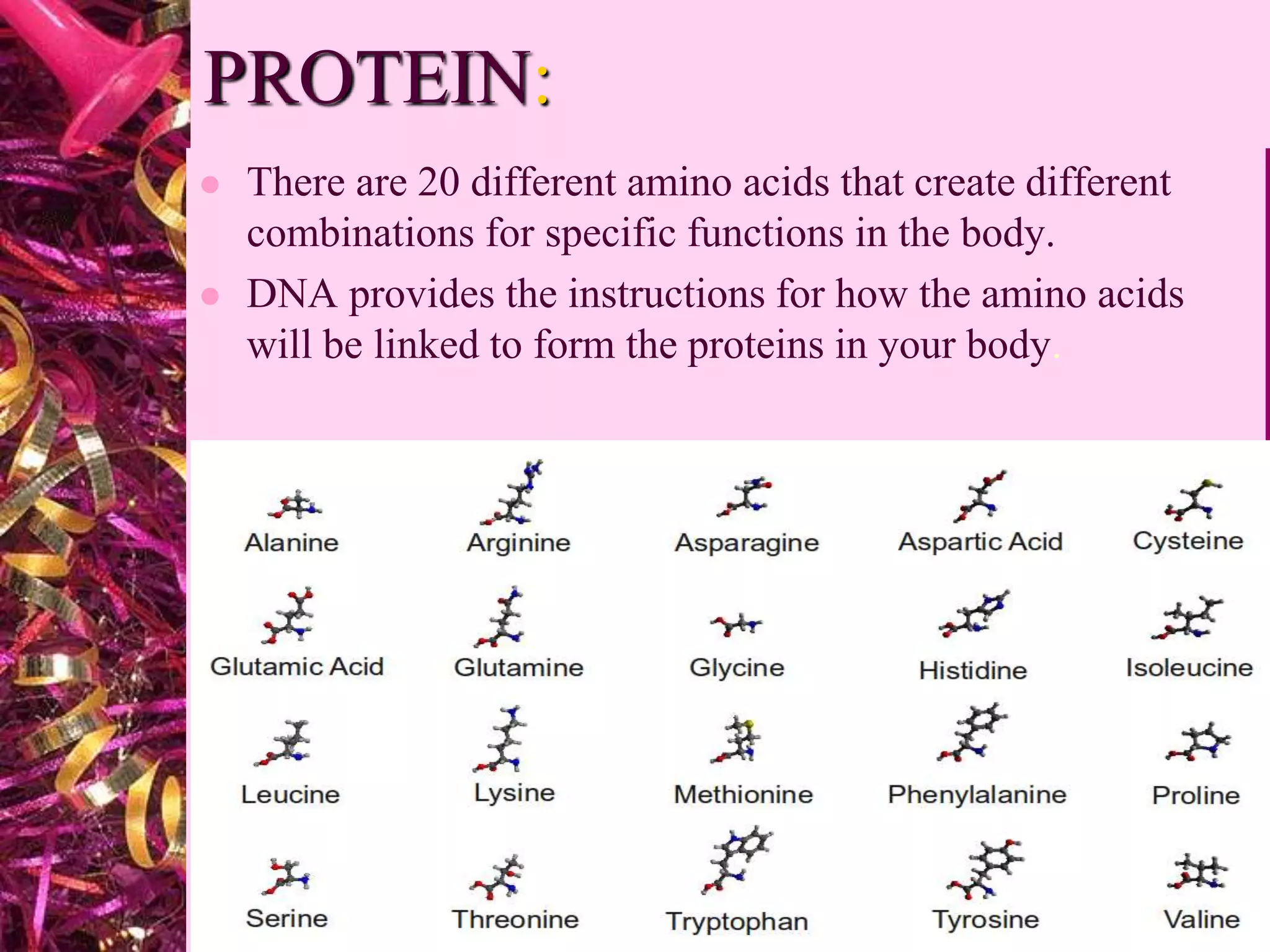
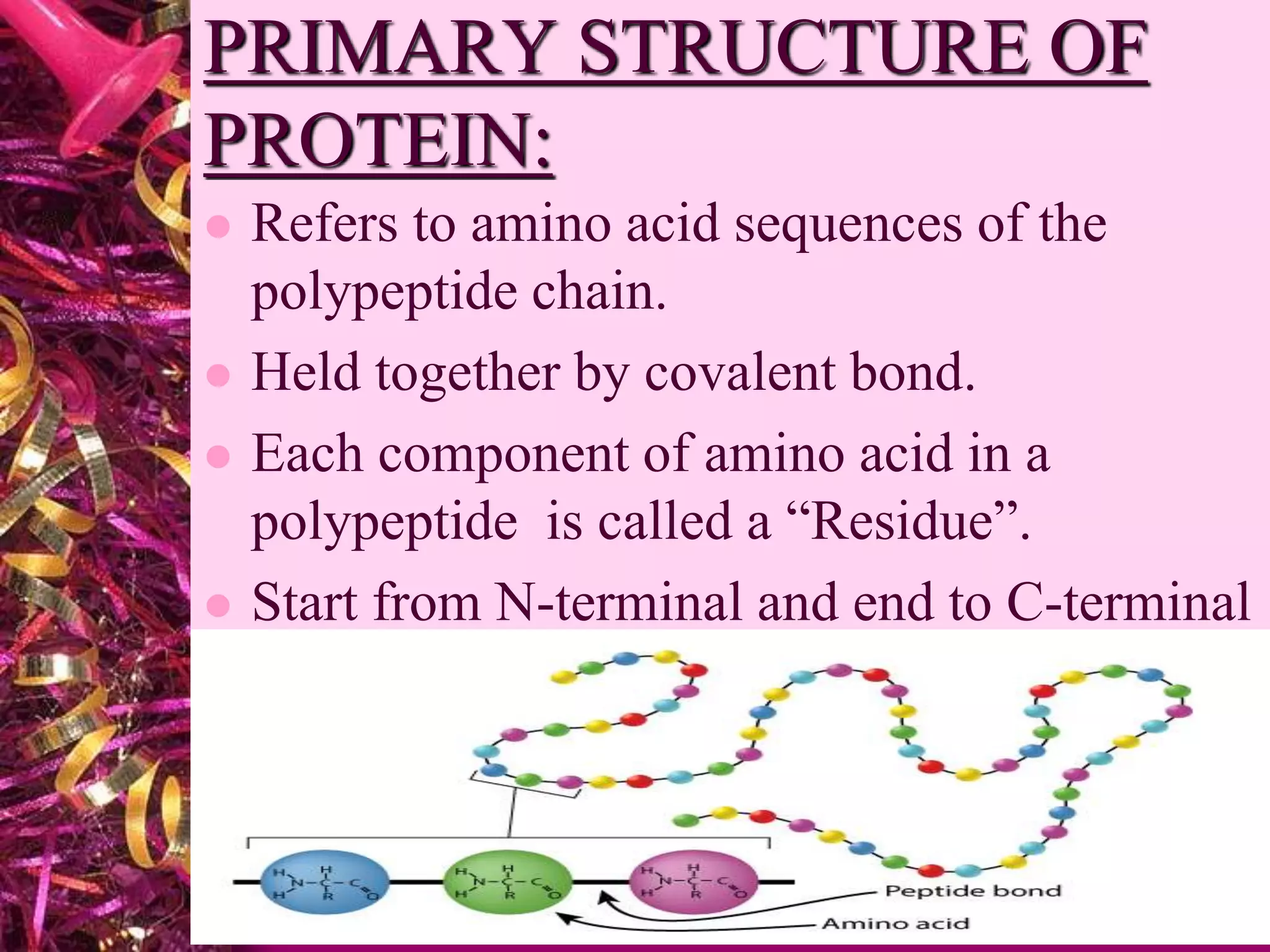
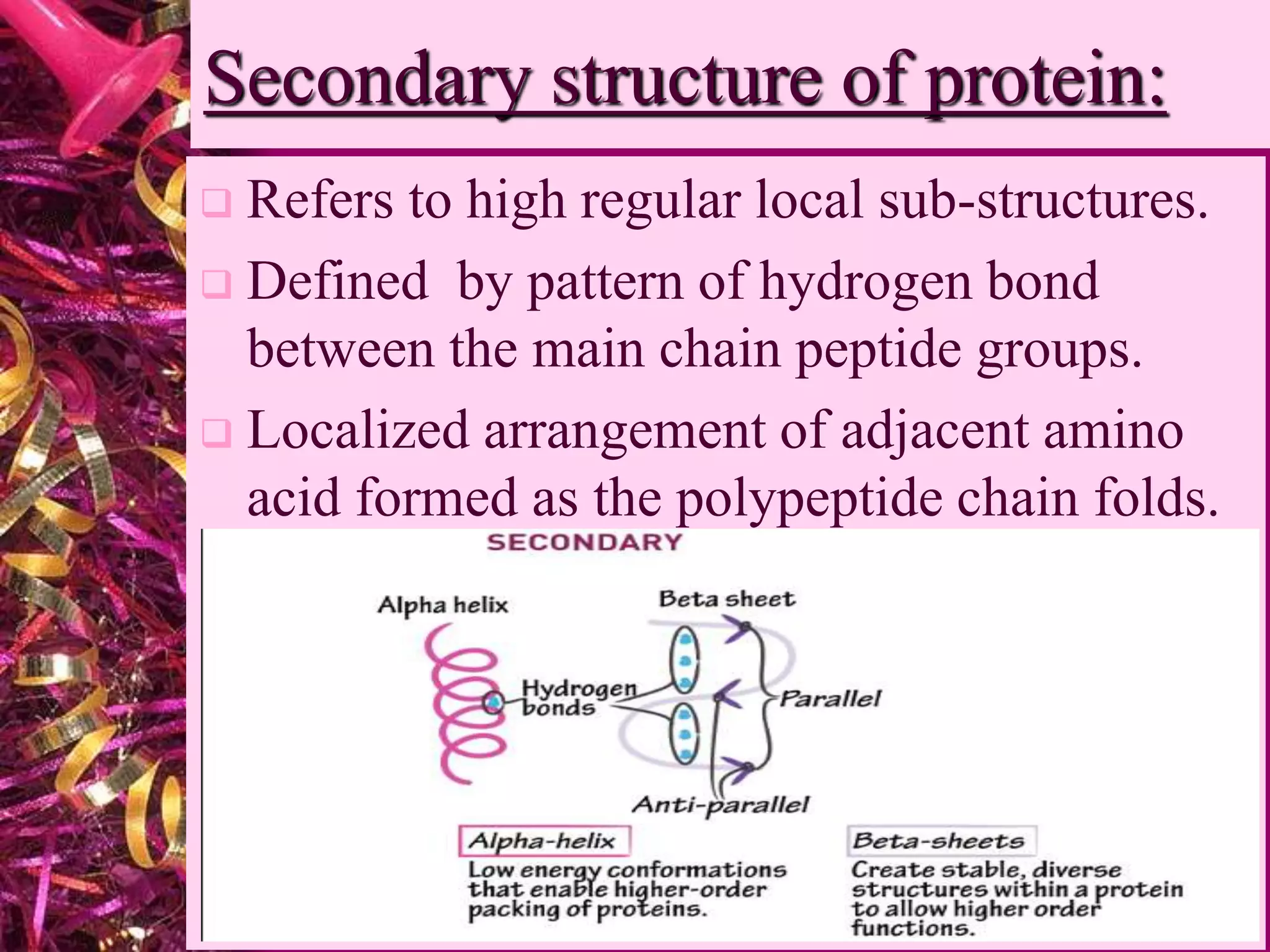
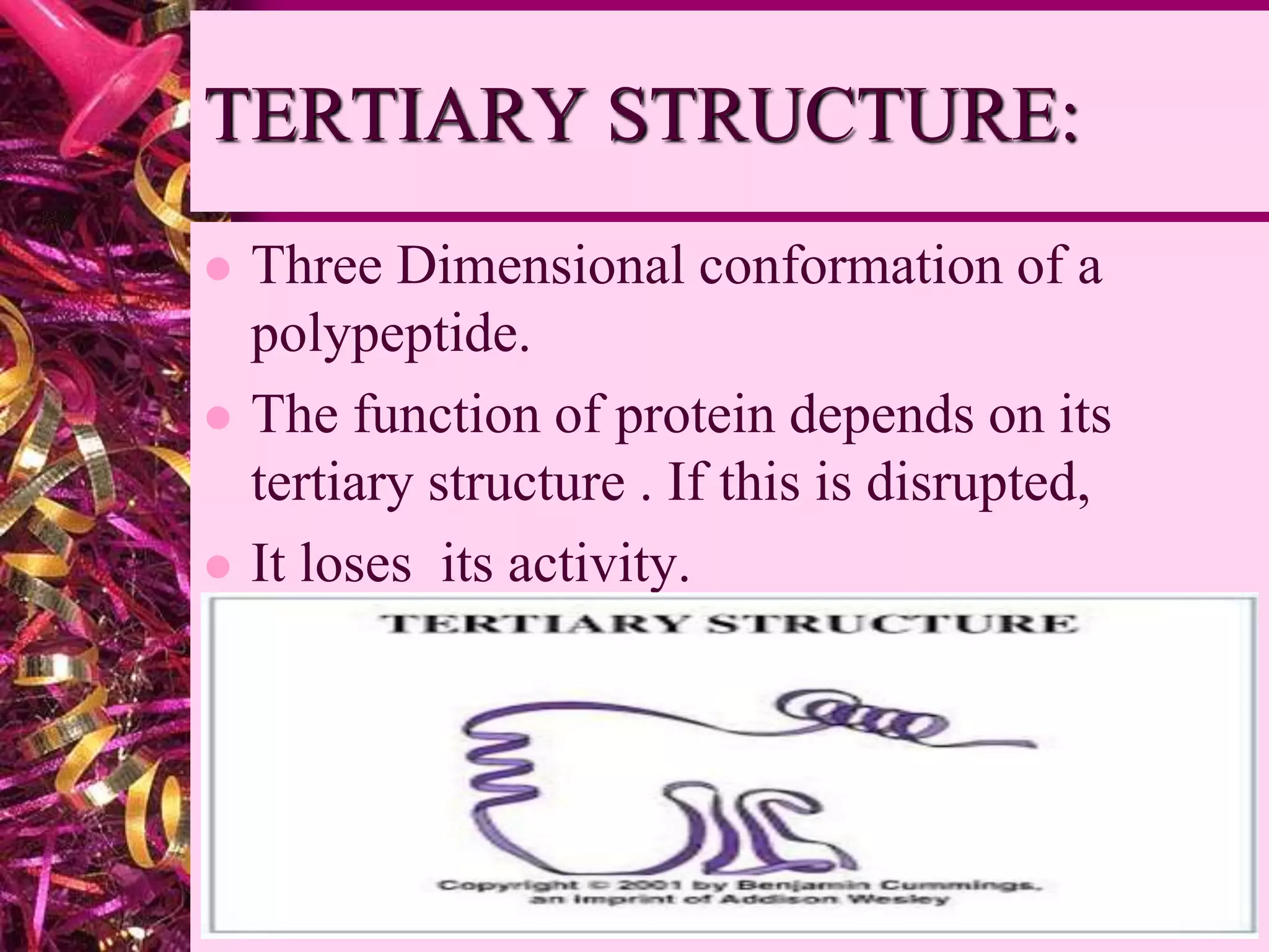
PROTEIN STRUCTURAL
ANALYSIS:
Presented by:Khansa Asad
Roll no :80020
PROTEIN:
 Proteins are large biomolecules, or
macromolecules, consisting of one or more long
chains of amino acid residues. Differs from
carbohydrates and fats because of the
presence of nitrogen.
 The body has at least 30,000 types of
protein, each with a different job.
 The building blocks of all protein
molecules are amino acids
PROTEIN:
 There are 20 different amino acids that create different
combinations for specific functions in the body.
 DNA provides the instructions for how the amino acids
will be linked to form the proteins in your body.
LEVEL OF PROTEIN
STRUCTURE:
1-primary structure 2-secondary structure
3-Tertiary structure 4-Quaternary structure
PRIMARY STRUCTURE OF
PROTEIN:
 Refers to amino acid sequences of the
polypeptide chain.
 Held together by covalent bond.
 Each component of amino acid in a
polypeptide is called a “Residue”.
 Start from N-terminal and end to C-terminal
Secondary structure of protein:
 Refers to high regular local sub-structures.
 Defined by pattern of hydrogen bond
between the main chain peptide groups.
 Localized arrangement of adjacent amino
acid formed as the polypeptide chain folds.
Alpha Helix And Beeta Sheet:
Alpha helix:
 H-Bond from within a
strand.
 Smaller in size.
 3.6 residues per turn.
 Road like structure.
 Helical coiled.
Beeta sheets:
 H-Bond from between two
or more strand.
 Larger in size.
 More than 3.6 residues per
turn.
 Sheet like structure.
 Fully extended.
Super secondary structure(Motif)
 Certain groupings of secondary structural
elements are called MOTIF.
TERTIARY STRUCTURE:
 Three Dimensional conformation of a
polypeptide.
 The function of protein depends on its
tertiary structure . If this is disrupted,
 It loses its activity.
DOMAINS:
 Polypeptide chains containing more than
200 residues usually fold into two or more
globular clusters known as Domains.
 Interaction
Stabilizing:
 Bonding interaction
Between the side chain of amino acid.
• Hydrogen Bond.
• Ionic bond.
• Disulphide bridges.
QUATERNARY STRUCTURE:
 Arrangement of polypeptide sub unit is
called Quaternary structure.
 sub-units are held together by
covalent interaction.
 Water soluble.
 Compact in shape and spherical.
LEVEL OF ORGANZITION:
PDB(Protein Data
Bank)Homepage:
PDB Structure summary:
Structural analysis of protein by
PDB:
Protein structural analysis

Protein structural analysis

  • 1.
  • 2.
    PROTEIN:  Proteins arelarge biomolecules, or macromolecules, consisting of one or more long chains of amino acid residues. Differs from carbohydrates and fats because of the presence of nitrogen.  The body has at least 30,000 types of protein, each with a different job.  The building blocks of all protein molecules are amino acids
  • 3.
    PROTEIN:  There are20 different amino acids that create different combinations for specific functions in the body.  DNA provides the instructions for how the amino acids will be linked to form the proteins in your body.
  • 4.
    LEVEL OF PROTEIN STRUCTURE: 1-primarystructure 2-secondary structure 3-Tertiary structure 4-Quaternary structure
  • 5.
    PRIMARY STRUCTURE OF PROTEIN: Refers to amino acid sequences of the polypeptide chain.  Held together by covalent bond.  Each component of amino acid in a polypeptide is called a “Residue”.  Start from N-terminal and end to C-terminal
  • 6.
    Secondary structure ofprotein:  Refers to high regular local sub-structures.  Defined by pattern of hydrogen bond between the main chain peptide groups.  Localized arrangement of adjacent amino acid formed as the polypeptide chain folds.
  • 7.
    Alpha Helix AndBeeta Sheet: Alpha helix:  H-Bond from within a strand.  Smaller in size.  3.6 residues per turn.  Road like structure.  Helical coiled. Beeta sheets:  H-Bond from between two or more strand.  Larger in size.  More than 3.6 residues per turn.  Sheet like structure.  Fully extended.
  • 8.
    Super secondary structure(Motif) Certain groupings of secondary structural elements are called MOTIF.
  • 9.
    TERTIARY STRUCTURE:  ThreeDimensional conformation of a polypeptide.  The function of protein depends on its tertiary structure . If this is disrupted,  It loses its activity.
  • 10.
    DOMAINS:  Polypeptide chainscontaining more than 200 residues usually fold into two or more globular clusters known as Domains.  Interaction Stabilizing:  Bonding interaction Between the side chain of amino acid. • Hydrogen Bond. • Ionic bond. • Disulphide bridges.
  • 11.
    QUATERNARY STRUCTURE:  Arrangementof polypeptide sub unit is called Quaternary structure.  sub-units are held together by covalent interaction.  Water soluble.  Compact in shape and spherical.
  • 12.
  • 13.
  • 15.
  • 16.
    Structural analysis ofprotein by PDB: