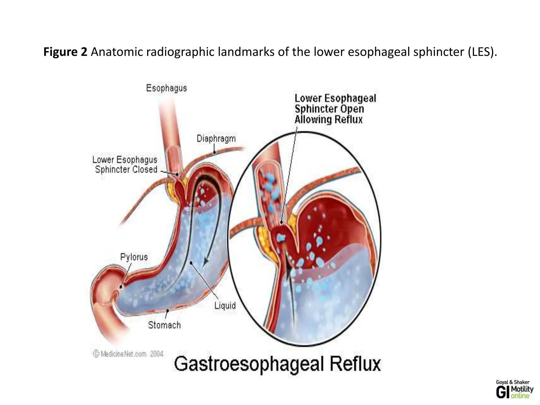
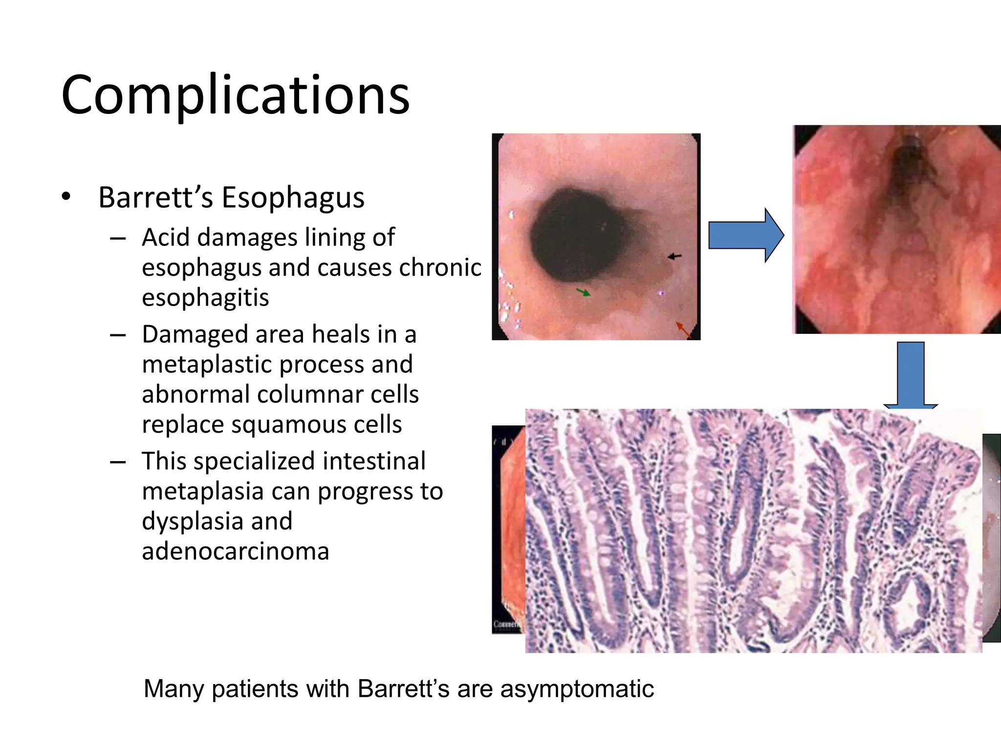
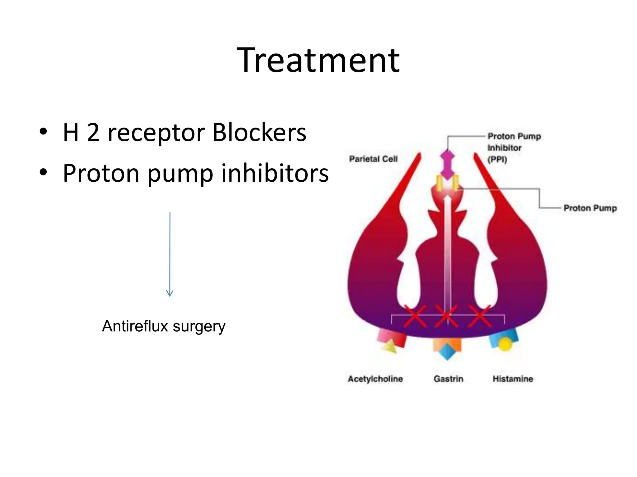
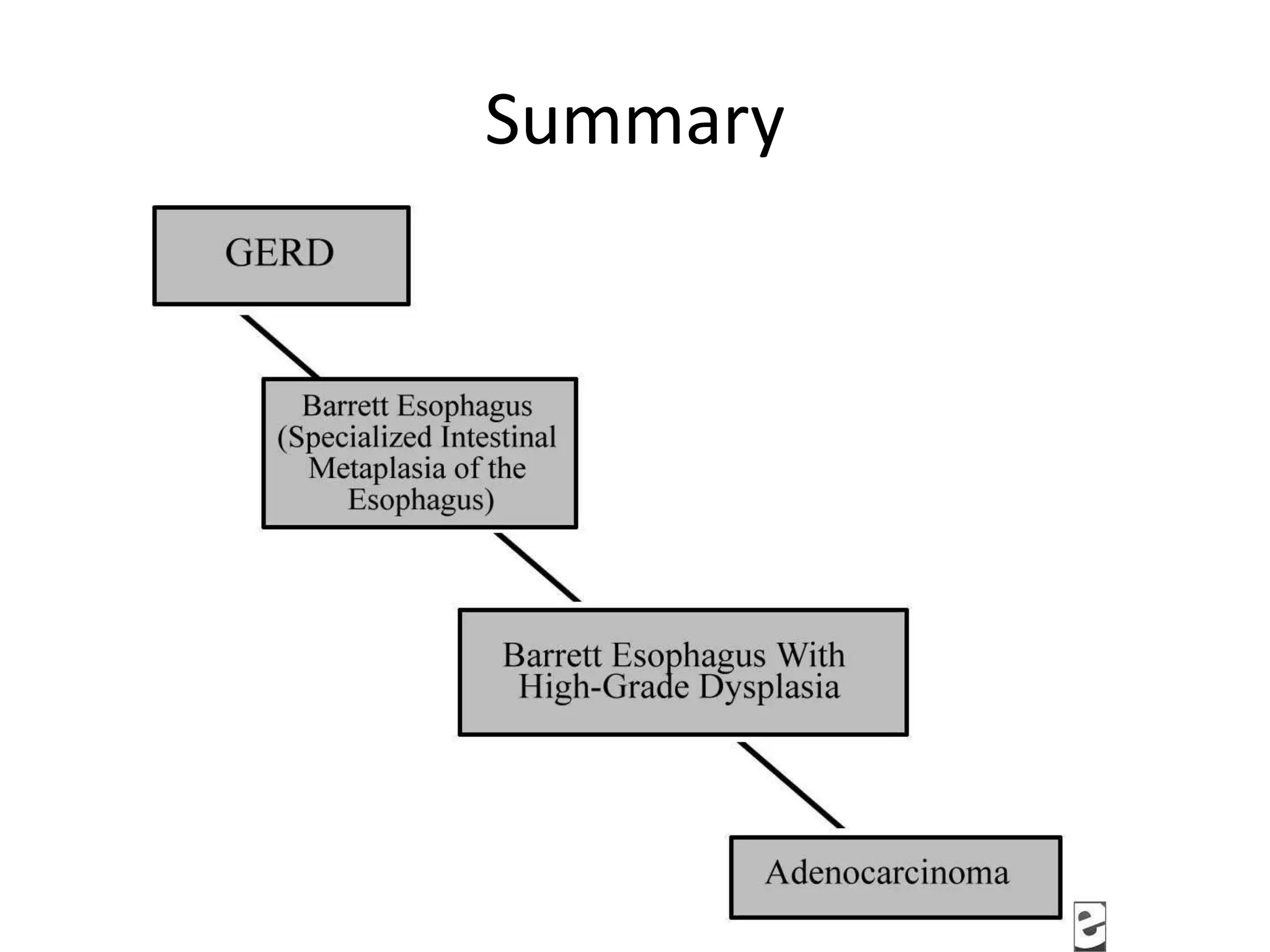
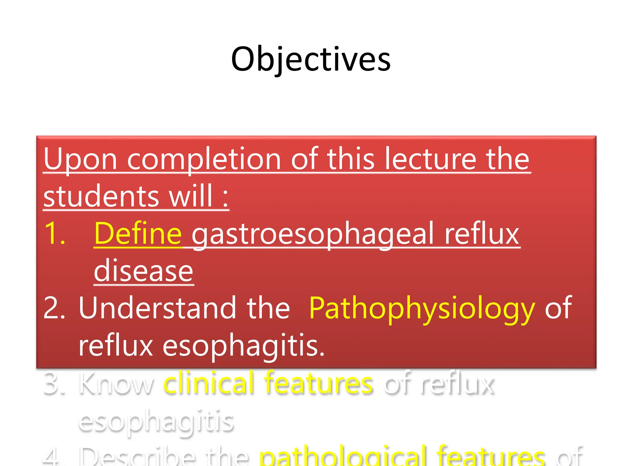
8 LECTURES
Gastro-esophageal reflux disease
Peptic Ulcer Disease
Inflammatory bowel disease-1
Malabsorption
Diarrhea
Colonic polyps and carcinoma-1
Inflammatory bowel disease-2
Colonic polyps and carcinoma-2
8 LECTURES
Gastro-esophageal reflux disease
Peptic Ulcer Disease
Revision
Next
lecture
Objectives
Upon completion of this lecture the
students will :
1. Define gastroesophageal reflux
disease
2. Understand the Pathophysiology of
reflux esophagitis.
3. Know clinical features of reflux
esophagitis
Figure 2 Anatomic radiographic landmarks of the lower esophageal sphincter (LES).
Gastroesophageal Reflux Disease (GERD)
• Gastroesophageal reflux is a normal
physiologic phenomenon experienced
intermittently by most people, particularly
after a meal.
• Gastroesophageal reflux disease (GERD)
occurs when the amount of gastric juice that
refluxes into the esophagus exceeds the
normal limit, causing symptoms with or
without associated esophageal mucosal injury.
Definition
• American College of
Gastroenterology (ACG)
– Symptoms OR mucosal damage
produced by the abnormal reflux
of gastric contents into the
esophagus
– Often chronic and relapsing
– May see complications of GERD in
patients who lack typical
symptoms
Physiologic vs Pathologic
• Physiologic GERD
– Postprandial
– Short lived
– Asymptomatic
– No nocturnal symptomes
• Pathologic GERD
– Symptoms
– Mucosal injury
– Nocturnal symptomes
Epidemiology
• About 44% of the US adult population have
heartburn at least once a month
• 14% of Americans have symptoms weekly
• 7% have symptoms daily
GERD
Pathophysiology
• Abnormal lower esophageal sphincter
• or
• Increase abdominal pressure
GERD
Pathophysiology
A. Abnormal lower esophageal sphincter
1. Functional (frequent transient LES relaxation)
2. Mechanical (hypotensive LES)
3. Foods (eg, coffee, alcohol),
4. Medications (eg, calcium channel blockers),
5. Location .......... hiatal hernia
• or
B. Increase abdominal pressure
The most
common cause
of (GERD).
decrease the
pressure of the
LES.
obesity
Pregnancy
increased gastric volume
Pathophysiology
• Primary barrier to
gastroesophageal reflux is
the lower esophageal
sphincter
• LES normally works in
conjunction with the
diaphragm
• If barrier disrupted, acid
goes from stomach to
esophagus
Clinical Manisfestations
• Most common symptoms
– Heartburn—retrosternal burning discomfort
– Regurgitation—effortless return of gastric
contents into the pharynx without nausea,
retching, or abdominal contractions
Atypical symptoms….coughing, chest pain,
and wheezing.
Diagnostic Evaluation
– If classic symptoms of heartburn and regurgitation
exist in the absence of “alarm symptoms” the
diagnosis of GERD can be made clinically and
treatment can be initiated
Esophagogastrodudenoscopy
• Endoscopy (with biopsy if needed)
– In patients with alarm signs/symptoms
– Those who fail a medication trial
– Those who require long-term tx
pH
• 24-hour pH monitoring
– Accepted standard for establishing or excluding
presence of GERD for those patients who do not
have mucosal changes
– Trans-nasal catheter or a wireless capsule shaped
device
Complications
• Erosive esophagitis
• Stricture
• Barrett’s esophagus
Complications
• Erosive esophagitis
– Responsible for 40-60% of GERD symptoms
– Severity of symptoms often fail to match severity
of erosive esophagitis
Esophagitis
basal zone hyperplasia, Elongation of lamina propria papillae
Eosinophils and neutrophils
Complications
• Esophageal stricture
– Result of healing of
erosive esophagitis
– May need dilation
Complications
• Barrett’s Esophagus
– Intestinal metaplasia of
the esophagus
– Associated with the
development of
adenocarcinoma
8-15%
Complications
• Barrett’s Esophagus
– Acid damages lining of
esophagus and causes chronic
esophagitis
– Damaged area heals in a
metaplastic process and
abnormal columnar cells
replace squamous cells
– This specialized intestinal
metaplasia can progress to
dysplasia and
adenocarcinoma
Many patients with Barrett’s are asymptomatic
Treatment
• H 2 receptor Blockers
• Proton pump inhibitors
Antireflux surgery
Summary
Objectives
Upon completion of this lecture the
students will :
1. Define gastroesophageal reflux
disease
2. Understand the Pathophysiology of
reflux esophagitis.
3. Know clinical features of reflux
esophagitis
Gastro-esophageal reflux disease
Peptic Ulcer Disease
Upon completion of this lecture the students will :
1. Understand the Pathophysiology of acute and
chronic peptic ulcer
2. Know the possible causes of gastric and
duodenal ulcers with emphasis on most
common causes (H pylori and drugs)
3. Recognize the gross and microscopic features
of peptic ulcer
4. Recognize the clinical features and
consequences of acute and chronic peptic ulcer
Objectives
Ulcer
1. Peptic ulcer
2. Stress ulcers (acute gastric ulcers)
Ulcer: a breach in the mucosa of
the alimentary tract extending
through muscularis mucosa into
submucosa or deeper.
Pathophysiology
Aggressive
Factors
Defensive
Factors
Aggressive Factors
H. pylori
Drugs (NSAIDs)
Acid
pepsin
Bile salts
Defensive Factors
Mucus
bicarbonate
Blood flow,
cell renewal
Prostaglandins
Phospholipid
Pathophysiology
imbalance
Aggressive
Factors
Defensive
Factors
Aggressive Factors
H. pylori
Drugs (NSAIDs)
Acid
pepsin
Bile salts
Defensive Factors
Mucus
bicarbonate
Blood flow,
cell renewal
Prostaglandins
Phospholipid
Pathophysiology
imbalance
Aggressive
Factors
Defensive
Factors
Acute peptic ulcers
Pathophysiology
• As part of an acute gastritis
• As a complication of a severe stress response
• As a result of extreme hyperacidity.
Acute peptic ulcers
• As part of an acute gastritis (acute response to an
irritant 'chemical' injury by drugs or alcohol
• As a complication of a severe stress response
(severe burns (Curling's ulcer), major trauma or
cerebrovascular accidents (cushing ulcer )
• As a result of extreme hyperacidity (Zollinger-
Ellison syndrome).
Pathophysiology
Chronic peptic ulcer
Peptic Ulcer Disease
Locations
• May occur in any portion of the GI tract exposed to
acidic gastric juices
• 98% located in first portion of duodenum or
stomach, ratio = 4:1
• Esophagus……. as a result of GERD or acid secretion by
ectopic gastric mucosa.
• Gastric mucosa within a Meckel diverticulum can result in
peptic ulceration of adjacent mucosa.
Peptic Ulcer Disease
Gastric ulcers
 The mucosal defences against acid attack consist of:
1. Mucus-bicarbonate barrier
2. The surface epithelium.
Pathophysiology
Peptic Ulcer Disease
Gastric ulcers
 The mucosal defences against acid attack consist of:
1. Mucus-bicarbonate barrier
2. The surface epithelium.
Duodeno-gastric reflux ( bile )
1. NSAIDs (blocking the synthesis of the prostaglandins)
2. H. pylori infection, ( cytotoxins and ammonia)
Thus peptic ulcers in the stomach, breakdown of mucosal defence is much
more important than excessive acid production.
Pathophysiology
Peptic Ulcer Disease
Duodenal ulcers
Increased production of acid assumes more importance in the pathogenesis of
duodenal ulceration
H. pylori-infected individuals secrete 2-6 times as much acid as non-infected
controls
Helicobacter Pylori does not colonise normal duodenal epithelium
Helicobacter is involved in duodenal ulceration because there is gastric
metaplasia in response to excess acid. Gastric metaplasia paves the way for
colonisation by Helicobacter
Pathophysiology
Increased production of acid Helicobacter P
+
= Duodenal ulcers
Peptic Ulcer Disease
Pathophysiology
Gastric
ulcers
Duodenal
ulcers
H. pylori
Duodeno-gastric reflux ( bile )
NSAIDs
Hyperacidity
H pylori infection of the pyloric antrum is present in nearly all patients with chronic
duodenal ulcer and approximately 75% of patients with chronic gastric ulcer.
Although more than 70% of individuals with PUD are infected by H. pylori, fewer than
20% of H. pylori–infected individuals develop peptic ulcer.
Morphology
• Gross
• usually less than 20 mm in diameter but
they may > 100 mm in diameter.
• The classic peptic ulcer is a round to oval,
sharply punched-out defect
• In contrast, heaped-up margins are more
characteristic of cancers
• Microscopy
• the base consists of necrotic tissue and
polymorph exudate overlying inflamed
granulation tissue which merges with
mature fibrous (scar) tissue.
Clinical features
• Epigastric pain (the most common symptom)
– Gnawing or burning sensation
– Occurs 2-3 hours after meals
– Relieved by food or antacids
– Patient awakens with pain at night.
Some present with complications such as iron deficiency
anemia, frank hemorrhage, or perforation.
Therapy
Current therapies for PUD are aimed at
I. H. pylori eradication
II. Acid suppression
a) Proton pump inhibitors
b) H2 blockers
Upon completion of this lecture the students will :
1. Understand the Pathophysiology of acute and
chronic peptic ulcer
2. Know the possible causes of gastric and
duodenal ulcers with emphasis on most
common causes (H pylori and drugs)
3. Recognize the gross and microscopic features
of peptic ulcer
4. Recognize the clinical features and
consequences of acute and chronic peptic ulcer
Objectives

Lecture note on GERD and peptic ulcer.ppt

  • 1.
    8 LECTURES Gastro-esophageal refluxdisease Peptic Ulcer Disease Inflammatory bowel disease-1 Malabsorption Diarrhea Colonic polyps and carcinoma-1 Inflammatory bowel disease-2 Colonic polyps and carcinoma-2
  • 2.
    8 LECTURES Gastro-esophageal refluxdisease Peptic Ulcer Disease Revision Next lecture
  • 3.
    Objectives Upon completion ofthis lecture the students will : 1. Define gastroesophageal reflux disease 2. Understand the Pathophysiology of reflux esophagitis. 3. Know clinical features of reflux esophagitis
  • 4.
    Figure 2 Anatomicradiographic landmarks of the lower esophageal sphincter (LES).
  • 5.
    Gastroesophageal Reflux Disease(GERD) • Gastroesophageal reflux is a normal physiologic phenomenon experienced intermittently by most people, particularly after a meal. • Gastroesophageal reflux disease (GERD) occurs when the amount of gastric juice that refluxes into the esophagus exceeds the normal limit, causing symptoms with or without associated esophageal mucosal injury.
  • 6.
    Definition • American Collegeof Gastroenterology (ACG) – Symptoms OR mucosal damage produced by the abnormal reflux of gastric contents into the esophagus – Often chronic and relapsing – May see complications of GERD in patients who lack typical symptoms
  • 7.
    Physiologic vs Pathologic •Physiologic GERD – Postprandial – Short lived – Asymptomatic – No nocturnal symptomes • Pathologic GERD – Symptoms – Mucosal injury – Nocturnal symptomes
  • 8.
    Epidemiology • About 44%of the US adult population have heartburn at least once a month • 14% of Americans have symptoms weekly • 7% have symptoms daily
  • 9.
    GERD Pathophysiology • Abnormal loweresophageal sphincter • or • Increase abdominal pressure
  • 10.
    GERD Pathophysiology A. Abnormal loweresophageal sphincter 1. Functional (frequent transient LES relaxation) 2. Mechanical (hypotensive LES) 3. Foods (eg, coffee, alcohol), 4. Medications (eg, calcium channel blockers), 5. Location .......... hiatal hernia • or B. Increase abdominal pressure The most common cause of (GERD). decrease the pressure of the LES. obesity Pregnancy increased gastric volume
  • 11.
    Pathophysiology • Primary barrierto gastroesophageal reflux is the lower esophageal sphincter • LES normally works in conjunction with the diaphragm • If barrier disrupted, acid goes from stomach to esophagus
  • 12.
    Clinical Manisfestations • Mostcommon symptoms – Heartburn—retrosternal burning discomfort – Regurgitation—effortless return of gastric contents into the pharynx without nausea, retching, or abdominal contractions Atypical symptoms….coughing, chest pain, and wheezing.
  • 13.
    Diagnostic Evaluation – Ifclassic symptoms of heartburn and regurgitation exist in the absence of “alarm symptoms” the diagnosis of GERD can be made clinically and treatment can be initiated
  • 14.
    Esophagogastrodudenoscopy • Endoscopy (withbiopsy if needed) – In patients with alarm signs/symptoms – Those who fail a medication trial – Those who require long-term tx
  • 15.
    pH • 24-hour pHmonitoring – Accepted standard for establishing or excluding presence of GERD for those patients who do not have mucosal changes – Trans-nasal catheter or a wireless capsule shaped device
  • 16.
    Complications • Erosive esophagitis •Stricture • Barrett’s esophagus
  • 17.
    Complications • Erosive esophagitis –Responsible for 40-60% of GERD symptoms – Severity of symptoms often fail to match severity of erosive esophagitis
  • 18.
    Esophagitis basal zone hyperplasia,Elongation of lamina propria papillae Eosinophils and neutrophils
  • 19.
    Complications • Esophageal stricture –Result of healing of erosive esophagitis – May need dilation
  • 20.
    Complications • Barrett’s Esophagus –Intestinal metaplasia of the esophagus – Associated with the development of adenocarcinoma 8-15%
  • 21.
    Complications • Barrett’s Esophagus –Acid damages lining of esophagus and causes chronic esophagitis – Damaged area heals in a metaplastic process and abnormal columnar cells replace squamous cells – This specialized intestinal metaplasia can progress to dysplasia and adenocarcinoma Many patients with Barrett’s are asymptomatic
  • 22.
    Treatment • H 2receptor Blockers • Proton pump inhibitors Antireflux surgery
  • 23.
  • 24.
    Objectives Upon completion ofthis lecture the students will : 1. Define gastroesophageal reflux disease 2. Understand the Pathophysiology of reflux esophagitis. 3. Know clinical features of reflux esophagitis
  • 25.
  • 26.
    Upon completion ofthis lecture the students will : 1. Understand the Pathophysiology of acute and chronic peptic ulcer 2. Know the possible causes of gastric and duodenal ulcers with emphasis on most common causes (H pylori and drugs) 3. Recognize the gross and microscopic features of peptic ulcer 4. Recognize the clinical features and consequences of acute and chronic peptic ulcer Objectives
  • 27.
    Ulcer 1. Peptic ulcer 2.Stress ulcers (acute gastric ulcers) Ulcer: a breach in the mucosa of the alimentary tract extending through muscularis mucosa into submucosa or deeper.
  • 28.
  • 29.
    Aggressive Factors H. pylori Drugs(NSAIDs) Acid pepsin Bile salts Defensive Factors Mucus bicarbonate Blood flow, cell renewal Prostaglandins Phospholipid Pathophysiology imbalance Aggressive Factors Defensive Factors
  • 30.
    Aggressive Factors H. pylori Drugs(NSAIDs) Acid pepsin Bile salts Defensive Factors Mucus bicarbonate Blood flow, cell renewal Prostaglandins Phospholipid Pathophysiology imbalance Aggressive Factors Defensive Factors
  • 31.
    Acute peptic ulcers Pathophysiology •As part of an acute gastritis • As a complication of a severe stress response • As a result of extreme hyperacidity.
  • 32.
    Acute peptic ulcers •As part of an acute gastritis (acute response to an irritant 'chemical' injury by drugs or alcohol • As a complication of a severe stress response (severe burns (Curling's ulcer), major trauma or cerebrovascular accidents (cushing ulcer ) • As a result of extreme hyperacidity (Zollinger- Ellison syndrome). Pathophysiology
  • 33.
    Chronic peptic ulcer PepticUlcer Disease Locations • May occur in any portion of the GI tract exposed to acidic gastric juices • 98% located in first portion of duodenum or stomach, ratio = 4:1 • Esophagus……. as a result of GERD or acid secretion by ectopic gastric mucosa. • Gastric mucosa within a Meckel diverticulum can result in peptic ulceration of adjacent mucosa.
  • 34.
    Peptic Ulcer Disease Gastriculcers  The mucosal defences against acid attack consist of: 1. Mucus-bicarbonate barrier 2. The surface epithelium. Pathophysiology
  • 35.
    Peptic Ulcer Disease Gastriculcers  The mucosal defences against acid attack consist of: 1. Mucus-bicarbonate barrier 2. The surface epithelium. Duodeno-gastric reflux ( bile ) 1. NSAIDs (blocking the synthesis of the prostaglandins) 2. H. pylori infection, ( cytotoxins and ammonia) Thus peptic ulcers in the stomach, breakdown of mucosal defence is much more important than excessive acid production. Pathophysiology
  • 36.
    Peptic Ulcer Disease Duodenalulcers Increased production of acid assumes more importance in the pathogenesis of duodenal ulceration H. pylori-infected individuals secrete 2-6 times as much acid as non-infected controls Helicobacter Pylori does not colonise normal duodenal epithelium Helicobacter is involved in duodenal ulceration because there is gastric metaplasia in response to excess acid. Gastric metaplasia paves the way for colonisation by Helicobacter Pathophysiology Increased production of acid Helicobacter P + = Duodenal ulcers
  • 37.
    Peptic Ulcer Disease Pathophysiology Gastric ulcers Duodenal ulcers H.pylori Duodeno-gastric reflux ( bile ) NSAIDs Hyperacidity H pylori infection of the pyloric antrum is present in nearly all patients with chronic duodenal ulcer and approximately 75% of patients with chronic gastric ulcer. Although more than 70% of individuals with PUD are infected by H. pylori, fewer than 20% of H. pylori–infected individuals develop peptic ulcer.
  • 38.
    Morphology • Gross • usuallyless than 20 mm in diameter but they may > 100 mm in diameter. • The classic peptic ulcer is a round to oval, sharply punched-out defect • In contrast, heaped-up margins are more characteristic of cancers • Microscopy • the base consists of necrotic tissue and polymorph exudate overlying inflamed granulation tissue which merges with mature fibrous (scar) tissue.
  • 39.
    Clinical features • Epigastricpain (the most common symptom) – Gnawing or burning sensation – Occurs 2-3 hours after meals – Relieved by food or antacids – Patient awakens with pain at night. Some present with complications such as iron deficiency anemia, frank hemorrhage, or perforation.
  • 40.
    Therapy Current therapies forPUD are aimed at I. H. pylori eradication II. Acid suppression a) Proton pump inhibitors b) H2 blockers
  • 41.
    Upon completion ofthis lecture the students will : 1. Understand the Pathophysiology of acute and chronic peptic ulcer 2. Know the possible causes of gastric and duodenal ulcers with emphasis on most common causes (H pylori and drugs) 3. Recognize the gross and microscopic features of peptic ulcer 4. Recognize the clinical features and consequences of acute and chronic peptic ulcer Objectives