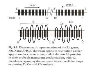
This presentation aims to help medicine undergraduates and post graduates in the department of Pathology and Department of transfusion medicine for better understanding of various blood grouping systems, sub groups, RBC antigens and corresponding antibodies. It also covers the practical aspect of blood grouping and cross matching.
Kell blood group system most important blood group system following to ABO and Rh blood group system, particularly RhD as far as immunogenicity is concerned and Its clinical importance.
ABO blood group system was decover by Karal landsteine
which contain A, B, and o antigen on the surface of BC, WBC,s platatelet and other body tissue cells except brain cell, and anti A, antiB and Anti Ab natural occuring antibodies in plasma of B,A, and O blood group individual respectively
Although individual humans (and all diploid organisms) can only have two alleles for a given gene, multiple alleles may exist at the population level.
“Three or more kinds of gene which occupy the same locus are referred to as multiple alleles.”
Rh typing and its technique , BLOOD TYPING , Rhesus (Rh) typing , procedures of rh typing, process of Rh typing, Test limitations, Sources of Error in Rh Antigen Typing, False positive reactions' reason, False negative reactions' reasons
Title: Sense of Smell
Presenter: Dr. Faiza, Assistant Professor of Physiology
Qualifications:
MBBS (Best Graduate, AIMC Lahore)
FCPS Physiology
ICMT, CHPE, DHPE (STMU)
MPH (GC University, Faisalabad)
MBA (Virtual University of Pakistan)
Learning Objectives:
Describe the primary categories of smells and the concept of odor blindness.
Explain the structure and location of the olfactory membrane and mucosa, including the types and roles of cells involved in olfaction.
Describe the pathway and mechanisms of olfactory signal transmission from the olfactory receptors to the brain.
Illustrate the biochemical cascade triggered by odorant binding to olfactory receptors, including the role of G-proteins and second messengers in generating an action potential.
Identify different types of olfactory disorders such as anosmia, hyposmia, hyperosmia, and dysosmia, including their potential causes.
Key Topics:
Olfactory Genes:
3% of the human genome accounts for olfactory genes.
400 genes for odorant receptors.
Olfactory Membrane:
Located in the superior part of the nasal cavity.
Medially: Folds downward along the superior septum.
Laterally: Folds over the superior turbinate and upper surface of the middle turbinate.
Total surface area: 5-10 square centimeters.
Olfactory Mucosa:
Olfactory Cells: Bipolar nerve cells derived from the CNS (100 million), with 4-25 olfactory cilia per cell.
Sustentacular Cells: Produce mucus and maintain ionic and molecular environment.
Basal Cells: Replace worn-out olfactory cells with an average lifespan of 1-2 months.
Bowman’s Gland: Secretes mucus.
Stimulation of Olfactory Cells:
Odorant dissolves in mucus and attaches to receptors on olfactory cilia.
Involves a cascade effect through G-proteins and second messengers, leading to depolarization and action potential generation in the olfactory nerve.
Quality of a Good Odorant:
Small (3-20 Carbon atoms), volatile, water-soluble, and lipid-soluble.
Facilitated by odorant-binding proteins in mucus.
Membrane Potential and Action Potential:
Resting membrane potential: -55mV.
Action potential frequency in the olfactory nerve increases with odorant strength.
Adaptation Towards the Sense of Smell:
Rapid adaptation within the first second, with further slow adaptation.
Psychological adaptation greater than receptor adaptation, involving feedback inhibition from the central nervous system.
Primary Sensations of Smell:
Camphoraceous, Musky, Floral, Pepperminty, Ethereal, Pungent, Putrid.
Odor Detection Threshold:
Examples: Hydrogen sulfide (0.0005 ppm), Methyl-mercaptan (0.002 ppm).
Some toxic substances are odorless at lethal concentrations.
Characteristics of Smell:
Odor blindness for single substances due to lack of appropriate receptor protein.
Behavioral and emotional influences of smell.
Transmission of Olfactory Signals:
From olfactory cells to glomeruli in the olfactory bulb, involving lateral inhibition.
Primitive, less old, and new olfactory systems with different path
MANAGEMENT OF ATRIOVENTRICULAR CONDUCTION BLOCK.pdfJim Jacob Roy
Cardiac conduction defects can occur due to various causes.
Atrioventricular conduction blocks ( AV blocks ) are classified into 3 types.
This document describes the acute management of AV block.
Flu Vaccine Alert in Bangalore Karnatakaaddon Scans
As flu season approaches, health officials in Bangalore, Karnataka, are urging residents to get their flu vaccinations. The seasonal flu, while common, can lead to severe health complications, particularly for vulnerable populations such as young children, the elderly, and those with underlying health conditions.
Dr. Vidisha Kumari, a leading epidemiologist in Bangalore, emphasizes the importance of getting vaccinated. "The flu vaccine is our best defense against the influenza virus. It not only protects individuals but also helps prevent the spread of the virus in our communities," he says.
This year, the flu season is expected to coincide with a potential increase in other respiratory illnesses. The Karnataka Health Department has launched an awareness campaign highlighting the significance of flu vaccinations. They have set up multiple vaccination centers across Bangalore, making it convenient for residents to receive their shots.
To encourage widespread vaccination, the government is also collaborating with local schools, workplaces, and community centers to facilitate vaccination drives. Special attention is being given to ensuring that the vaccine is accessible to all, including marginalized communities who may have limited access to healthcare.
Residents are reminded that the flu vaccine is safe and effective. Common side effects are mild and may include soreness at the injection site, mild fever, or muscle aches. These side effects are generally short-lived and far less severe than the flu itself.
Healthcare providers are also stressing the importance of continuing COVID-19 precautions. Wearing masks, practicing good hand hygiene, and maintaining social distancing are still crucial, especially in crowded places.
Protect yourself and your loved ones by getting vaccinated. Together, we can help keep Bangalore healthy and safe this flu season. For more information on vaccination centers and schedules, residents can visit the Karnataka Health Department’s official website or follow their social media pages.
Stay informed, stay safe, and get your flu shot today!
HOT NEW PRODUCT! BIG SALES FAST SHIPPING NOW FROM CHINA!! EU KU DB BK substit...GL Anaacs
Contact us if you are interested:
Email / Skype : kefaya1771@gmail.com
Threema: PXHY5PDH
New BATCH Ku !!! MUCH IN DEMAND FAST SALE EVERY BATCH HAPPY GOOD EFFECT BIG BATCH !
Contact me on Threema or skype to start big business!!
Hot-sale products:
NEW HOT EUTYLONE WHITE CRYSTAL!!
5cl-adba precursor (semi finished )
5cl-adba raw materials
ADBB precursor (semi finished )
ADBB raw materials
APVP powder
5fadb/4f-adb
Jwh018 / Jwh210
Eutylone crystal
Protonitazene (hydrochloride) CAS: 119276-01-6
Flubrotizolam CAS: 57801-95-3
Metonitazene CAS: 14680-51-4
Payment terms: Western Union,MoneyGram,Bitcoin or USDT.
Deliver Time: Usually 7-15days
Shipping method: FedEx, TNT, DHL,UPS etc.Our deliveries are 100% safe, fast, reliable and discreet.
Samples will be sent for your evaluation!If you are interested in, please contact me, let's talk details.
We specializes in exporting high quality Research chemical, medical intermediate, Pharmaceutical chemicals and so on. Products are exported to USA, Canada, France, Korea, Japan,Russia, Southeast Asia and other countries.
The prostate is an exocrine gland of the male mammalian reproductive system
It is a walnut-sized gland that forms part of the male reproductive system and is located in front of the rectum and just below the urinary bladder
Function is to store and secrete a clear, slightly alkaline fluid that constitutes 10-30% of the volume of the seminal fluid that along with the spermatozoa, constitutes semen
A healthy human prostate measures (4cm-vertical, by 3cm-horizontal, 2cm ant-post ).
It surrounds the urethra just below the urinary bladder. It has anterior, median, posterior and two lateral lobes
It’s work is regulated by androgens which are responsible for male sex characteristics
Generalised disease of the prostate due to hormonal derangement which leads to non malignant enlargement of the gland (increase in the number of epithelial cells and stromal tissue)to cause compression of the urethra leading to symptoms (LUTS
TEST BANK for Operations Management, 14th Edition by William J. Stevenson, Ve...kevinkariuki227
TEST BANK for Operations Management, 14th Edition by William J. Stevenson, Verified Chapters 1 - 19, Complete Newest Version.pdf
Factory Supply Best Quality Pmk Oil CAS 28578–16–7 PMK Powder in Stockrebeccabio
Factory Supply Best Quality Pmk Oil CAS 28578–16–7 PMK Powder in Stock
Telegram: bmksupplier
signal: +85264872720
threema: TUD4A6YC
You can contact me on Telegram or Threema
Communicate promptly and reply
Free of customs clearance, Double Clearance 100% pass delivery to USA, Canada, Spain, Germany, Netherland, Poland, Italy, Sweden, UK, Czech Republic, Australia, Mexico, Russia, Ukraine, Kazakhstan.Door to door service
Hot Selling Organic intermediates
These simplified slides by Dr. Sidra Arshad present an overview of the non-respiratory functions of the respiratory tract.
Learning objectives:
1. Enlist the non-respiratory functions of the respiratory tract
2. Briefly explain how these functions are carried out
3. Discuss the significance of dead space
4. Differentiate between minute ventilation and alveolar ventilation
5. Describe the cough and sneeze reflexes
Study Resources:
1. Chapter 39, Guyton and Hall Textbook of Medical Physiology, 14th edition
2. Chapter 34, Ganong’s Review of Medical Physiology, 26th edition
3. Chapter 17, Human Physiology by Lauralee Sherwood, 9th edition
4. Non-respiratory functions of the lungs https://academic.oup.com/bjaed/article/13/3/98/278874
Anti ulcer drugs and their Advance pharmacology ||
Anti-ulcer drugs are medications used to prevent and treat ulcers in the stomach and upper part of the small intestine (duodenal ulcers). These ulcers are often caused by an imbalance between stomach acid and the mucosal lining, which protects the stomach lining.
||Scope: Overview of various classes of anti-ulcer drugs, their mechanisms of action, indications, side effects, and clinical considerations.
Report Back from SGO 2024: What’s the Latest in Cervical Cancer?bkling
Are you curious about what’s new in cervical cancer research or unsure what the findings mean? Join Dr. Emily Ko, a gynecologic oncologist at Penn Medicine, to learn about the latest updates from the Society of Gynecologic Oncology (SGO) 2024 Annual Meeting on Women’s Cancer. Dr. Ko will discuss what the research presented at the conference means for you and answer your questions about the new developments.
Pulmonary Thromboembolism - etilogy, types, medical- Surgical and nursing man...VarunMahajani
Disruption of blood supply to lung alveoli due to blockage of one or more pulmonary blood vessels is called as Pulmonary thromboembolism. In this presentation we will discuss its causes, types and its management in depth.
Explore natural remedies for syphilis treatment in Singapore. Discover alternative therapies, herbal remedies, and lifestyle changes that may complement conventional treatments. Learn about holistic approaches to managing syphilis symptoms and supporting overall health.
Ethanol (CH3CH2OH), or beverage alcohol, is a two-carbon alcohol
that is rapidly distributed in the body and brain. Ethanol alters many
neurochemical systems and has rewarding and addictive properties. It
is the oldest recreational drug and likely contributes to more morbidity,
mortality, and public health costs than all illicit drugs combined. The
5th edition of the Diagnostic and Statistical Manual of Mental Disorders
(DSM-5) integrates alcohol abuse and alcohol dependence into a single
disorder called alcohol use disorder (AUD), with mild, moderate,
and severe subclassifications (American Psychiatric Association, 2013).
In the DSM-5, all types of substance abuse and dependence have been
combined into a single substance use disorder (SUD) on a continuum
from mild to severe. A diagnosis of AUD requires that at least two of
the 11 DSM-5 behaviors be present within a 12-month period (mild
AUD: 2–3 criteria; moderate AUD: 4–5 criteria; severe AUD: 6–11 criteria).
The four main behavioral effects of AUD are impaired control over
drinking, negative social consequences, risky use, and altered physiological
effects (tolerance, withdrawal). This chapter presents an overview
of the prevalence and harmful consequences of AUD in the U.S.,
the systemic nature of the disease, neurocircuitry and stages of AUD,
comorbidities, fetal alcohol spectrum disorders, genetic risk factors, and
pharmacotherapies for AUD.
1. The Rh Blood
Group System
Course: Adv. Blood Banking
Reference:
Harmening DM (2012). Modern blood banking and transfusion
practices, 7th ed. Philadelphia: F.A. Davis Company. Chap7
2. Introduction
• The term Rh refers to a specific red blood cell
antigen (D) and to a complex blood group
system currently composed of 61 different
antigenic specificities.
• Although Rh antibodies were among the first
to be described, researchers continue to
explore the complexities of the Rh blood
group system from its serology to its mode of
inheritance, genetic control, and the
biochemical structure of the Rh antigens
3. • Rh is the second in importance only to ABO
blood group system in terms of transfusion, as
the Rh system antigens are very immunogenic.
• Unlike ABO antibodies that are routinely
found in individuals who lack the
corresponding antigen, Rh antibodies are
produced only after exposure to foreign red
blood cells.
• Once present, they can produce significant
hemolytic disease of the fetus and newborn
(HDFN) as well as hemolytic transfusion
reactions
4. • The terms Rh-positive and Rh-negative are
used to describe the presence or absence of
the D antigen.
• Rh-positive indicates that an individual’s red
blood cells possess one particular Rh antigen,
the D antigen.
• Rh-negative indicates that the red blood cells
lack the D antigen.
5. History
• In 1939 Levine and Stetson described a hemolytic
transfusion reaction in an obstetrical patient.
• After delivering a stillborn infant, the woman required
transfusions.
• Her husband, who had the same ABO type, was
selected as her donor. After transfusion, the recipient
demonstrated classic symptoms of an acute hemolytic
transfusion reaction (AHTR).
• Subsequently, an antibody was isolated from the
mother’s serum that reacted at both 37oC and 20°C
with the father’s RBCs.
• It was postulated that the fetus and the father
possessed a common factor that the mother lacked
6. • While the mother carried the fetus, she was exposed to
this factor and subsequently produced an antibody
that showed positive reactivity when tested against the
transfused RBCs from the father.
• The antibody specificity was not identified, but it was
found to react with 80% of cells tested and was
suspected to be a maternal antibody to a factor on the
fetal cells.
• A year later Landsteiner and Wiener described an
antibody made by guinea pigs and rabbits when they
were transfused with rhesus macaque monkey RBCs.
• The antibody agglutinated 85% of human RBCs and
was named anti-Rh after the rhesus monkey
7. • A subsequent investigation by Levine and coworkers
demonstrated that the agglutinin causing the hemolytic
transfusion reaction, and the antibody described by
Landsteiner and Wiener appeared to define the same blood
group.
• Many years later it was recognized that the two antibodies
were different.
• However, the name Rh was retained for the human
produced antibody, and anti-rhesus formed by the animals
was renamed anti-LW in honor of those first reporting it
• By the mid-1940s, five antigens were defined in the Rh
system.
• In the 1980s, molecular testing further defined the
structure of the RH genes. The Rh blood group system
continues to be explored, and at present, over 60 different
Rh antigen specificities have been identified.
8. Terminology
• There are four terminologies used to describe the
Rh system.
• Two are based on postulated genetic theories of
Rh inheritance.
• The third common terminology used describes
only the presence or absence of a given antigen.
• The fourth was established by the International
Society of Blood Transfusion (ISBT) Committee on
Terminology for Red Cell Surface Antigens.
9. • It is important to distinguish between phenotype
and genotype before exploring the Rh
nomenclature.
• The phenotype of a given RBC is defined by the
serologic detection of antigens using specific
antisera.
• Therefore an Rh phenotype represents the results
for serologic testing of RBC for D, C, c, E, and e
antigens. A genotype is an individual’s actual
genetic makeup.
• An RH genotype refers to the actual RH genes
inherited by the individual from his parents.
Serologic results may not exactly correspond with
the genetic expression.
10. Fisher-Race: DCE Terminology
• In the mid-1940s Fisher and Race defined the
five common Rh antigens and postulated that
the antigens of the system were produced by
three closely linked genes (D/d, C/c, and E/e)
• Each gene was responsible for producing one
antigen on the RBC surface.
• Each antigen and corresponding gene were
given the same letter designation.
• Fisher and Race named the antigens of the
system D, d, C, c, E, and e
11.
12. • According to the Fisher-Race theory, an individual
inherits a set of RH genes from each parent (i.e.,
one D or d, one C or c, and one E or e).
• The combination of genes inherited from one
parent is called a haplotype. For example, if one
parent has the genes D, C, and e, then the
haplotype is written as DCe.
• The pairing of maternal and paternal haplotypes
determines the offspring’s genotype
• The genotype is written as two haplotypes
separated by a / (i.e., DCe/Dce).
16. Fisher-Race: The DCE Terminology
• According to Fisher-Race proposal, each person
inherits a set of Rh genes from each parent (one
D or d, one C or c, and one E or e)
• The combination of the inherited haplotypes
determines one’s genotype and dictates one’s
phenotype
• The designation “d” does not represent an Ag
but the absence of D Ag
• In rare instances, a person may fail to express C
or c, E or e, or CcEe;
17. • The genotype for the Rh + exhibiting a
deletion phenotype may be –De, -DE,
CD- or cD-or -D-
• The person expressing no Rh Ags is said
to be Rhnull phenotype is - -/- - .
• Weakened expression of all Ags also
been reported; Rhmod
18. Wiener: Rh-Hr Terminology
• In his early work defining the Rh antigens, Wiener
believed there was one gene responsible for
defining Rh that produced an agglutinogen
containing three Rh factors.
• The agglutinogen may be considered the
phenotypic expression of the haplotype. Each
factor is an antigen recognized by an antibody.
• The original Wiener nomenclature named the
five common Rh antigens as Rho, rh’, rh’’, hr’, and
hr’’, but these terms are no longer used in favor
of a modified form of Wiener notation.
22. Rosenfield & Coworkers:
Alphanumeric Terminology
• It was proposed in 1960 in which a
number is assigned to each Ag
• DCEce: D is assigned Rh1, C Rh2, E Rh3,
c Rh4, e Rh5
• For the RBCs that type D+, C+, E+, c-, e-,
Rosenfield designation is Rh: 1, 2, 3, -4, -5
23.
24.
25.
26.
27.
28.
29.
30.
31. Proposed Genetic Pathways of the Rh System
• There are two closely linked genes located on
chromosome 1 control the expression of Rh
• RHD gene codes for presence or absence of D
polypeptides
• RHCE gene codes for either RHCe, RHcE, RHce, or
RHCE
• Another gene RHAG that resides on chromosome 6
and produces an Rh-associated GPs that forms
complexes with the Rh polypeptides within the
membrane
32. Proposed Genetic Pathways of the Rh System
• Rh-associated glycoprotein (RHAG gene)
must be present for the successful
expression of the Rh Ags and thus called
a coexpressor gene
• The genes are inherited as codominant
alleles. Offspring inherit one Rh haplotype
from each parent
• Rh Ags are transmembrane polypeptides
and are an integral part of the RBC
membrane traversing it 12 times
33.
34.
35.
36.
37.
38.
39.
40. Weak D: Variations of D Antigen Expression
• When Rh-positive RBC samples are typed for the D antigen,
they are expected to show strong positive reactivity with anti-D
reagents.
• However, some individuals have RBCs that possess variations in
the quantity of D antigen or the specificity of D antigen
epitopes, resulting in weakened expression of the D antigen
when tested with serologic methods.
• Serologic weak D is noted when initial anti-D testing is negative,
or less than or equal to 2+ strong, but detectable at the indirect
antiglobulin testing (IAT) phase.
• Advances in Rh testing methodology and reagents have resulted
in detecting more weak D types without the need for IAT, but
there are still circumstances in which weak D testing is
necessary.
• Individuals with RBCs carrying weaker D antigen (historically
called Du type) can produce anti-D if they are missing epitopes
of the D antigen
41. • For many years, the generic term weak D was used to
identify individuals whose D was not detectable at
immediate spin, without defining the nature of the
weakened expression.
• Now it is known that weak D can arise from several
mechanisms. Weak D types can be separated into three
categories: position effect, quantitative, and partial-D
antigen (missing one or more alleles).
• It has been shown that up to 2% of individuals with
European ancestry possess some altered form of D
antigen.
• Altered D antigen occurs more often in individuals of
African descent, but the exact prevalence is not known
• Finally, to further complicate matters, there are rare
individuals who possess D epitopes on their RhCE
protein.
42. Weak D or Du Variants
• Rh + cells when typed react strongly with anti-D
reagents
• With certain RBCs, antiglobulin testing is needed to
demonstrate the presence of D Ag. These are called
Du
Genetic Weak D: The D Ags are complete but low in
number due to mutations in the Rh polypeptides. Rare in
whites but frequent in blacks
C Trans: The allele carrying D is trans to that carrying C
as in Dce/dCe. This position effect results in weakened
expression of the D Ag
Partial D (D mosaic): One or more of the D epitopes is
either missing and/or is altered
43. • The anti-D made by individuals expressing
partial D can cause HDNB & HTRxs
• Once anti-D is identified, Rh – is used for
transfusion.
• Del is a phenotype occurring in individuals
whose RBCs possess an extremely low
number of D antigen sites that most reagent
anti-D are unable to detect.
• Adsorbing and eluting anti-D from the
individual’s red cells is often the only way to
detect the D antigen.
44. Determination of D status
• Blood for Transfusion is considered +ve if either the D or the
weak D test is positive
• All Rh –ve results must be confirmed by Du testing????
• For transfusion recipients, the application of the Du test is
controversial. Those with genetic weak D and D trans have
complete D Ag & thus can be transfused safely with Rh +
blood without fear of production of anti-D. On the other hand,
the number of recipients with homozygous D mosaic is so
low and their chance to be sensitized is so low too. This
doesn’t Justify the use of Rh –ve blood for them
• Policy regarding transfusion of weak D recipients is
established individually within each transfusion service
45. • In obstetric patients, determining the D or weak
D status is critical
• All Rh –ve and weak D +ve obstetric patients
are candidates for RhIg (Rh Immune globulin) to
prevent developing anti-D
• When the mother is Rh-ve and the NB is
negative, the weak D status of the NB must be
determined
• When cells are coated heavily with IgG anti-D,
they may give negative results with anti-D.
Elution and identification of the Abs are needed
to verify the Rh status of the NB
Rh Antibodies
46. Rh Antibodies
• Rh antibodies are mostly IgG reacting optimally at 37 C
• They can cross the placenta and cause HDNB. It is often
severe bc the Rh Ags are well developed on fetal cells
• Rh Ags are highly immunogenic: D > c > E > C > e
• Less than 0.1 mL of Rh +ve cells can stimulate the IR
• Rh Abs are predominantly of subclass IgG1 & IgG3.
However, they don’t bind complement!!!!!!!!! Why?
• Rh Abs persists for years. Taking good history is essential
• They appear in primary exposure (IgM) within 120 days
and within 2-7 days after secondary exposure
• In Rh-mediated HTRxs, DAT is usually positive while Ab
screen may be negative
47. Rh Antibodies
• In Rh-mediated HTRxs result in extravascular
destruction of immunoglobulin coated RBCs.
• The transfusion recipient may have an
unexplained fever, a mild bilirubin elevation, and a
decrease in hemoglobin and haptoglobin.
• To prevent susceptible (D-negative) mothers from
forming anti-D, Rh-immune globulin, a purified
preparation of IgG anti-D, is given to D-negative
woman during pregnancy and following delivery of
a D-positive fetus. No effort has been made to
develop immune globulin C, c, E, e
48. Rh Antigen Typing Reagents
• Saline anti-D (IgM): It is of low-protein based & thus can be
used to type IgG coated RBCs. Disadvantages: Low
availability, costly, long incubation, & not used for Du
• High-protein anti-D: Due to the presence of potentiators and
high protein concentration, it may give false positive results.
Control is a must!!! It requires short incubation and used for
Du and slide method
• Chemically modified IgG anti-D: A disulfide bond is broken. It
is of lower protein content, Used for slide & tube methods and
for Du
• Monoclonal Abs and Blend IgG-IgM anti-D: Can be used for
slide, tube, microwell, automation, & Du testing. They don’t
transmit infectious diseases
49. Rh Antigen Typing Reagents
• Monoclonal Abs and Blend IgG-IgM anti-D:
• D Ag is composed of many epitopes and the
monoclonal Rh antibodies have a narrow
specificity, monoclonal anti-D reagents are
usually a combination of monoclonal anti-D
reagents from several different clones to ensure
reactivity with a broad spectrum of Rh-positive
RBCs.
• Some companies also blend IgM and IgG anti-D
to maximize visualization of reactions at
immediate spin testing and to allow IAT for weak
D antigen with the same reagent
50.
51. Rh Deficiency Syndrome: Rhnull & Rhmod
• The individuals who lack all Rh-Ags are said to
have Rhnull syndrome, produced by two
mechanisms:
1. Regulator-Type: Mutation in the RHAG gene (no
expression) while the RHD & RHCE genes are normal
2. Amorphic-Type: There is a mutation in each of the
RHCE genes and deletion of the RHD gene while the
RHAG gene is normal
These individuals demonstrate a mild compensated mild
HA, retics, stomato, low Hb, low Haptoglobin, increased
Hb F. For transfusion, only Rhnull blood is given
• Rhmod is a severely reduced expression of all Rh
Ags with less severe symptoms due to suppression of the
RHAG gene
52. Unusual and rare phenotypes
• Cw: It is rare and found in 2% of population
• f (ce)
• rh1 (Ce)
• G: present on most D & C positive RBCs. Obstetric
pts with anti-G are candidates for RhIg
• Rh17 (Hr0), 23, 33, 32, ……etc
• V & VS
• E Varients
• Deletion: D- that lacks Ee & Cc
Several reports described individuals who were typed Rh+ but produced Anti-D that reacted with all D-positive cells but not there own
Wiener & Unger postulated that individuals lacking one or more epitopes of the total D antigen can made Abs to the missing fractions if exposed to RBCs with complete D Ags
When anti-D sera from these individuals reacted with the RBCs from D+ people producing anti-D, & categories were recognized; I……VII